

 PDF
PDF ePub
ePub Citation
 Print
Print


Abstract
Purpose
To evaluate surgical results after the reconstruction of isolated medial orbital wall fractures.
Methods
We performed a retrospective chart review of patients with isolated medial orbital wall fractures who underwent reconstruction using the transcaruncular approach from March 2012 to October 2013. Computed tomography (CT) was performed before and after surgery. Postoperative follow-ups were conducted at 1 week, 1 month, and 3 months. Diplopia, ocular motility, postoperative complication, and exophthalmometry were recorded at each follow-up visit. Preoperative and postoperative enophthalmos was quantified using Hertel exophthalmometry and a quantitative method for the area of the circular sector under the chord (CA)/orbital area (OA) ratio at the CT scan. Patients were divided into either the incomplete or complete reduction groups based on the degree of reduction observed on postoperative CT.
Results
We evaluated 55 patients (42 males, 13 females) with an average age of 36 years. Five of 55 patients with preoperative enophthalmos of more than 2 mm obtained good symmetry after surgery. Diplopia at primary gaze was resolved in 9 of 9 patients and 2 patients had residual diplopia on lateral gaze. The difference of exophthalmometry and CA-to OA ratio between before and after reconstruction was not significantly changed in either the incomplete or complete groups.
References
1. Mathog RH. Management of orbital blow-out fractures. Otolaryngol Clin North Am. 1991; 24:79–91.

2. Dulley B, Fells P. Long-term follow up of orbital blowout fracture-with or without surgery. Mod Probl Ophthalmol. 1975; 14:467–70.
3. Jin HR, Shin SO, Choo MJ, Choi YS. Relationship between the extent of fracture and the degree of enophthalmos in isolated blowout fractures of the medial orbital wall. J Oral Maxillofac Surg. 2000; 58:617–20.
4. Thiagarajah C, Kersten RC. Medial wall fracture: an update. Craniomaxillofac Trauma Reconstr. 2009; 2:135–9.
5. Nowinski D, Messo E, Hedlund A. Treatment of orbital fractures: evaluation of surgical techniques and materials for reconstruction. J Craniofac Surg. 2010; 21:1033–7.
6. Lee CS, Yoon JS, Lee SY. Combined transconjunctival and transcaruncular approach for repair of large medial orbital wall fractures. Arch Ophthalmol. 2009; 127:291–6.
7. Mun GH, Song YH, Bang SI. Endoscopically assisted transconjunctival approach in orbital medial wall fractures. Ann Plast Surg. 2002; 49:337–43.
8. Wu W, Yan W, Cannon PS, Jiang AC. Endoscopic transethmoidal and transconjunctival inferior fornix approaches for repairing the combined medial wall and orbital floor blowout fractures. J Craniofac Surg. 2011; 22:537–42.
9. Wu W, Jing W, Selva D, et al. Endoscopic transcaruncular repair of large medial orbital wall fractures near the orbital apex. Ophthalmology. 2013; 120:404–9.
10. Musch DC, Frueh BR, Landis JR. The reliability of Hertel exophthalmometry. Observer variation between physician and lay readers. Ophthalmology. 1985; 92:1177–80.
11. Campi I, Vannucchi GM, Minetti AM, et al. A quantitative method for assessing the degree of axial proptosis in relation to orbital tissue involvement in Graves’ orbitopathy. Ophthalmology. 2013; 120:1092–8.
12. Manson PN, Grivas A, Rosenbaum A, et al. Studies on enophthalmos: II. The measurement of orbital injuries and their treatment by quantitative computed tomography. Plast Reconstr Surg. 1986; 77:203–14.
13. Manson PN, Clifford CM, Su CT, et al. Mechanisms of global support and posttraumatic enophthalmos: I. The anatomy of the ligament sling and its relation to intramuscular cone orbital fat. Plast Reconstr Surg. 1986; 77:193–202.
14. Lee CS, Yoon JS, Lee SY. Combined transconjunctival and transcaruncular approach for repair of large medial orbital wall fractures. Arch Ophthalmol. 2009; 127:291–6.
Figure 1.
Measurements of orbital parameters on axial computed tomography. CA = cord area; OA = oribital area; R = radius. A: Medial wall of the ethmoidal lamina papyracea; B, C: The point of intersecting the globe with the triangle; D: Lateral anterior zygomatic emergence; H1: Height of orbital triangle; H2: Height of triangle; α: The triangle.
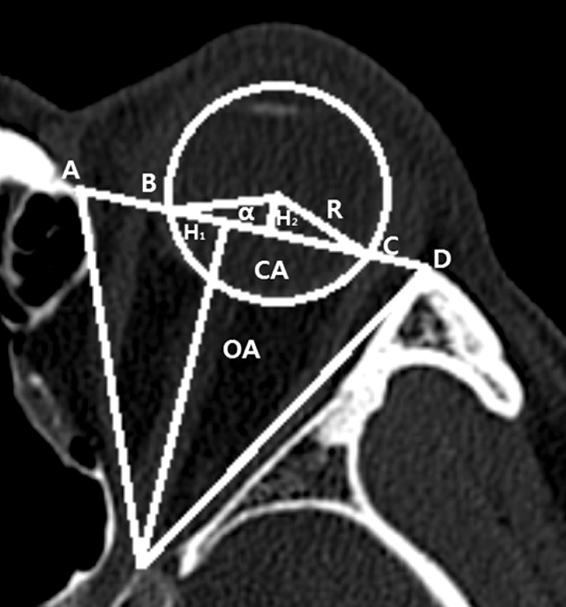
Figure 2.
Group 1: incomplete reduction (blow out fracture at med. Wall, Lt.). (A) Preoperative axial CT image. (B) Postoperative axial CT image.
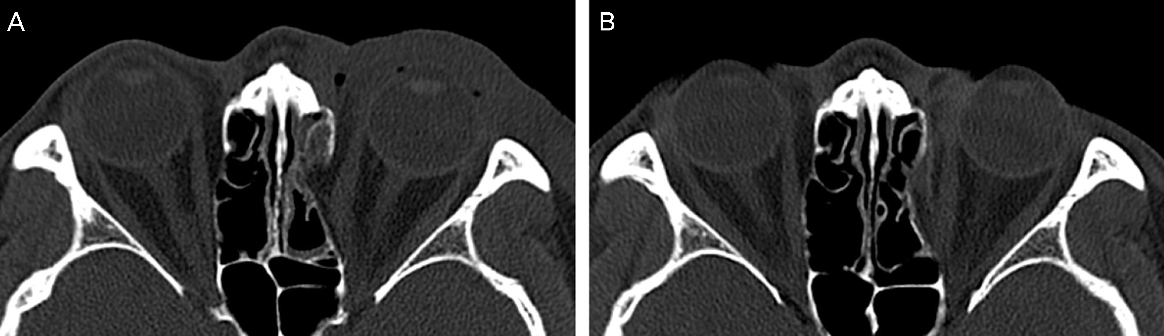
Figure 3.
Group 2: complete reduction (blow out fracture at med. Wall, Rt.). (A) Preoperative axial CT image. (B) Postoperative axial CT image: orbital soft-tissue herniation is decreased.
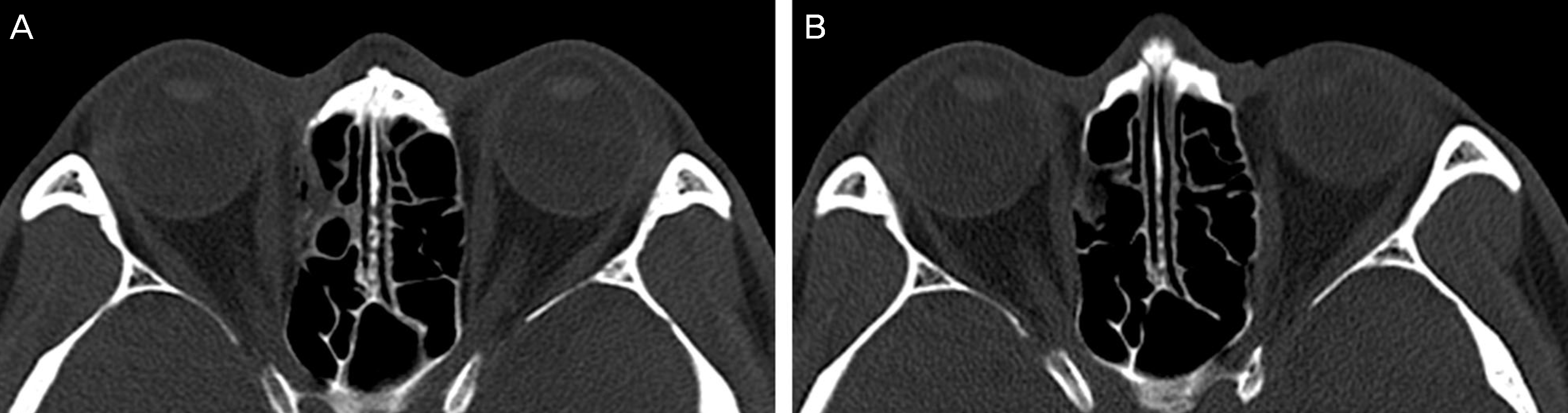
Table 1.
Patients demographics
| Age (years) | Male | Female | Total |
|---|---|---|---|
| <10 | 1 | 0 | 1 |
| 10-20 | 8 | 0 | 8 |
| 20-30 | 11 | 3 | 14 |
| 30-40 | 11 | 2 | 13 |
| 40-50 | 5 | 1 | 6 |
| 50-60 | 5 | 3 | 8 |
| 60-70 | 1 | 1 | 2 |
| >70 | 0 | 3 | 3 |
| Total | 42 | 13 | 55 |
Table 2.
Causes of fracture
| Causes | No. of patients (%) |
|---|---|
| Assault | 20 (36.4) |
| Slip down | 17 (30.9) |
| Sports | 7 (12.7) |
| Other causes | 6 (10.9) |
| Traffic accident | 5 (9) |
| Total | 55 (100) |
Table 3.
Enophthalmos improvement 3 months after surgery
Table 4.
Preoperative and posoperative values of enophthalmos measured by exophthalmometry
Table 5.
Preoperative and posoperative values of enophthalmos measured by CA-to-OA ratio
 XML Download