

 PDF
PDF ePub
ePub Citation
 Print
Print


Abstract
Methods
Thirty-two patients who visited our hospital between February 2013 and November 2013 for pinguecula were enrolled in the study. The clinical characteristics were evaluated by the location, size, shape, elevation, color, vascularization and the grade of pingueculae.
Results
Ninety-eight pingueculae were found in the 32 patients, 58 (59.18%) pingueculae on the nasal side, and 40 (40.82%) pingueculae on the temporal side. The mean grade of pingueculae of the nasal side was 1.19 ± 0.40 and on the temporal side was 1.15 ± 0.43. Compared with the temporal side, pingueculae on the nasal side were more frequent (p = 0.032). The size, color, shape and vascularization of nasal and temporal pingueculae were not significantly differentiated. Medical history, tear film break-up time, Schirmer test, history of contact lens wearing, refractive surgery, occupational activity and residence were not correlated with the grade of pingueculae. However, ocular surface disease index score was correlated with the grade of nasal pingueculae (p = 0.01).
References
1. Dong N, Li W, Lin H, et al. Abnormal epithelial differentiation and tear film alteration in pinguecula. Invest Ophthalmol Vis Sci. 2009; 50:2710–5.

2. Fotouhi A, Hashemi H, Khabazkhoob M, Mohammad K. Prevalence and risk factors of pterygium and pinguecula: the Tehran Eye Study. Eye (Lond). 2009; 23:1125–9.
3. Viso E, Gude F, driguez-Ares MT. Prevalence of pinguecula and pterygium in a general population in Spain. Eye (Lond). 2011; 25:350–7.
4. Asokan R, Venkatasubbu RS, Velumuri L, et al. Prevalence and associated factors for pterygium and pinguecula in a South Indian population. Ophthalmic Physiol Opt. 2012; 32:39–44.
5. Rezvan F, Hashemi H, Emamian MH, et al. The prevalence and determinants of pterygium and pinguecula in an urban population in Shahroud, Iran. Acta Med Iran. 2012; 50:689–96.
6. Lee SY, Choi O. A clinical study of the pinguecula. J Korean Ophthalmol Soc. 1980; 21:35–41.
7. Perkins ES. The association between pinguecula, sunlight and cataract. Ophthalmic Res. 1985; 17:325–30.
8. Mimura T, Usui T, Mori M, et al. Pinguecula and contact lenses. Eye (Lond). 2010; 24:1685–91.
9. Mimura T, Obata H, Usui T, et al. Pinguecula and diabetes mellitus. Cornea. 2012; 31:264–8.
10. Mimura T, Usui T, Obata H, et al. Severity and determinants of pinguecula in a hospital-based population. Eye Contact Lens. 2011; 37:31–5.
11. Shin KH, Kwon JW. Clinical features of pinguecula in NorthWestern Gyeonggi Province. J Korean Ophthalmol Soc. 2013; 54:691–5.
12. Oh HJ, Park YG, Yoon KC. Changes of ocular surface and tear film in patients with pinguecula and pterygium. J Korean Ophthalmol Soc. 2006; 47:717–24.
13. Oguz H, Karadede S, Bitiren M, et al. Tear functions in patients with pinguecula. Acta Ophthalmol Scand. 2001; 79:262–5.
14. Miller KL, Walt JG, Mink DR, et al. Minimal cilinically important difference for the ocular surface disease index. Arch Ophthalmol. 2010; 128:94–101.
Figure 1.
Grading system of pinguecula. No pinguecula (A) is categorized into Grade P (0), mild and moderate pinguecula (B) is categorized into Grade P (1), severe pinguecula (C) is categorized into Grade P (2). Grade P = Grade of pingueculae.
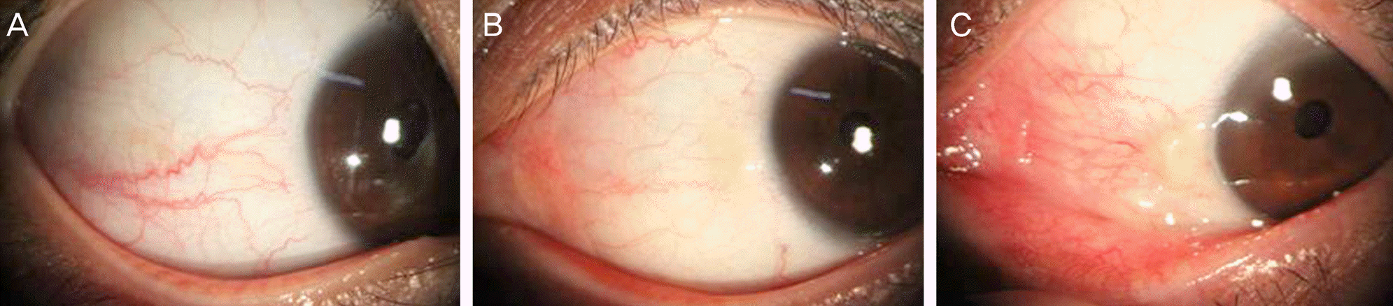
Table 1.
Clinical characteristics of patients with pinguecula of 20 to 39 years of age
| Patients (n = 32) | Correlation with grade of nasal pinguecula (p-value) | Correlation with grade of temporal pinguecula (p-value) | |
|---|---|---|---|
| Age (years) | 32.81 ± 4.84 | ||
| Sex (Male/Female) | 6/26 | ||
| Systemic disease | |||
| Thyroid disease* (%) | 1 (3.1) | 0.94 | 0.38 |
| Occupational activity† | 0.79 | 0.30 | |
| Indoor (%) | 27 (84.4) | ||
| Outdoor (%) | 5 (15.6) | ||
| Residence (years)† | 0.83 | 0.42 | |
| Urban‡ | 22.75 ± 12.7 | ||
| Rural§ | 10.06 ± 12 | ||
| History | |||
| Contact lens wear* (%) | 18 (56.2) | 0.13 | 1.00 |
| Refractive surgery* (%) | 9 (28.1) | 0.13 | 0.19 |
| Tear film and meibomian gland findings | |||
| Tear film break-up time† (sec) | 4.75 ± 2.41 | 0.82 | 0.22 |
| Schirmer test† (mm) | 9.12 ± 6.62 | 0.70 | 0.27 |
| Ocular surface disease index score † | 26.00 ± 16.87 | 0.01 | 0.38 |
| Grade of meibomian gland dysfunction† | 0.56 ± 0.61 | 0.75 | 0.63 |
Table 2.
Profile of pinguecula by location
| Nasal | Temporal | p-value | |
|---|---|---|---|
| Number by location (%) | 58 (59.18) | 40 (40.82) | |
| Grade | 1.19 ± 0.4 | 1.18 ± 0.39 | 0.86* |
| P (1) | 47 (81.03) | 33 (82.5) | |
| P (2) | 11 (18.97) | 7 (17.5) | |
| Size | |||
| Horizontal diameter (mm) | 2.43 ± 0.93 | 2.83 ± 1.36 | 0.09* |
| Vertical diameter (mm) | 2.13 ± 0.65 | 2.21 ± 1.6 | 0.72* |
| Average diameter (mm) | 2.28 ± 0.69 | 2.52 ± 1.1 | 0.18* |
| Color | 0.69 † | ||
| White (%) | 17 (29.31) | 14 (35) | |
| Yellowish white (%) | 27 (46.55) | 19 (47.5) | |
| Yellow (%) | 14 (24.14) | 7 (17.5) | |
| Brown (%) | 0 | 0 | |
| Elevation | 0.03 † | ||
| Flat (%) | 12 (20.69) | 18 (45) | |
| Slight elevation (%) | 26 (44.83) | 14 (35) | |
| Noticeable elevation (%) | 20 (34.48) | 8 (20) | |
| Vascularization | 0.13 † | ||
| Mild (%) | 28 (48.27) | 26 (65) | |
| Moderate (%) | 21 (36.21) | 7 (17.5) | |
| Severe (%) | 9 (15.52) | 7 (17.5) | |
| Shape | 0.11 † | ||
| Triangular (%) | 13 (22.41) | 14 (35) | |
| Circular (%) | 15 (25.86) | 4(10) | |
| Oval (%) | 30 (51.73) | 22 (55) | |
 XML Download