

 PDF
PDF ePub
ePub Citation
 Print
Print


Abstract
Case summary
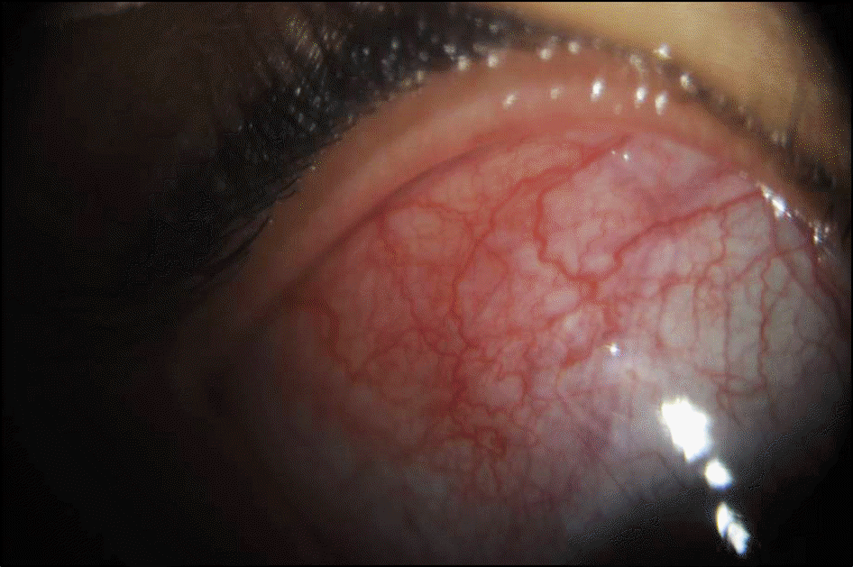
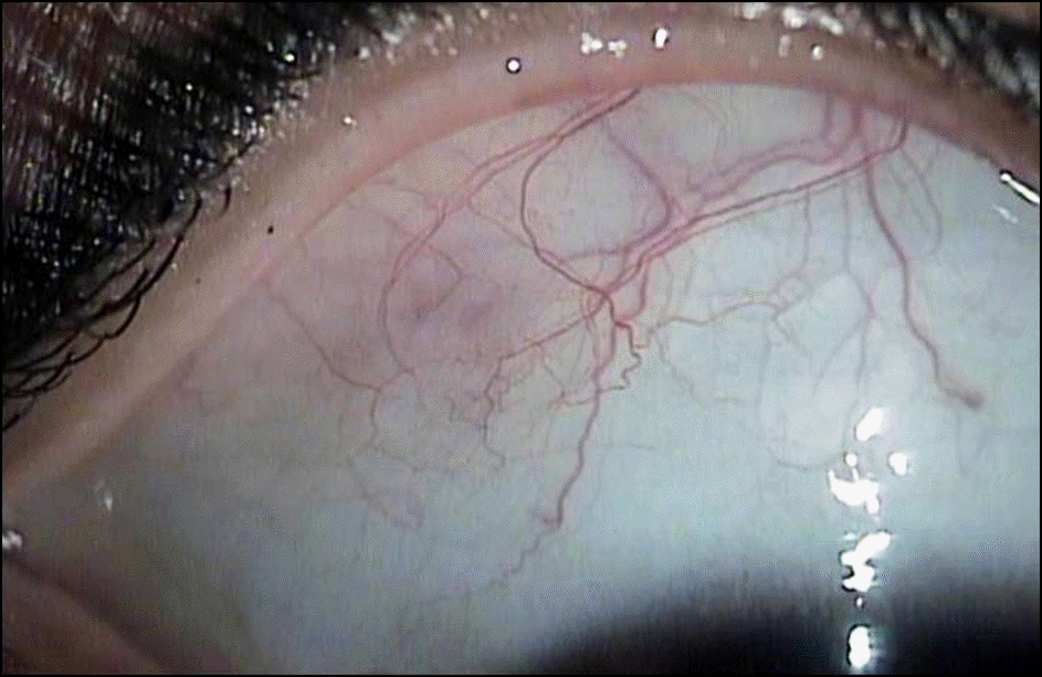
A 40-year-old female was referred to the outpatient clinic because of right episcleritis that was unchanged during the month of treatment. Her headache persisted, and slit lamp examination showed tortuous congestion of engorged episcleral vessels with swelling in the superior-temporal region of the right eye, but fundus and radiological studies showed normal findings. Serological tests were reactive for venereal disease research laboratory test, treponema pallidum hemagglutination assay test, and fluorescent treponemal antibody absorption test. Under the suspicion of persistent syphilis infection, cerebrospinal fluid examination was performed, and the diagnosis of neurosyphilis with episcleritis was diagnosed. Treatment consisted of intravenous injections of 5 million IU penicillin G potassium every 4 hours for 14 days. The ocular inflammation resolved within the first week of treatment and did not recur.
References
1. Tamesis RR, Foster CS. Ocular syphilis. Ophthalmology. 1990; 97:1281–7.

2. Margo CE, Hamed LM. Ocular syphilis. Surv Ophthalmol. 1992; 37:203–20.
3. Gaudio PA. Update on ocular syphilis. Curr Opin Ophthalmol. 2006; 17:562–6.
4. Park HJ. Clinical observation and statistical consideration of syph- ilis (2000-2007). Korean J Dermatol. 2008; 46:1344–52.
5. Kiss S, Damico FM, Young LH. Ocular manifestations and treat- ment of syphilis. Semin Ophthalmol. 2005; 20:161–7.
6. Marks R, Thomas-Kaskel AK, Schmidt D, Donauer J. Steroid re- fractory episcleritis as early manifestation of neurosyphilis. Eur J Med Res. 2006; 11:309–12.
7. Yoon KC, Im SK, Seo MS, Park YG. Neurosyphilitic episcleritis. Acta Ophthalmol Scand. 2005; 83:265–6.
8. Watson PG, Hayreh SS. Scleritis and episcleritis. Br J Ophthalmol. 1976; 60:163–91.
9. Casey R, Flowers CW Jr, Jones DD, Scott L. Anterior nodular scleritis secondary to syphilis. Arch Ophthalmol. 1996; 114:1015–6.
10. Deschenes J, Seamone CD, Baines MG. Acquired ocular syphilis: diagnosis and treatment. Ann Ophthalmol. 1992; 24:134–8.
11. Schlaegel TF Jr, Kao SF. A review (1970-1980) of 28 presumptive cases of syphilitic uveitis. Am J Ophthalmol. 1982; 93:412–4.
12. Aldave AJ, King JA, Cunningham ET Jr. Ocular syphilis. Curr Opin Ophthalmol. 2001; 12:433–41.
13. Parc CE, Chahed S, Patel SV, Salmon-Ceron D. Manifestations and treatment of ocular syphilis during an epidemic in France. Sex Transm Dis. 2007; 34:553–6.
 XML Download