

 PDF
PDF ePub
ePub Citation
 Print
Print


Abstract
Purpose
To report a case of a rapidly progressive endogenous endophthalmitis with subretinal abscess that involved the macula and was treated with early vitrectomy.
Case summary
A 42-year-old man with liver cirrhosis, hepatic cellular carcinoma and diabetes, who underwent regular fundus checkup for diabetic retinopathy presented with reduced vision, ocular pain in the left eye and headache. Indirect ophthalmoscopy showed subretinal abscess approximately five times the optic disc size and surrounding retinal hemorrhage in the nasal upper quadrant. A provisional diagnosis of bacterial endophthalmitis was made based on systemic disease and funduscopic findings. Treatment with topical and systemic empirical antibiotics was initiated along with intravitreal vancomycin and ceftazidime injection. Despite the treatment, after 24 hours the abscess size increased to approximately 10 times the optic disc size and began to involve the macula. The patient underwent diagnostic and therapeutic pars plana vitrectomy as well as vitreous and abscess content cultures. MRSA was found in a blood culture test. Five days postoperatively, the patient's vision and symptoms improved significantly and the residual lesion was cleared, with retinal scars.
Conclusions
In a patient with endogenous endophthalmitis with subretinal abscess, presence of macular invasion and rate of progression is important in determining the time and method of operation. In this case, early vitrectomy was a good choice to preserve macular structure and the patient's visual acuity.
References
1. Jackson TL, Eykyn SJ, Graham EM, Stanford MR. Endogenous bacterial endophthalmitis: a 17-year prospective series and review of267 reported cases. Surv Ophthalmol. 2003; 48:403–23.
2. Harris EW, D'Amico DJ, Bhisitkul R, et al. Bacterial subretinal abscess: a case report and review of the literature. Am J Ophthalmol. 2000; 129:778–85.

3. Webber SK, Andrews RA, Gillie RF, et al. Subretinal Pseudomonas abscess after lung transplantation. Br J Ophthalmol. 1995; 79:861–6.
4. Yarng SS, Hsieh CL, Chen TL. Vitrectomy for endogenous Klebsiella pneumoniae endophthalmitis with massive subretinal abscess. Ophthalmic Surg Lasers. 1997; 28:147–50.
5. Melo GB, Bispo PJ, Yu MC, et al. Microbial profile and antibiotic susceptibility of culture-positive bacterial endophthalmitis. Eye (Lond). 2011; 25:382–7.
6. Trigui A, Laabidi H, Khairallah M, Féki J. Retinal abscess: case re- port of an uncommon evolution. Int Ophthalmol. 2011; 31:327–31.
7. Chee SP, Ang CL. Endogenous Klebsiella endophthalmitis–a case series. Ann Acad Med Singapore. 1995; 24:473–8.
8. Sipperley JO, Shore JW. Septic retinal cyst in endogenous Klebsiella endophthalmitis. Am J Ophthalmol. 1982; 94:124–5.
9. Limaye SR, Goldberg MH. Septic submacular choroidal embolus associated with intravenous drug abuse. Ann Ophthalmol. 1982; 14:518–22.
10. Gregor RJ, Chong CA, Augsburger JJ, et al. Endogenous Nocardia asteroides subretinal abscess diagnosed by transvitreal fine-needle aspiration biopsy. Retina. 1989; 9:118–21.
11. Rimpel NR, Cunningham ET Jr, Howes EL Jr, Kim RY. Viridans group Streptococcus subretinal abscess. Br J Ophthalmol. 1999; 83:373–4.
12. Ku M, Jung JO, Lee DY, Nam DH. Endogenous Candida endoph- thalmitis with bilateral massive submacular abscess. J Korean Ophthalmol Soc. 2008; 49:1701–5.
13. Edwards JE Jr, Foos RY, Montgomerie JZ, Guze LB. Ocular mani- festations of Candida septicemia: review of seventy-six cases of hematogenous Candida endophthalmitis. Medicine (Baltimore). 1974; 53:47–75.
14. Results of the Endophthalmitis Vitrectomy Study. A randomized trial of immediate vitrectomy and of intravenous antibiotics for the treatment of postoperative bacterial endophthalmitis. Endophthalmitis Vitrectomy Study Group. Arch Ophthalmol. 1995; 113:1479–96.
15. Suh DS, Roh JH, Kim SD. Surgical management of infectious en- dophthalmitis : Early vitrectomy vs late vitrectomy. J Korean Ophthalmol Soc. 1998; 39:2418–25.
16. Doft BH, Kelsey SF, Wisniewski S, et al. Treatment of endoph- thalmitis after cataract extraction. Retina. 1994; 14:297–304.
17. Nelsen PT, Marcus DA, Bovino JA. Retinal detachment following endophthalmitis. Ophthalmology. 1985; 92:1112–7.
18. Verbraeken H. Treatment of postoperative endophthalmitis. Op- hthalmologica. 1995; 209:165–71.
Figure 1.
Serial fundus photographs 3 months before (A), at the first visit after symtom begin (B), 24 hours after intravitreal antibiotics injection (C), 5 days after vitrctomy (D). Some generalized microaneurysmmm and cotton wool spots are visible. (A) A round, elevated yellowish mound lesion about 5 times of optic disc-sized lesion with retinal hemorrhages (B) was worsened, increased size about 10 times of optic disc-sized, involving macula (C). Fundus photograph after vitrectomy shows flattened lesion covered with translucent yellowish membrane, surrounded laser photocoagulation.
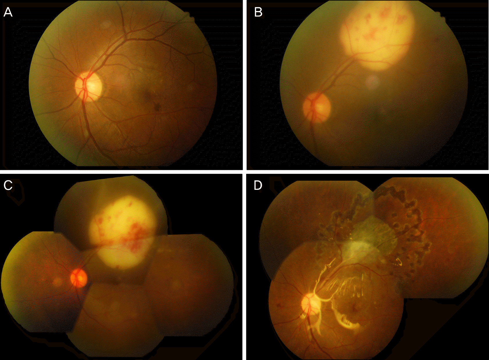
Figure 2.
Fluorescein angiography and indocyanin green angiography show masking effect (subretinal abscess) and early hyperfluorescence which is increased until late phase, active leakage around lesion and optic disc.

Figure 3.
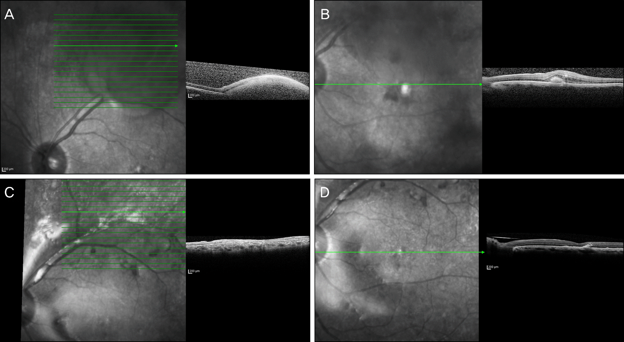
Optical coherence tomography (OCT) showed the localization of the mass, protruding into vitreal cavity underneath retinal pigment epithelium (A). Despite intravitreal antibiotics injection, abscess increased to about 10 times the size of the optic disc, began to involve the macula (B). After surgery, flattened retina without retinal detachment or subretinal fluids and restored continuity with adjacent normal tissues at abscess lesion (C), and macular (D).
 XML Download