Abstract
Purpose
To report a case of bilateral congenital lacrimal fistula that presented with repeated infection and inflammation after complete fistulectomy, which required an incision and drainage of pus from the operation site.
Case summary
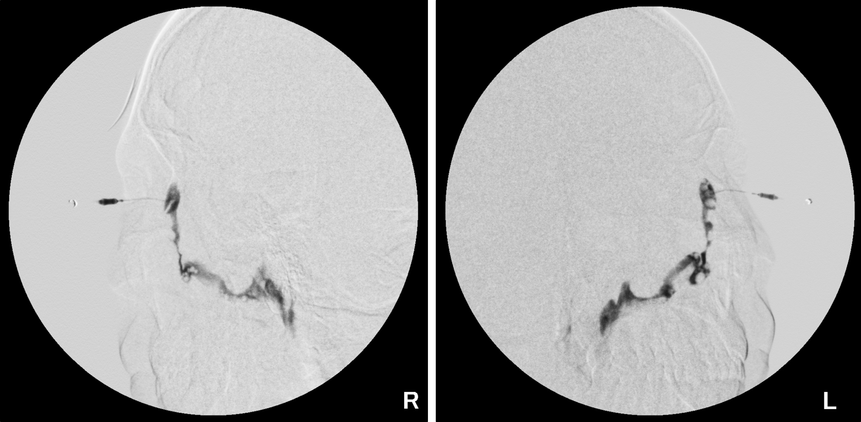
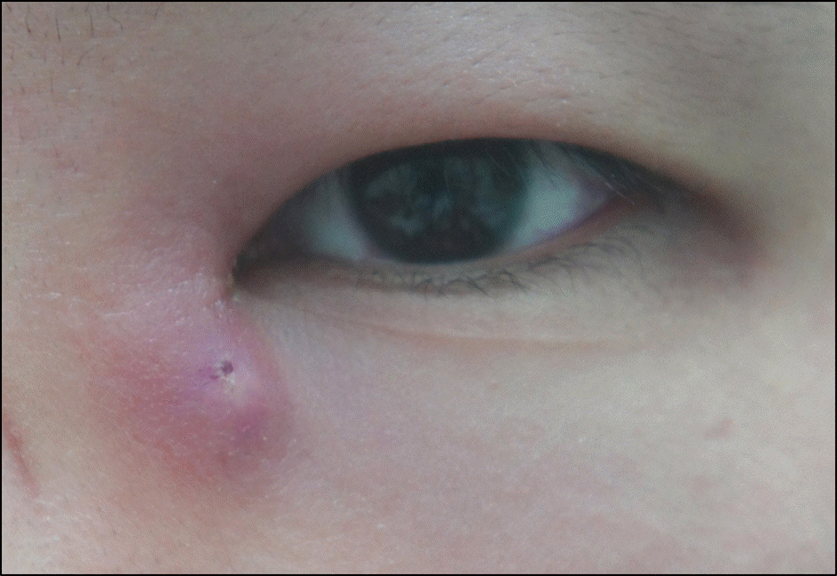
A 22-year-old male without any medical history presented with repeated erythematous swelling and inflammation, resulting in tenderness around the opening of congenital lacrimal fistula. The lacrimal fistula opening was located approximately 12 mm inferiorly apart from the medial canthus. The complete excision of lacrimal fistula was performed without any inter-operative events. However, 4 days postoperatively, the patient complained of discomfort and swelling, with purulent discharge from the bilateral operation site. There was no improvement although treatment with antibiotics, incision and drainage was performed. After 1 month, an additional incision and drainage was necessary due to inflammation in the left operation site. One month later, pus and purulent discharge were occurring from the right operation site, requiring an additional incision and drainage. At that time, Actinomyces israelii was identified on wound culture examination. One month later, an additional incision and drainage was performed due to repeated inflammation in the left operation site. In the present case, we hypothesized the opening site of congenital lacrimal fistula was relatively far apart from the medial canthus and played a role in atypical repeated inflammation and infection on the operation site.
Conclusions
In surgical treatment of congenital lacrimal fistula, careful preoperative observation of the location of the lacrimal fistula's opening site would be helpful in prediction of postoperative complication, such as wound infection and inflammation, as well as in educating and informing the patient.
References
1. Kim HY, Kim JS. Two cases of congenital fistula of the lacrimal sac. J Korean Ophthalmol Soc. 1983; 24:925–7.
2. Song BY, Kang HR, Kim S. The clinical evaluation of congenital lacrimal fistula. J Korean Ophthalmol Soc. 2004; 45:1603–8.
3. Zhuang L, Sylvester CL, Simons JP. Bilateral congenital lacrimal fistulae: a case report and review of the literature. Laryngoscope. 2010; 120(Suppl 4):S230.

4. Song BY, Ji YS, Wu MH, et al. The clinical manifestations and treatment results of congenital lacrimal fistula. J Korean Ophthalmol Soc. 2006; 47:871–6.
5. Jeong SK. Surgery for the congenital lacrimal fistula. J Korean Ophthalmol Soc. 2002; 43:801–4.
8. Vujancevic S, Meyer-Rüsenberg HW. [Therapy for actinomycosis in the lacrimal pathway]. Klin Monbl Augenheilkd. 2010; 227:568–74.
9. Yuksel D, Hazirolan D, Sungur G, Duman S. Actinomyces canal- iculitis and its surgical treatment. Int Ophthalmol. 2012; 32:183–6.
10. Briscoe D, Edelstein E, Zacharopoulos I, et al. Actinomyces canal- iculitis: diagnosis of a masquerading disease. Graefes Arch Clin Exp Ophthalmol. 2004; 242:682–6.
11. Ari S, Cingu K, Çahin A, Özkök A. Çaça I. The outcomes of surgi- cal treatment in fistulous dacryocystitis. Eur Rev Med Pharmacol Sci. 2013; 17:243–6.


 PDF
PDF ePub
ePub Citation
 Print
Print


 XML Download