

 PDF
PDF ePub
ePub Citation
 Print
Print


Abstract
Purpose
To analyze the structural changes in the β-zone of peripapillary atrophy (PPA-β) using cross-sectional image of the optic disc head from spectral-domain optical coherence tomography (SD-OCT) according to the optic disc shape.
Methods
One hundred thirty-seven eyes in 137 patients with glaucoma having PPA-β and 31 normal eyes (control group) were evaluated retrospectively. Cross-sectional images of the optic disc were taken using the Cirrus HD-OCT. We classified optic disc patterns into normal, focal, myopic, generalized enlargement and senile sclerotic appearance types and analyzed the shape of Bruch's membrane (BM), composition of retinal layer and retinal slope according to the optic disc shape.
Results
Among the 137 eyes with glaucoma, 54 eyes were focal disc type, 34 eyes were myopic disc type, 28 eyes were generalized enlargement disc type and 21 eyes were senile sclerotic disc type. The myopic disc group showed a noticeable difference compared to the other groups in terms of a higher percentage of BM defect type, the lowest retinal slope (70.6 ± 12.0°) and the earlier termination of retinal layers. The generalized enlargement disc group showed the highest percentage of curved BM type. Retinal slope angle increased with age and decreased with axial length.
References
1. Sommer A, Katz J, Quigley HA, et al. Clinically detectable nerve fiber atrophy precedes the onset of glaucomatous field loss. Arch Ophthalmol. 1991; 109:77–83.

2. Park KH, Tomita G, Liou SY, Kitazawa Y. Correlation between peripapillary atrophy and optic nerve damage in normal-tension glaucoma. Ophthalmology. 1996; 103:1899–906.
3. Jonas JB, Naumann GO. Parapapillary chorioretinal atrophy in normal and glaucoma eyes. II. Correlations. Invest Ophthalmol Vis Sci. 1989; 30:919–26.
4. Uchida H, Yamamoto T, Tomita G, Kitazawa Y. Peripapillary atro- phy in primary angle-closure glaucoma: a comparative study with primary open-angle glaucoma. Am J Ophthalmol. 1999; 127:121–8.
5. Tezel G, Kass MA, Kolker AE, Wax MB. Comparative optic disc analysis in normal pressure glaucoma, primary open-angle glauco- ma, and ocular hypertension. Ophthalmology. 1996; 103:2105–13.
6. Kono Y, Zangwill L, Sample PA, et al. Relationship between parapapillary atrophy and visual field abnormality in primary open-angle glaucoma. Am J Ophthalmol. 1999; 127:674–80.
7. Lee KY, Tomidokoro A, Sakata R, et al. Cross-sectional anatomic configurations of peripapillary atrophy evaluated with spectral do- main-optical coherence tomography. Invest Ophthalmol Vis Sci. 2010; 51:666–71.
8. Nassif N, Cense B, Park B, et al. In vivo high-resolution video-rate spectral-domain optical coherence tomography of the human retina and optic nerve. Opt Express. 2004; 12:367–76.
9. Manjunath V, Shah H, Fujimoto JG, Duker JS. Analysis of peri- papillary atrophy using spectral domain optical coherence tomography. Ophthalmology. 2011; 118:531–6.
10. Na JH, Moon BG, Sung KR, et al. Characterization of peripapillary atrophy using spectral domain optical coherence tomography. Korean J Ophthalmol. 2010; 24:353–9.
11. Geijssen HC, Greve EL. The spectrum of primary open angle glaucoma. I: Senile sclerotic glaucoma versus high tension glaucoma. Ophthalmic Surg. 1987; 18:207–13.
12. Geijssen HC, Greve EL. Focal ischaemic normal pressure glauco- ma versus high pressure glaucoma. Doc Ophthalmol. 1990; 75:291–301.
13. Drance SM, et al. What can we learn from the disc appearance about the risk factors in glaucoma? Can J Ophthalmol. 2008; 43:322–7.
14. Broadway DC, Nicolela MT, Drance SM. Optic disk appearances in primary open-angle glaucoma. Surv Ophthalmol. 1999; 43(Suppl 1):S223–43.
15. Nicolela MT, Drance SM. Various glaucomatous optic nerve ap- pearances: clinical correlations. Ophthalmology. 1996; 103:640–9.
16. Hayashi K, Tomidokoro A, Lee KY, et al. Spectral-domain optical coherence tomography of beta-zone peripapillary atrophy: influ- ence of myopia and glaucoma. Invest Ophthalmol Vis Sci. 2012; 53:1499–505.
17. Curcio CA, Saunders PL, Younger PW, Malek G. Peripapillary chorioretinal atrophy: Bruch's membrane changes and photo- receptor loss. Ophthalmology. 2000; 107:334–43.
18. Jonas JB, Nguyen XN, Gusek GC, Naumann GO. Parapapillary chorioretinal atrophy in normal and glaucoma eyes. I. Morphometric data. Invest Ophthalmol Vis Sci. 1989; 30:908–18.
19. Jonas JB, Fernández MC, Naumann GO. Glaucomatous para- papillary atrophy. Occurrence and correlations. Arch Ophthalmol. 1992; 110:214–22.
20. Jonas JB, Gründler AE. Correlation between mean visual field loss and morphometric optic disk variables in the open-angle glaucomas. Am J Ophthalmol. 1997; 124:488–97.
21. Park SC, De Moraes CG, Tello C, et al. In-vivo microstructural anatomy of beta-zone parapapillary atrophy in glaucoma. Invest Ophthalmol Vis Sci. 2010; 51:6408–13.
Figure 1.
Optic disc shape (A) Focal disc pattern, (B) Myopic disc pattern, (C) General enlargement disc pattern, (D) Senile sclerotic pattern.
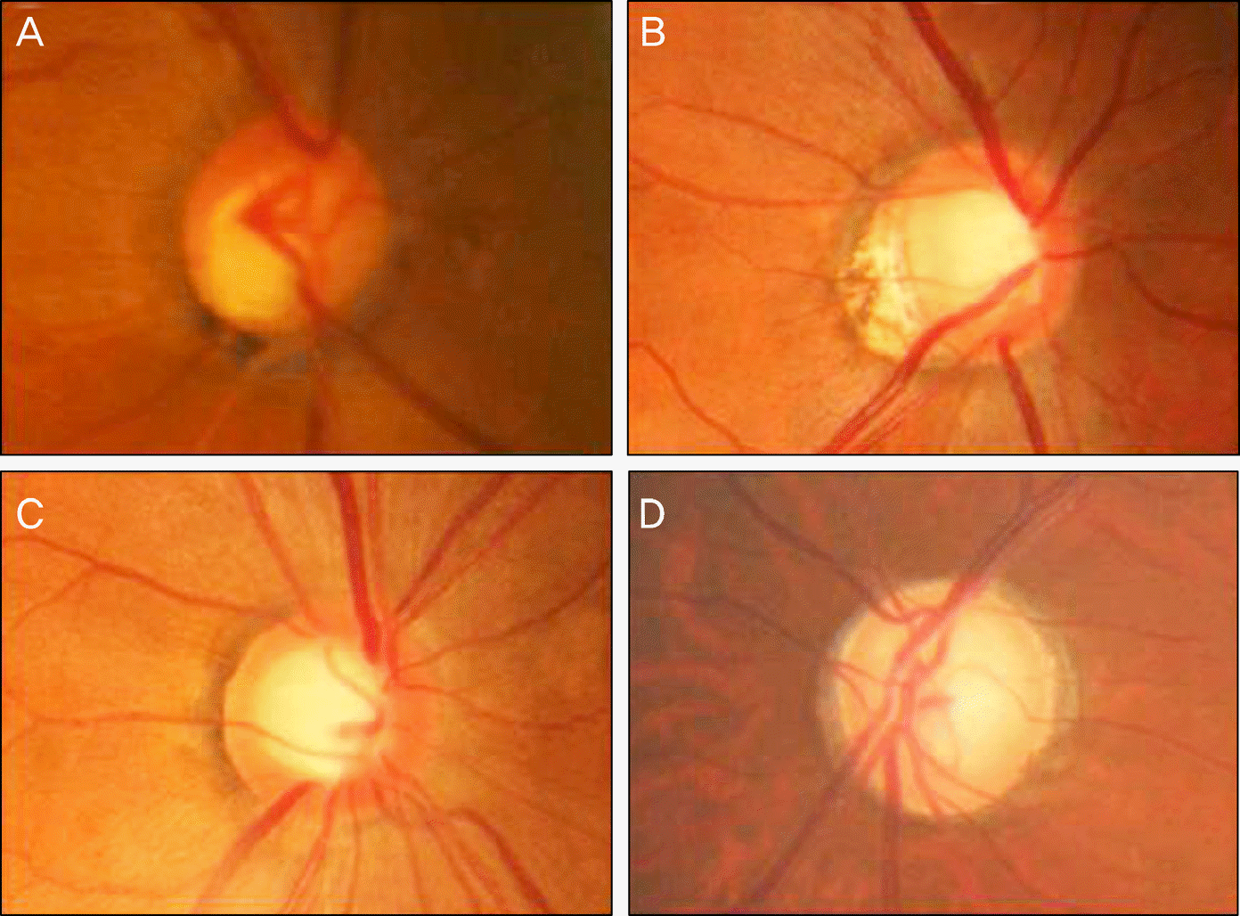
Figure 2.
B-scan images of peripapillary atrophy. Two vertical lines indicate the distal edge of PPA-ß (*) and optic disc edge (). Red line passes through the center of the optic disc. (A) straight Bruch's membrane, (B) curved Bruch's membrane, (C) Bruch's membrane defect. ELM = external limiting membrane; IPL = inner plexiform layer; IS/OS = inner and outer segment of photoreceptor; RNFL = retinal nerve fiber layer; RPE/BM = retinal pigment epithelium/Bruch's membrane complex.
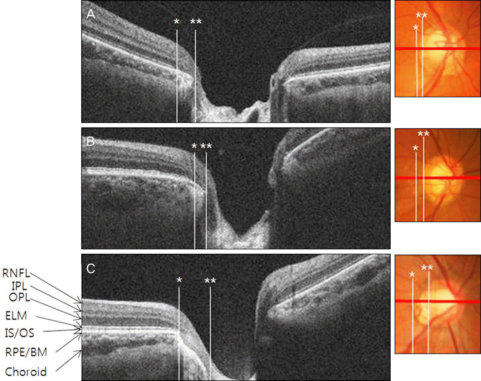
Figure 3.
Measurement of retinal slope around optic nerve head. The retinal slope () is defined as the angle between the optic nerve junction and the retinal pigment epithelium (RPE). GCL = ganglion cell layer; ELM = external limiting membrane; IPL = inner plexiform layer; IS/OS = inner and outer segment of photoreceptor; RNFL = retinal nerve fiber layer; RPE/BM = retinal pigment epithelium/Bruch's membrane complex.
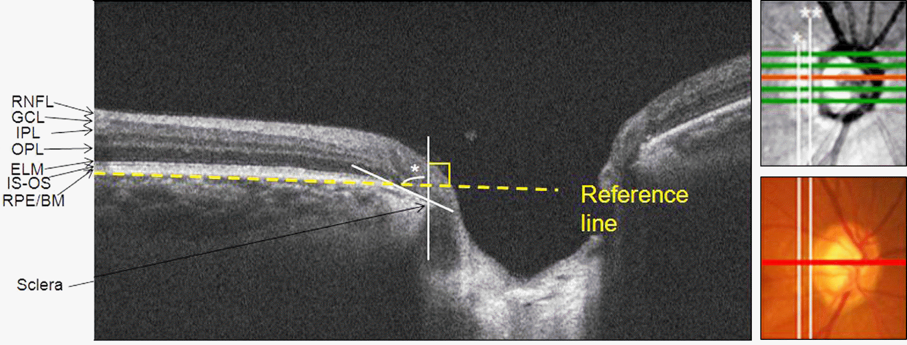
Figure 4.
Scatterplot shows the relationship between retinal slope and other influencing factors. With increasing age, retinal slope (A) increased. In contrast, as axial length increased, retinal slope (C) decreased. There was no correlation between the retinal slope and refractive error (B), MD (D), PSD (E). AXL = axial length; MD = mean deviation; PSD = pattern standard deviation; SE = spherical equivalent. Pearson correleation analysis was used.
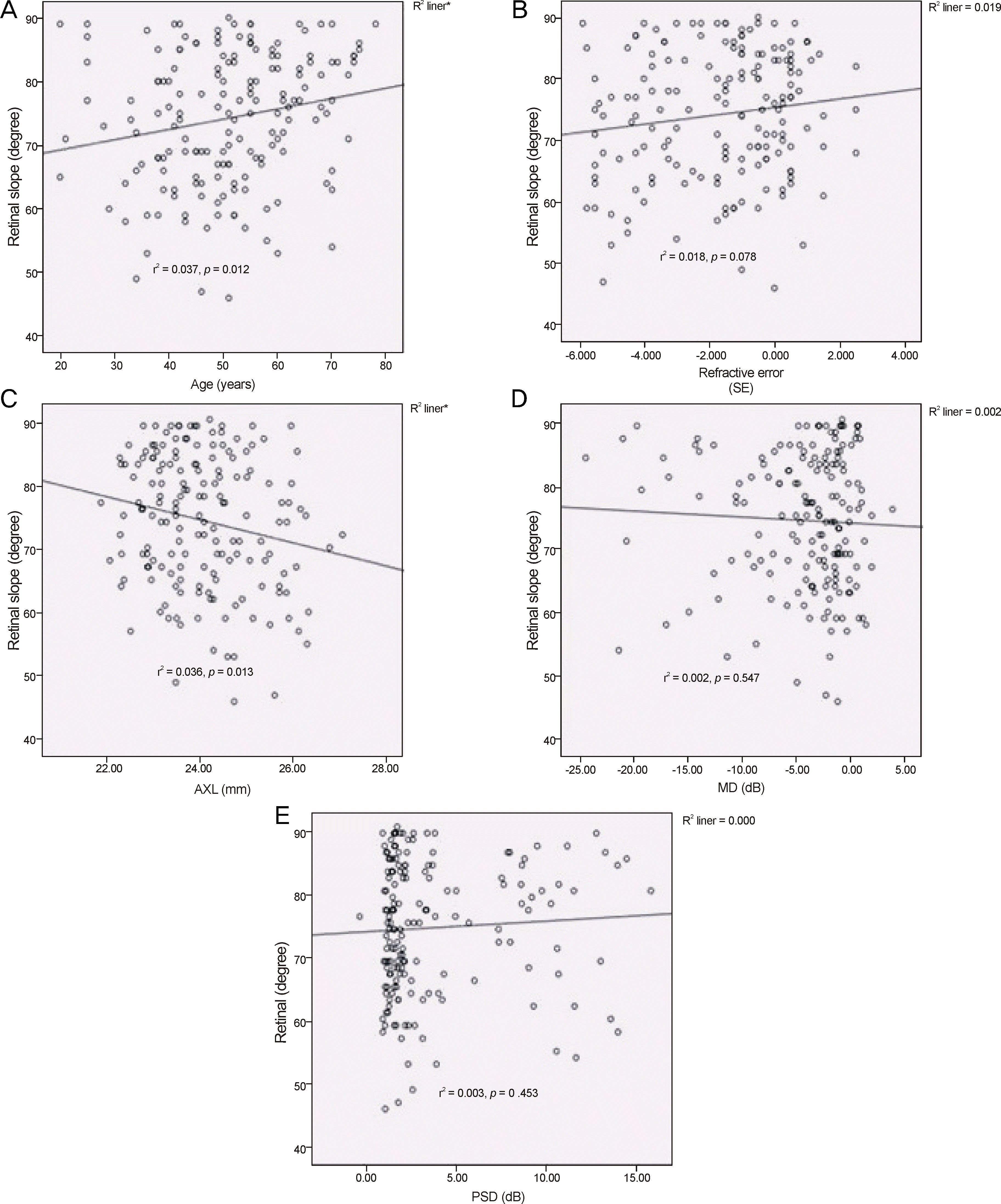
Table 1.
Demographic data of the patients with different disc types
| Normal (n = 31) | Focal (n = 54) | Myopic (n = 34) | G Enlarge (n = 28) | S Sclerotic (n = 21) | p-value | |
|---|---|---|---|---|---|---|
| Age (years) Sex (n) | 49.1 ± 10.9 | 50.2 ± 12.1 | 48.9 ± 15.0 | 53.2 ± 12.0 | 55.7 ± 11.2 | 0.223* |
| Male | 13 | 21 | 23 | 13 | 15 | 0.070† |
| Female Eyes (n) | 18 | 33 | 11 | 15 | 6 | |
| OD | 17 | 29 | 18 | 16 | 7 | 0.493† |
| OS | 14 | 25 | 16 | 12 | 14 | |
| SE (diopter) | −0.40 ± 1.43 | −1.60 ± 1.97 | −3.57 ± 1.88‡ | −0.77 ± 1.74 | −1.77 ± 1.81 | <0.001* |
| AXL (mm) | 23.70 ± 0.90 | 23.85 ± 0.98 | 24.97 ± 1.11‡ | 23.88 ± 0.84 | 23.97 ± 1.18 | <0.001* |
| MD (dB) | −2.48 ± 4.83 | −3.53 ± 3.90 | −4.55 ± 5.03 | −4.88 ± 4.21 | −9.44 ± 6.73‡ | <0.001* |
| PSD (dB) | 2.08 ± 1.76 | 3.40 ± 3.70 | 4.15 ± 3.38 | 3.41 ± 3.07 | 7.30 ± 4.12‡ | <0.001* |
Table 2.
Factors associated with each type of peripapillary atrophy classified according to PPA bed shape
| PPA bed | Age (years) | Sex | SE (diopter) | AXL (mm) | MD (dB) | PSD (dB) | |
|---|---|---|---|---|---|---|---|
| M | F | ||||||
| Straight BM (n = 49) | 53.7 ± 12.7 | 19 | 30 | −1.30 ± 1.85 | 23.79 ± 0.95 | −5.34 ± 6.15 | 4.12 ± 4.01 |
| Curved BM (n = 77) | 52.2 ± 10.5 | 40 | 37 | −1.07 ± 1.92 | 23.89 ± 1.06 | −4.04 ± 4.39 | 3.29 ± 3.08 |
| BM defect (n = 42) | 45.29 ± 14.0* | 26 | 16 | −3.15 ± 1.96* | 24.72 ± 1.09‡ | −4.37 ± 5.14 | 4.37 ± 3.90 |
| p-value | 0.002* | 0.084† | <0.001* | <0.001* | 0.378* | 0.226* | |
Table 3.
Prevalence of each type of peripapillary atrophy bed configuration according to optic disc pattern
| Normal (n = 31) | Focal (n = 54) | Myopic (n = 34) | G Enlarge (n = 28) | S Sclerotic (n = 21) | Total* (No.(%)) | |
|---|---|---|---|---|---|---|
| Straight BM | 8 (25.8) | 22 (40.7) | 6 (17.6) | 5 (17.9) | 8 (38.1) | 49 (29.2) |
| Curved BM | 15 (48.4) | 27 (50.0) | 2 (5.9) | 22 (78.6) | 11 (52.4) | 77 (45.8) |
| BM defect | 8(25.8) | 5 (9.3) | 26 (76.5) | 1 (3.6) | 2 (9.5) | 42 (25.0) |
| Total (n) | 31 (100) | 54 (100) | 34 (100) | 28 (100) | 21(100) | 168 (100) |
Table 4.
Retinal slope according to optic disc pattern
| Normal (n = 31) | Focal (n = 54) | Myopic (n = 34) | G Enlarge (n = 28) | S Sclerotic (n = 21) | p-value* | |
|---|---|---|---|---|---|---|
| Retinal slope (degree) | 72.4 ± 11.2 | 78.0 ± 8.3† | 70.6 ± 12.0† | 71.4 ± 10.2 | 77.0 ± 8.6 | 0.002 |
Table 5.
Prevalence of continuity of each retinal layer before the optic disc edge within peripapillary atrophy-β according to optic disc shape
| Normal (n = 31) | Focal (n = 54) | Myopic (n = 34) | G Enlarge (n = 28) | S Sclerotic (n = 21) | p-value* | |
|---|---|---|---|---|---|---|
| GCL | 9 (29%) | 10 (18.5%) | 0 (0%) | 7 (25%) | 3 (14.3%) | 0.022* |
| IPL | 15 (48.4%) | 30 (55.6%) | 2 (5.9%) | 11 (39.3%) | 14 (66.7%) | <0.001* |
| OPL | 12 (38.7%) | 29 (53.7%) | 2 (5.9%) | 13 (46.4%) | 8 (38.1%) | <0.001* |
| ELM | 3 (9.7%) | 8 (14.8%) | 0 (0%) | 3 (0%) | 4 (19%) | 0.159* |
| IS-OS | 1 (3.2%) | 3 (5.6%) | 0 (0%) | 2 (7.1%) | 0 (0%) | 0.448* |
| RPE/Bruch | 23 (74.2%) | 47 (87.0%) | 8 (23.5%) | 24 (85.7%) | 18 (85.7%) | <0.001* |
Values are presented as the numbers of subjects (percentage of total group).
GCL = ganglion cell layer; IPL = inner plexiform layer; OPL = outer plexiform layer; ELM = external limiting membrane; IS-OS = inner and outer segment of photoreceptor; RPE/Bruch = retinal pigment epithelium/Bruch's membrane complex.
Table 6.
Prevalence of continuity of each retinal layer before optic disc edge within peripapillary atrophy-β according to PPA bed shape
| Straight BM (n = 49) | Curved BM (n = 77) | BM defect (n = 42) | p-value | |
|---|---|---|---|---|
| GCL | 16 (32.7%) | 13 (16.9%) | 0 (0%) | <0.001* |
| IPL | 35 (71.4%) | 34 (44.2%) | 3 (7.1%) | <0.001* |
| OPL | 32 (65.3%) | 32 (41.6%) | 0 (0%) | <0.001* |
| ELM | 12 (24.5%) | 6 (7.8%) | 0 (0%) | <0.001* |
| IS-OS | 4 (8.2%) | 2 (5.6%) | 0 (0%) | 0.092 |
| RPE/Bruch | 49 (100%) | 71 (92.2%) | 0 (0%) | <0.001* |
Values are presented as the numbers of subjects (percentage of total group).
BM = Bruch's membrance; GCL = ganglion cell layer; IPL = inner plexiform layer; OPL = outer plexiform layer; ELM = external limiting membrane; IS-OS = inner and outer segment of photoreceptor; RPE/Bruch = retinal pigment epithelium/Bruch's membrane complex.
 XML Download