Abstract
Purpose
To report the distribution of ocular higher-order aberrations in candidates for refractive surgery measured by Zywave® II aberrometer.
Methods
The present study included 232 eyes of 116 subjects. Ocular aberration data were obtained by measurements per eye using Zywave® II aberrometer. The mean Zywave spherical equivalent (SE) and higher order aberrations (HOAs) calculated in the central 6-mm zone and expressed as root mean square (RMS) values were analyzed. Correlation analysis was performed to assess the association between ocular HOAs and gender, age, SE refractive error, or central corneal thickness (CCT) and investigate the aberration symmetry between right and left eyes.
Results
The average SE was −4.67 ± 1.83 diopters (D). The mean RMS values of total HOA, 3rd, 4th or 5th summated HOAs, coma, trefoil and spherical aberration (SA) were 0.421 ± 0.201 gm, 0.346 ± 0.206 gm, 0.202 ± 0.105 gm, 0.087 ± 0.048 gm, 0.241 ± 0.172 gm, 0.225 ± 0.154 gm and 0.136 ± 0.102 gm, respectively. There was no significant differences of the mean total HOA, summated HOAs, coma, trefoil and SA between genders, age and refractive errors, but the 3rd order trefoil was strongly related with myopia (r = −0.900, p = 0.008). There was symmetry of ocular aberrations between both eyes and the ocular aberrations were not correlated with CCT.
References
1. Chalita MR, Chavala S, Xu M, Krueger RR. Wavefront analysis in post-LASIK eyes and its correlation with visual symptoms, re- fraction, and topography. Ophthalmology. 2004; 111:447–53.
2. Yamane N, Miyata K, Samejima T, et al. Ocular higher-order aber- rations and contrast sensitivity after conventional laser in situ keratomileusis. Invest Ophthalmol Vis Sci. 2004; 45:3986–90.
3. Seiler T, Kaemmerer M, Mierdel P, Krinke HE. Ocular optical aberrations after photorefractive keratectomy for myopia and my- opic astigmatism. Arch Ophthalmol. 2000; 118:17–21.
4. Kim TW, Wee WR, Lee JH, Kim MK. The changes of contrast sen- sitivity and glare in laser keratomileusis and laser epithelial kerato- mileusis with VISX S4. J Cataract Refract Surg. 2007; 23:355–61.
5. Lee SW, Choi TH, Lee HB. Comparison of wavefront guided cus- tomized ablation vs. conventional ablation. J Korean Ophthalmol Soc. 2003; 44:2607–14.
6. Oh SJ, Lee IS, Lee YG, et al. Comparison of higher-order aberrations (HOAs) between wavefront-guided laser in situ keratomileusis and laser epithelial keratomileusis. J Korean Ophthalmol Soc. 2004; 45:1652–8.
7. Born M, Wolf E. Principles of optics. 7th ed.New York, NY: Cambridge University Press;1999:p. 523–6.
8. Wang L, Koch DD. Ocular higher-order aberrations in individuals screened for refractive surgery. J Cataract Refract Surg. 2003; 29:1896–903.

9. Porter J, Guirao A, Cox IG, Williams DR. Monochromatic aberrations of human eye in a large population. J Opt Soc Am A Opt Image Sci Vis. 2001; 18:1793–803.
10. Castejón-Mochón JF, López-Gil N, Benito A, Artal P. Ocular wave-front aberrations statistics in a normal young population. Vision Res. 2002; 42:1611–7.
11. Salmon TO, van de Pol C. Normal-eye Zernike coefficients and root-mean-square wavefront errors. J Cataract Refract Surg. 2006; 32:2064–74.

12. Prakash G, Sharma N, Choudhary V, Titiyal JS. Higher-order aber- rations In young refractive surgery candidates in India; establish- ment of normal values and comparison with white and Chinese Asian populations. J Cataract Refract Surg. 2008; 34:1306–11.
13. Wei RH, Lim L, Chan WK, Tan DT. Higher order ocular aberrations in eyes with myopia in a Chinese population. J Refract Surg. 2006; 22:695–702.

14. Cantú R, Rosales MA, Tepichin E, et al. Whole eye wavefront aberrations in Mexican male subjects. J Refract Surg. 2004; 20:685–8.

15. Lim KL, Fam HB. Ethnic differences in high-order aberrations: spherical aberration in the South East Asian Chinese eye. J Cataract Refract Surg. 2009; 35:2144–8.
16. Cerviño A, Hosking SL, Ferrer-Blaso T, et al. A pilot study on the differences in wavefront aberrations between two ethnic groups of young generally myopic subjects. Ophthal Physiol Opt. 2008; 28:532–7.

17. Chun DH, Choi TH, Lee HB. Age and spherical equivalent related changes in wavefront aberrations. J Korean Ophthalmol Soc. 2004; 45:266–72.
18. Yum JH, Choi SK, Kim JH, Lee DH. Comparison of aberrations in Korean normal eyes measured with two different aberrometers. J Korean Ophthalmol Soc. 2009; 50:1789–94.

19. Durrie DS, Stahl ED. Comparing wavefront devices. Kruger RR, Applegate RA, MacRae SM, editors. Wavefront Customized Visual Corrections: The Quest for Super Vision II. Thorofare, NJ: SLACK Inc;2004. 1:chap. 21.
20. Jeong JH, Kim MJ, Tchah HW. Clinical comparison of laser ray tracing aberrometer and Shack-Hartmann aberrometer. J Korean Ophthalmol Soc. 2006; 47:1911–9.
21. Mirshahi A, Bühren J, Gerhardt D, Kohnen T. In vivo and in vitro repeatability of Hartmann-Shack aberrometry. J Cataract Refract Surg. 2003; 29:2295–301.

22. Hament WJ, Nabar VA, Nuijts RM. Repeatability and validity of Zywave aberrometer measurements. J Cataract Refract Surg. 2002; 28:2135–41.

23. Thibos LN, Hong X, Bradley A, Cheng X. Statistical variation of aberration structure and image quality in a normal population of healthy eyes. J Opt Soc Am A Opt Image Sci Vis. 2002; 19:2329–48.

24. Thibos LN, Bradley A, Hong X. A statistical model of the aberration structure of normal, well-corrected eyes. Ophthalmic Physiol Opt. 2002; 22:427–33.

25. Reilly CD, Blair MA. Gender and wavefront higher order aberrations: Do the genders see the world differently? Nepal J Ophthalmol. 2009; 1:85–9.

26. Kuroda T, Fujikado T, Nonomiya S, et al. Effect of aging on ocular light scatter and higher order aberrations. J Refract Surg. 2002; 18:S598–602.

27. Berrio E, Tabernero J, Artal P. Optical aberrations and alignment of the eye with age. J Vis. 2010; 10:pii: 34.

28. Amano S, Amano Y, Yamagami S, et al. Age-related changes in corneal and ocular higher-order wavefront aberrations. Am J Ophthalmol. 2004; 137:988–92.

29. Cheng X, Bradley A, Hong X, Thibos LN. Relationship between refractive error and monochromatic aberrations of eye. Optom Vis Sci. 2003; 80:43–9.
30. Lee SJ, Kim HJ, Joo CK. Change of High-order Aberration after Wavefront-guided LASIK and LASEK. J Korean Ophthalmol Soc. 2005; 46:1848–54.
Figure 1.
(A) Error bar showing inter-subject averages and standard deviations for all higher order true Zernike coefficients (6-mm pupils). (B) Error bar showing inter-subject averages and standard deviations of all absolute values for higher order Zernike coefficients (6-mm pupil).
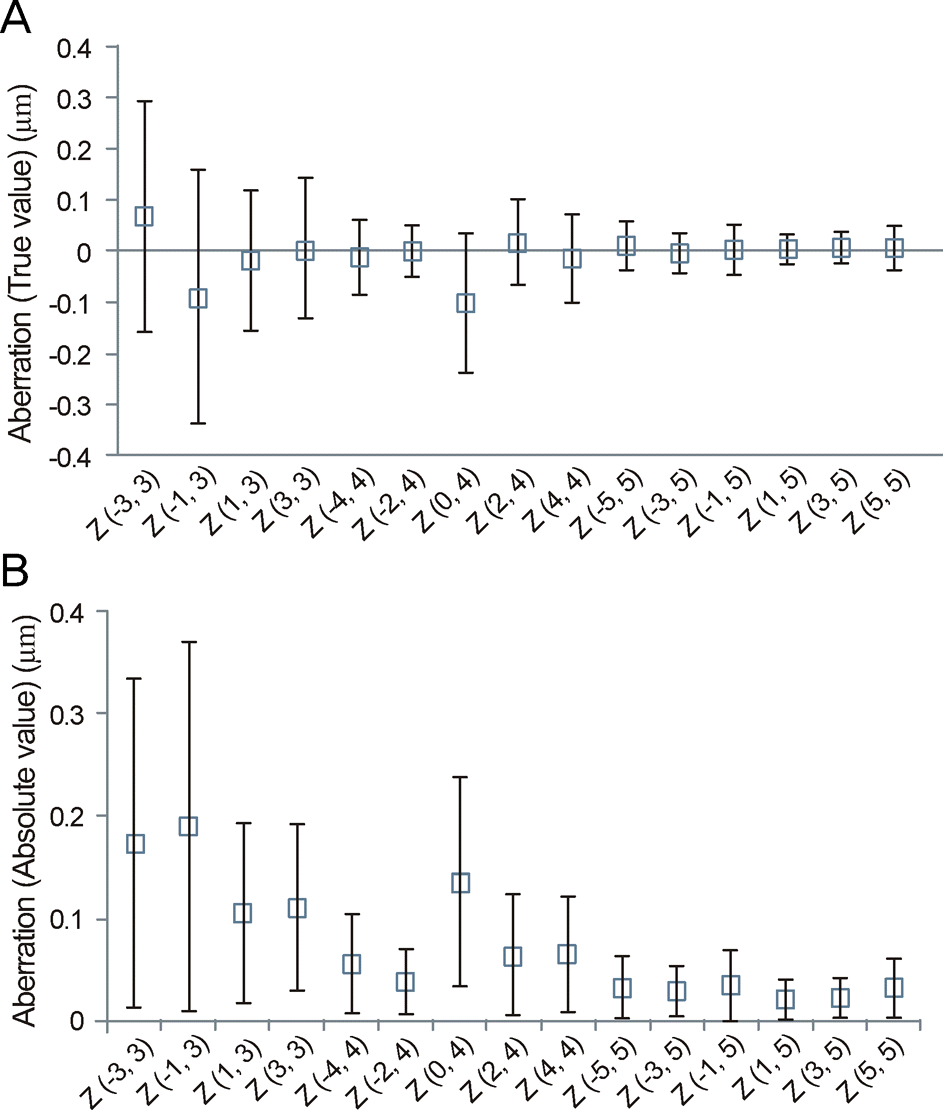
Figure 2.
Graph of summated RMS value (horizontal line) ± 1 standard deviation (ends of vertical lines) for 3rd- to 5th-order aberrations (RMS = root mean square; Sph Abb = spherical aberration).
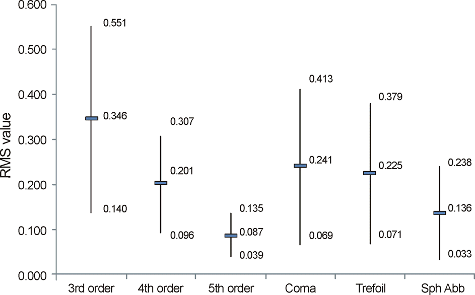
Figure 3.
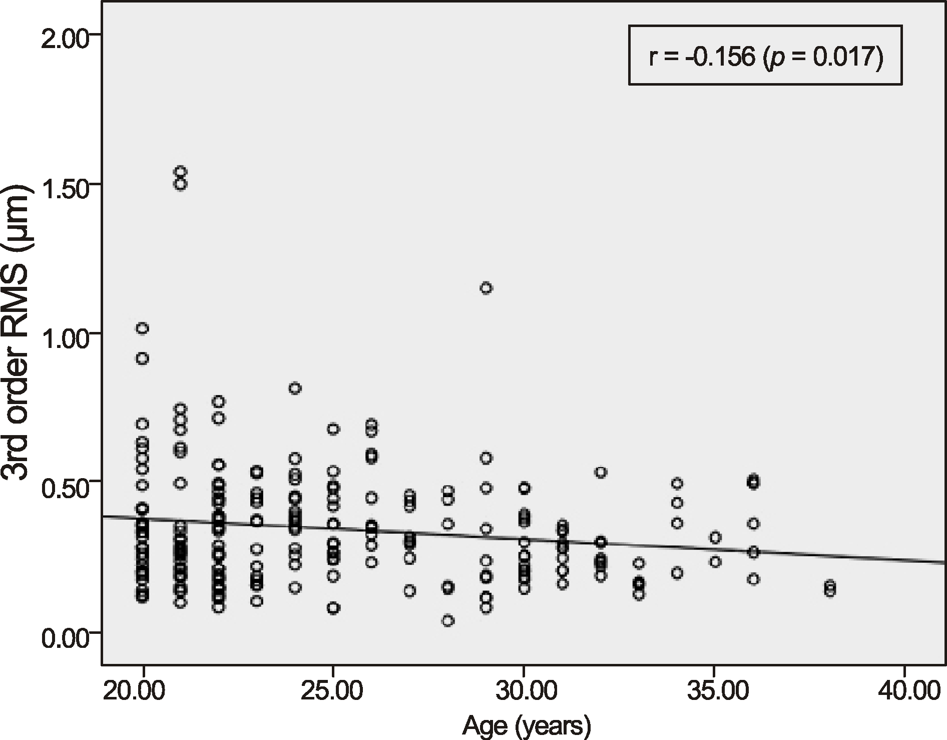
Scattergrams of total higher order aberration (third to fifth order) as a function of age; RMS = root mean square.
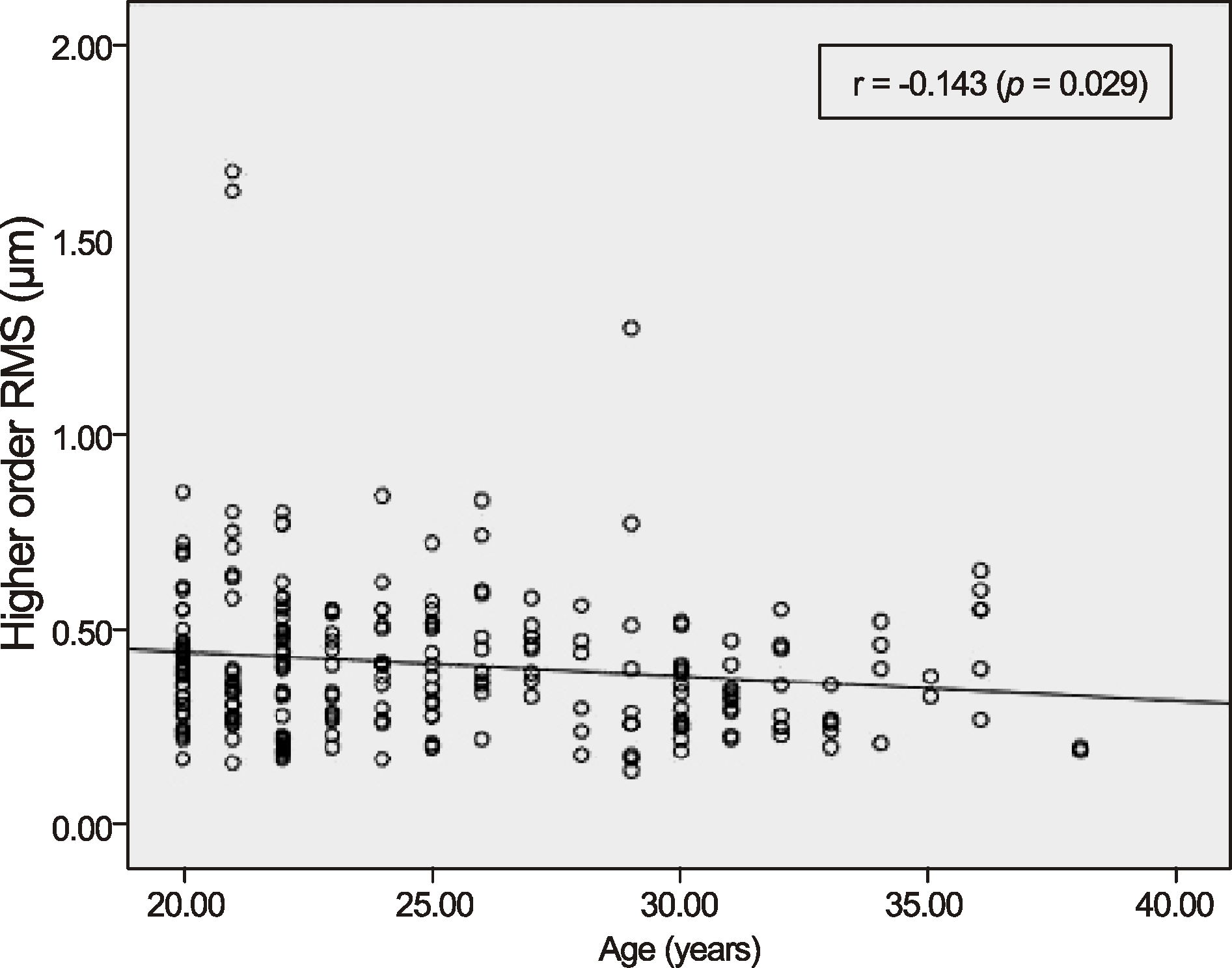
Figure 5.
Scattergram of 3rd trefoil as a function of spherical refractive error; RMS = root mean square.
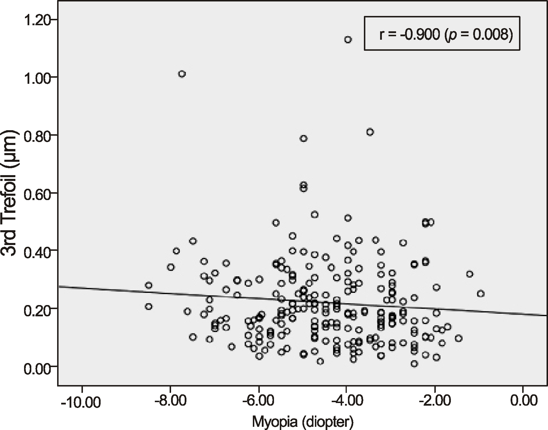
Table 1.
Signed and absolute values of the Zernike coefficients in the study population
|
Mean ± SD (μm) |
Range (μm) |
|||
|---|---|---|---|---|
| Zernicke Coefficient* | Signed value | Absolute value | Minimum | Maximum |
| 3rd order | ||||
| Z(-3,3) | 0.067 ± 0.23 | 0.172 ± 0.16 | −1.09 | 0.81 |
| Z-1,3) | −0.089 ± 0.25 | 0.190 ± 0.18 | −1.29 | 0.55 |
| Z(1,3) | −0.019 ± 0.14 | 0.105 ± 0.09 | −0.41 | 0.41 |
| Z(3,3) | 0.005 ± 0.14 | 0.110 ± 0.08 | −0.35 | 0.32 |
| 4th order | ||||
| Z(-4,4) | −0.012 ± 0.17 | 0.056 ± 0.05 | −0.37 | 0.21 |
| Z(-2,4) | −0.001 ± 0.05 | 0.038 ± 0.03 | −0.13 | 0.12 |
| Z(0,4) | −0.101 ± 0.14 | 0.136 ± 0.10 | −0.53 | 0.23 |
| Z(2,4) | 0.019 ± 0.08 | 0.064 ± 0.06 | −0.17 | 0.38 |
| Z(4,4) | −0.014 ± 0.09 | 0.065 ± 0.06 | −0.39 | 0.32 |
| 5th order | ||||
| Z(-5,5) | 0.010 ± 0.05 | 0.032 ± 0.04 | −0.14 | 0.32 |
| Z(-3,5) | −0.004 ± 0.04 | 0.030 ± 0.02 | −0.13 | 0.1 |
| Z(-1,5) | 0.004 ± 0.04 | 0.035 ± 0.03 | −0.3 | 0.17 |
| Z(1,5) | 0.004 ± 0.03 | 0.022 ± 0.02 | −0.08 | 0.08 |
| Z(3,5) | 0.007 ± 0.03 | 0.023 ± 0.02 | −0.07 | 0.11 |
| Z(5,5) | 0.006 ± 0.04 | 0.032 ± 0.03 | −0.22 | 0.14 |
Table 2.
Ocular aberrations of the subjects
Table 3.
Correlation between age and higher order aberrations
Table 4.
Correlation between spherical equivalent and higher order aberrations
Table 5.
Comparison of HOAs in present patient population with those in other studies
| Parameter | Current Study (Korea) | Prakash et al12 (India) | Wei et al13 (Singapore) | Salmon and Van de Pol11 (USA)* |
|---|---|---|---|---|
| Sample size | 232 | 412 | 166 | 2,560 |
| Ethnicity | Korean | Indian | Chinese | Mixed |
| Aberrometric principle and instrument used | Hartmann Shack, Zywave II | Hartmann Shack, Zywave | Hartmann Shack, Zywave | Hartmann-Shack, multiple aberrometers |
| HOA RMS | 0.421 ± 0.20 | 0.36 ± 0.266 | 0.49 ± 0.16 | 0.33 ± 0.13 |
| Summated RMS of 3rd to 5th order μm) | ||||
| 3rd order† | 0.346 ± 0.206 | 0.23 ± 0.15 | 0.37 ± 0.16 | 0.25 ± 0.12 |
| 4th order† | 0.202 ± 0.105 | 0.17 ± 0.09 | 0.29 ± 0.11 | 0.169 ± 0.09 |
| 5th order† | 0.087 ± 0.048 | 0.08 ± 0.38 | 0.08 ± 0.04 | 0.067 ± 0.03 |
| Ratio of means of 3rd:4th:5th order | 1:0.58:0.25 | 1:0.7:0.3 | 1:0.78:0.002 | 1:0.68:0.3 |
| Difference (%) of 3rd-5th order means; | ||||
| Korean versus other populations‡ | ||||
| 3rd order† | 33.5 | −6.9 | 27.7 | |
| 4th order† | 15.8 | −43.5 | 16.3 | |
| 5th order† | 8.0 | 8.0 | 22.9 | |
| Major HOAs | ||||
| Coma† | 0.241 ± 0.17 | 0.14 ± 0.10 | 0.27 ± 0.14 | NS |
| Trefoil† | 0.225 ± 0.154 | 0.16 ± 0.12 | NS | NS |
| Spherical aberration† | 0.136 ± 0.102 | 0.03 ± 0.05 | 0.22 ± 0.14 | 0.13 ± 0.09 |


 PDF
PDF ePub
ePub Citation
 Print
Print


 XML Download