

 PDF
PDF ePub
ePub Citation
 Print
Print


Abstract
Purpose
To investigate the effect of curcumin, known to inhibit hypoxia-inducible factor-1, on retinal neovascularization in a mouse model of oxygen-induced retinopathy (OIR).
Methods
OIR was induced by exposing C57BL/6 mice on postnatal day 7 (P7) to 75% hyperoxia for 5 days, followed by 5 days in a room with normal oxygen level. Curcumin was administered intraperitoneally once a day for 5 days from P12 or intravitreally once on P13. Mice retinas on P17 were analyzed for retinal neovascularization, which was compared between curcumin-treated and control mice.
Results
After intraperitoneal and intravitreal administration of curcumin, qualitative assessment of retinal neovascularization of flat-mounted retina showed no significant difference compared to control retinas. Quantitative assessment of retinal neovascularization also showed no significant difference between curcumin-treated and control mice.
Conclusions
Both intraperitoneal and intravitreal administration of curcumin did not reduce retinal neovascularization in an OIR mouse model. Further investigation including development of new formulations is required for the use of curcumin as an anti-angiogenic agent for retinal neovascularization.
References
1. Lim LS, Mitchell P, Seddon JM. . Age-related macular degeneration. Lancet. 2012; 379:1728–38.

2. Antonetti DA, Klein R, Gardner TW. Diabetic retinopathy. N Engl J Med. 2012; 366:1227–39.
3. Sapieha P, Joyal JS, Rivera JC. . Retinopathy of prematurity: understanding ischemic retinal vasculopathies at an extreme of life. J Clin Invest. 2010; 120:3022–32.
4. Smith LE, Shen W, Perruzzi C. . Regulation of vascular endothelial growth factor-dependent retinal neovascularization by insulin-like growth factor-1 receptor. Nat Med. 1999; 5:1390–5.
5. Smith LE, Kopchick JJ, Chen W. . Essential role of growth hormone in ischemia-induced retinal neovascularization. Science. 1997; 276:1706–9.
6. Chen J, Connor KM, Aderman CM, Smith LE. Erythropoietin deficiency decreases vascular stability in mice. J Clin Invest. 2008; 118:526–33.
7. Drixler TA, Borel Rinkes IH, Ritchie ED. . Angiostatin inhibits pathological but not physiological retinal angiogenesis. Invest Ophthalmol Vis Sci. 2001; 42:3325–30.
8. Stellmach V, Crawford SE, Zhou W, Bouck N. Prevention of ischemia-induced retinopathy by the natural ocular antiangiogenic agent pigment epithelium-derived factor. Proc Natl Acad Sci USA. 2001; 98:2593–7.
9. Forsythe JA, Jiang BH, Iyer NV. . Activation of vascular endothelial growth factor gene transcription by hypoxia-inducible factor 1. Mol Cell Biol. 1996; 16:4604–13.
10. Fu S, Kurzrock R. Development of curcumin as an epigenetic agent. Cancer. 2010; 116:4670–6.
11. Binion DG, Otterson MF, Rafiee P. Curcumin inhibits VEGF- mediated angiogenesis in human intestinal microvascular endothelial cells through COX-2 and MAPK inhibition. Gut. 2008; 57:1509–17.
12. Bae MK, Kim SH, Jeong JW. . Curcumin inhibits hypoxia-induced angiogenesis via down-regulation of HIF-1. Oncol Rep. 2006; 15:1557–62.
13. Mrudula T, Suryanarayana P, Srinivas PN, Reddy GB. Effect of curcumin on hyperglycemia-induced vascular endothelial growth factor expression in streptozotocin-induced diabetic rat retina. Biochem Biophys Res Commun. 2007; 361:528–32.
14. Wang L, Li C, Guo H. . Curcumin inhibits neuronal and vascular degeneration in retina after ischemia and reperfusion injury. PLoS One. 2011; 6:e23194.
15. Grossniklaus HE, Kang SJ, Berglin L. Animal models of choroidal and retinal neovascularization. Prog Retin Eye Res. 2010; 29:500–19.
16. Smith LE, Wesolowski E, McLellan A. . Oxygen-induced retinopathy in the mouse. Invest Ophthalmol Vis Sci. 1994; 35:101–11.
17. Penn JS, Tolman BL, Lowery LA. Variable oxygen exposure causes preretinal neovascularization in the newborn rat. Invest Ophthalmol Vis Sci. 1993; 34:576–85.
18. Connor KM, Krah NM, Dennison RJ. . Quantification of oxygen-induced retinopathy in the mouse: a model of vessel loss, vessel regrowth and pathological angiogenesis. Nat Protoc. 2009; 4:1565–73.
19. Kim SJ, Jin J, Kim YJ. . Retinal proteome analysis in a mouse model of oxygen-induced retinopathy. J Proteome Res. 2012; 11:5186–203.
20. Mohanty C, Das M, Sahoo SK. Emerging role of nanocarriers to increase the solubility and bioavailability of curcumin. Expert Opin Drug Deliv. 2012; 9:1347–64.
21. Dujic J, Kippenberger S, Hoffmann S. . Low concentrations of curcumin induce growth arrest and apoptosis in skin keratinocytes only in combination with UVA or visible light. J Invest Dermatol. 2007; 127:1992–2000.
22. Penn JS, McCollum GW, Barnett JM. . Angiostatic effect of penetrating ocular injury: role of pigment epithelium-derived factor. Invest Ophthalmol Vis Sci. 2006; 47:405–14.
23. Stitt AW, Graham D, Gardiner TA. Ocular wounding prevents pre-retinal neovascularization and upregulates PEDF expression in the inner retina. Mol Vis. 2004; 10:432–8.
Figure 1.
Fluorescence micrographs of flat-mounted retinas from the mouse model of oxygen-induced retinopathy (OIR). (A) Control OIR retina treated with intraperitoneal PBS injection. (B) OIR retina treated with intraperitoneal curcumin (100 mg/kg) injection. (C) Control OIR retina treated with intravitreal PBS injection. (D) OIR retina treated with intra- vitreal curcumin (1 μg) injection.

Figure 2.
Quantitative assessment of retinal neovascularization in the mouse model of oxygen-induced retinopathy treated with intraperitoneal curcumin injection. (A) The average vascular lumens per section for each group are presented as the means ± SEM. Note that there is no significant difference in the retinal neovascularization between the control and curcu- min-treated eyes. Representative photomicrographs of OIR retinas treated with intraperitoneal PBS (B) and intraperitoneal curcumin (100 mg/kg) injection (C).
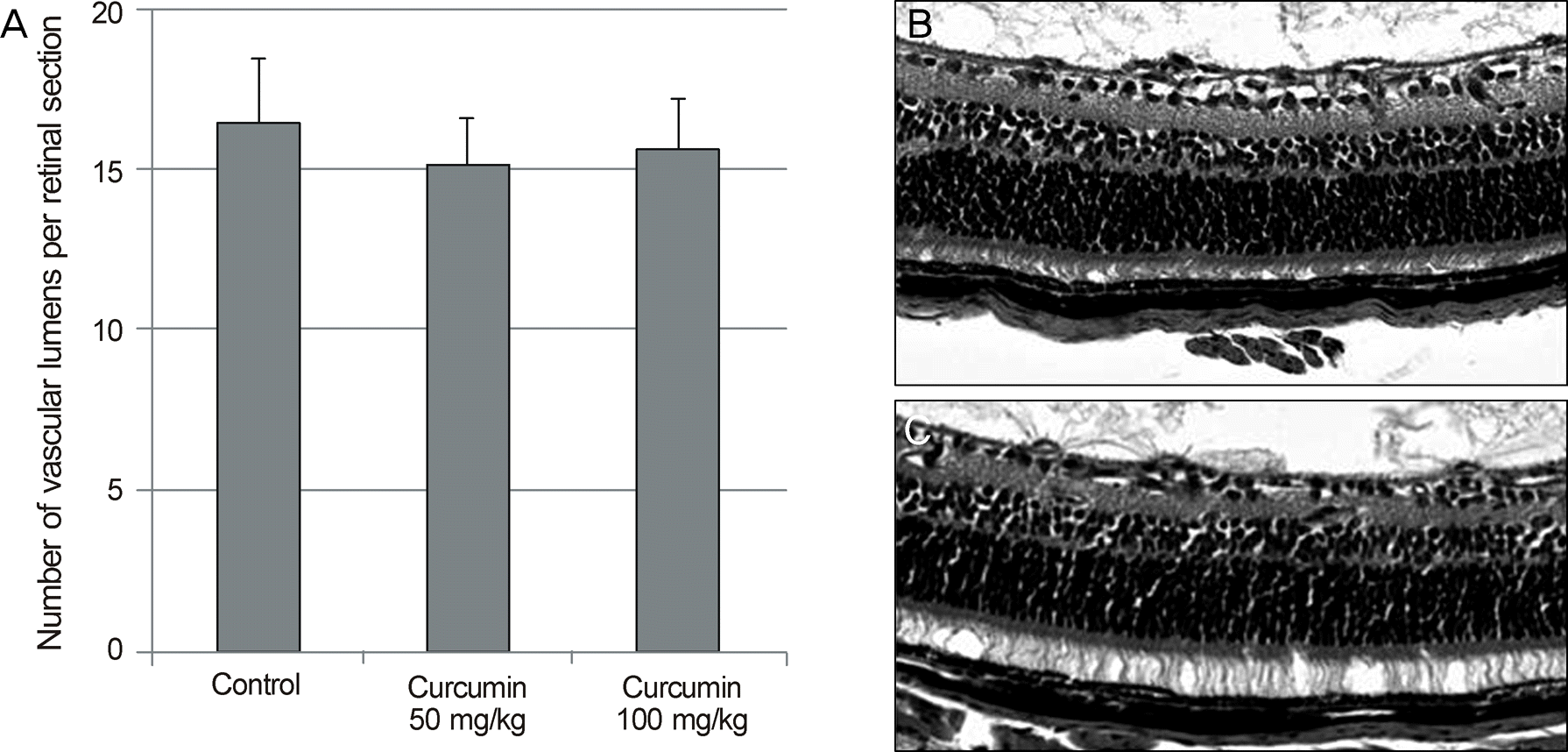
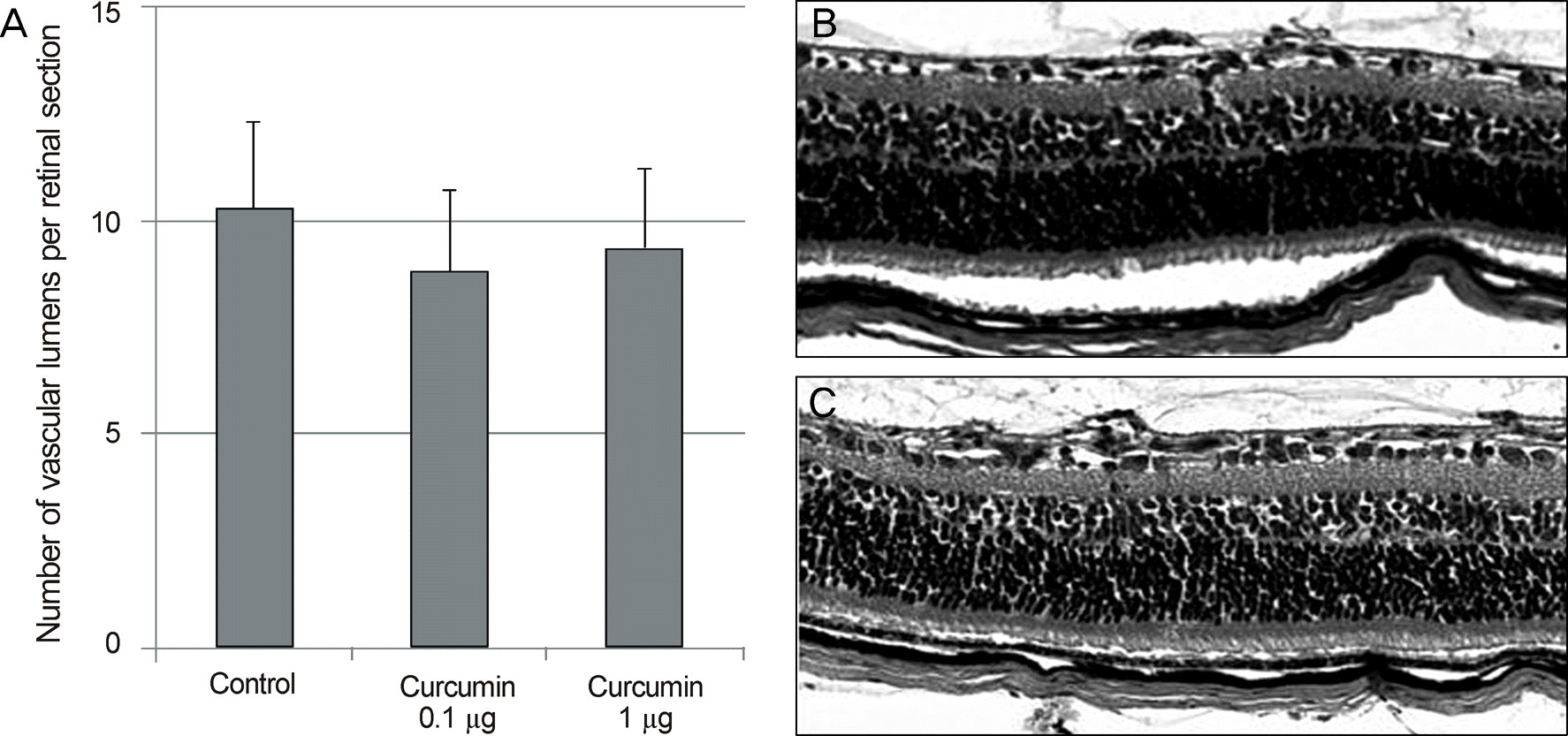
Figure 3.
Quantitative assessment of retinal neovascularization in the mouse model of oxygen-induced retinopathy treated with intravitreal curcumin injection. (A) The average vascular lumens per section for each group are presented as the means ± SEM. Note that there is no significant difference in the retinal neovascularization between the control and curcumin treated eyes. Representative photomicrographs of OIR retinas treated with intravitreal PBS (B) and intraperitoneal curcumin (1 μg) injection (C).
 XML Download