

 PDF
PDF ePub
ePub Citation
 Print
Print


Abstract
Purpose
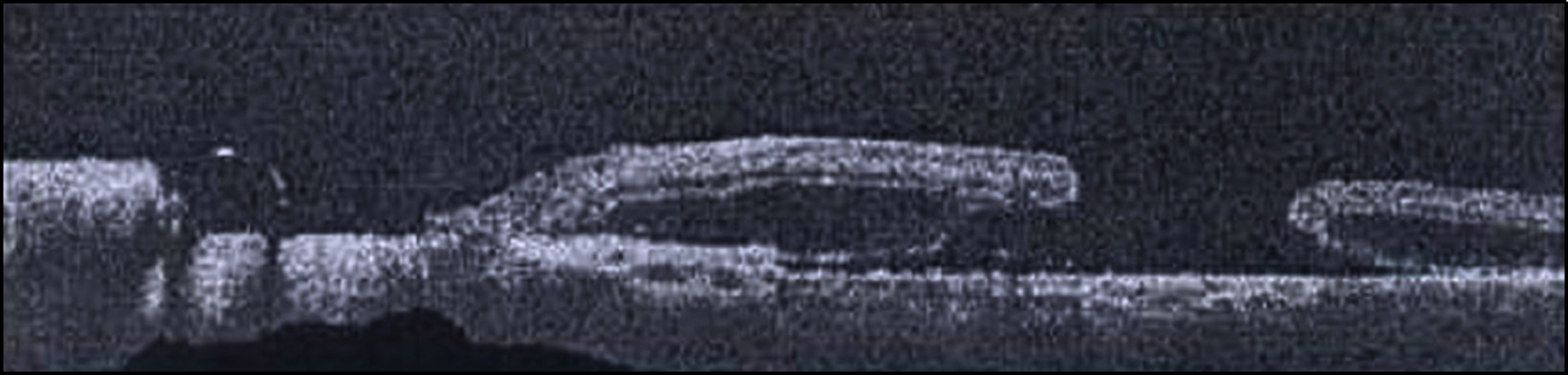
To report a case of macular hole after surgery in macular serous detachment associated with optic disc pit in a child, which was treated with silicone oil as an intraocular substitute.
Case summary
A 12-year-old boy was referred for examination due to visual disturbance in his left eye for the last 1 week. Corrected visual acuity at presentation was 0.08 in the left eye. The anterior part of the left eye was normal on slit lamp examination. Funduscopic examination revealed optic disc pit associated with macular detachment. The patient was treat-ed with vitrectomy, internal limiting membrane (ILM) peeling and gas tamponade. One week after treatment, the patient presented with central visual disturbance and showed a full thickness macular hole in the left eye. Extensive ILM peeling and silicone oil instillation were performed and after 2 months, silicone oil removal was performed. The macular hole ap-peared to be closed and visual acuity improved to 0.2. Recurrence was not observed until 20 months after treatment.
References
1. Theodossiadis PG, Grigoropoulos VG, Emfietzoglou J, et al. Optical coherence tomography study of vitreoretinal interface in full thickness macular hole associated with optic disk pit maculopathy. Eur J Ophthalmol. 2007; 17:272–6.

2. Bechmann M, Mueller AJ, Gandorfer A, et al. Macular hole sur-gery in an eye with an optic pit. Am J Ophthalmol. 2001; 132:263–4.
3. Hong JH, Kim YY. A case of vitrectomy without Laser for serous macular detachment associated with optic disc pit. J Korean Ophthalmol Soc. 2011; 52:1114–8.
4. Sengün A, Batioglu F, Akbatur H, Atmaca L. Vitreoretinal surgery of retinal detachment and macular hole associated with optic nerve pit: an optical coherence tomography study. Eur J Ophthalmol. 2004; 14:355–7.
5. Ryu JW, Ra H, Lee WK. A case of surgically treated serous macular detachment associated with optic disc pit. J Korean Ophthalmol Soc. 2010; 51:155–8.
6. Imamura Y, Zweifel SA, Fujiwara T, et al. High resolution optical coherence tomography findings in optic pit maculopathy. Retina. 2010; 30:1104–12.
7. Ivanovska-Adjievska B, Boskurt S, Semiz F, et al. Treatment of idi-opathic macular hole with silicone oil tamponade. Clin Ophthalmol. 2012; 6:1449–54.
8. Bonnet M. Serous macular detachment associated with optic nerve pits. Graefes Arch Clin Exp Ophthalmol. 1991; 229:526–32.
9. Krivoy D, Gentile R, Liebmann JM. Imaging congenital optic disc pits and associated maculopathy using optical coherence tomography. Arch Ophthalmol. 1996; 114:165–70.
10. Kuhn F, Kover F, Szabo I, Mester V. Intracranial migration of sili-cone oil from an eye with optic pit. Graefes Arch Clin Exp Ophthalmol. 2006; 244:1360–2.


 XML Download