

 PDF
PDF ePub
ePub Citation
 Print
Print


Abstract
Purpose
To investigate the visualization of cystoid macular edema (CME) using noninvasive retromode imaging by a new scanning laser ophthalmoscope (SLO) and compare to previous imaging modalities.
Methods
The authors of the present study retrospectively reviewed the medical records of 21 eyes of 20 patients with CME due to various etiologies. All eyes were examined with fundus camera, fluorescein angiography (TRC-50EX, Topcon, Tokyo, Japan), SLO (F-10, Nidek, Gamagori, Japan), and spectral-domain optical coherence tomography (OCT) (3D OCT-1000, Topcon, Tokyo, Japan). In the present study the SLO was used in the retro-mode with an infrared laser.
Results
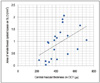
Previous fundus photography could not detect CME adequately although SLO retro-mode could show numerous oval or polygonal cystoid spaces more readily. Furthermore, each individual small cystoid space could be detected and the area of each cystoid space could be measured. The area of the largest cystoid space showed a correlation with its height, as measured with OCT (R = 0.606, p = 0.004). The area of the whole foveal cystoid space showed a correlation with central macular thickness, as measured with OCT (R = 0.493, p = 0.023).
Figures and Tables
Figure 1
(A) Confocal mode: Images consist primarily of direct reflex from the vitreomacular interface. (B) Indirect mode with a ring aperture: Direct reflex from the vitreomacular interface is blocked with a central stop. More multiply scattered light from intraretinal structure is collected by the detector. (C) Retro-mode: The opening of the ring aperture is restricted and is deviated laterally from the confocal light path. Multiply scattered light from only one direction is collected by the detector.
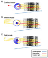
Figure 2
(A) The largest cystoid space is selected on retro-mode image and the area of the cystoid space can be measured using the loaded software in F-10. (B) The height of the selected cystoid space can measured using the loaded software in 3D-OCT.
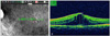
Figure 3
A case of 29-year-old woman with idiopathic cystoid macular edema. (A) No cystoid spaces are detected on fundus photography. (B) Late-phase fluorescein angiography shows many cystoids spaces. (C) Scanning laser ophthalmoscope (SLO) retro-mode shows numerous polygonal cystoid spaces.
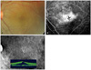
Figure 4
A case of 64-year-old woman with central retinal vein occulsion. (A) No cystoid spaces are detected on fundus photography. (B) Late phase fluorescein angiography cannot show cystoid spaces obviously due to leakage from vessels and choroidal fluorescence. (C) Scanning laser ophthalmoscope (SLO) retro-mode shows numerous fine small cysts in the macular area.
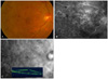
Figure 5
A case of 46-year-old man with vitreous hemorrhage due to proliferative diabetic retinopathy. (A) No cystoid spaces are detected on fundus photography. (B) Late-phase fluorescein angiography cannot show cystoids spaces obviously due to media opacity caused by vitreous hemorrhage. (C) Scanning laser ophthalmoscope (SLO) retro-mode shows numerous cysts despite media opacity.
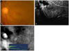
Figure 6
The area of the largest cystoid space measured with a scanning laser ophthalmoscope (SLO) retro-mode shows a correlation with its height, as measured with optical coherence tomography (OCT) (R = 0.606; p = 0.004).


References
1. Rotsos TG, Moschos MM. Cystoid macular edema. Clin Ophthalmol. 2008. 2:919–930.
2. Drolsum L, Haaskjold E. Causes of decreased visual acuity after cataract extraction. J Cataract Refract Surg. 1995. 21:59–63.
3. Ray S, D'Amico DJ. Pseudophakic cystoid macular edema. Semin Ophthalmol. 2002. 17:167–180.
4. Cunha-Vaz JG, Travassos A. Breakdown of the blood-retinal barriers and cystoid macular edema. Surv Ophthalmol. 1984. 28:485–492.
5. Brar M, Yuson R, Kozak I, et al. Correlation between morphologic features on spectral-domain optical coherence tomography and angiographic leakage patterns in macular edema. Retina. 2010. 30:383–389.
6. Jittpoonkuson T, Garcia PM, Rosen RB. Correlation between fluorescein angiography and spectral-domain optical coherence tomography in the diagnosis of cystoid macular edema. Br J Ophthalmol. 2010. 94:1197–1200.
7. Yamaike N, Tsujikawa A, Ota M, et al. Three-dimensional imaging of cystoid macular edema in retinal vein occlusion. Ophthalmology. 2008. 115:355–362.
8. Kashani AH, Keane PA, Dustin L, et al. Quantitative subanalysis of cystoid spaces and outer nuclear layer using optical coherence tomography in age-related macular degeneration. Invest Ophthalmol Vis Sci. 2009. 50:3366–3373.
9. Ikeda T, Sato K, Katano T, Hayashi Y. Examination of patients with cystoid macular edema using a scanning laser ophthalmoscope with infrared light. Am J Ophthalmol. 1998. 125:710–712.
10. Beausencourt E, Remky A, Elsner AE, et al. Infrared scanning laser tomography of macular cysts. Ophthalmology. 2000. 107:375–385.
11. Remky A, Beausencourt E, Hartnett ME, et al. Infrared imaging of cystoid macular edema. Graefes Arch Clin Exp Ophthalmol. 1999. 237:897–901.
12. Manivannan A, Kirkpatrick JN, Sharp PF, Forrester JV. Clinical investigation of an infrared digital scanning laser ophthalmoscope. Br J Ophthalmol. 1994. 78:84–90.
13. Beckman C, Bond-Taylor L, Lindblom B, Sjöstrand J. Confocal fundus imaging with a scanning laser ophthalmoscope in eyes with cataract. Br J Ophthalmol. 1995. 79:900–904.
14. Woon WH, Fitzke FW, Bird AC, Marshall J. Confocal imaging of the fundus using a scanning laser ophthalmoscope. Br J Ophthalmol. 1992. 76:470–474.
15. Yoshida A, Ishiko S, Akiba J, et al. Radiating retinal folds detected by scanning laser ophthalmoscopy using a diode laser in a dark-field mode in idiopathic macular holes. Graefes Arch Clin Exp Ophthalmol. 1998. 236:445–450.
16. Yamamoto M, Tsujikawa A, Mizukami S, et al. Cystoid macular edema in polypoidal choroidal vasculopathy viewed by a scanning laser ophthalmoscope: CME in PCV viewed by SLO. Int Ophthalmol. 2008. 10. 15. [Epub ahead of print].
17. Yamamoto M, Mizukami S, Tsujikawa A, et al. Visualization of cystoid macular oedema using a scanning laser ophthalmoscope in the retro-mode. Clin Experiment Ophthalmol. 2010. 38:27–36.
18. Tanaka Y, Shimada N, Ohno-Matsui K, et al. Retromode retinal imaging of macular retinoschisis in highly myopic eyes. Am J Ophthalmol. 2010. 149:635–640.
19. Ishiko S, Akiba J, Horikawa Y, Yoshida A. Detection of drusen in the fellow eye of Japanese patients with age-related macular degeneration using scanning laser ophthalmoscopy. Ophthalmology. 2002. 109:2165–2169.
 XML Download