

 PDF
PDF ePub
ePub Citation
 Print
Print


Abstract
Case summary
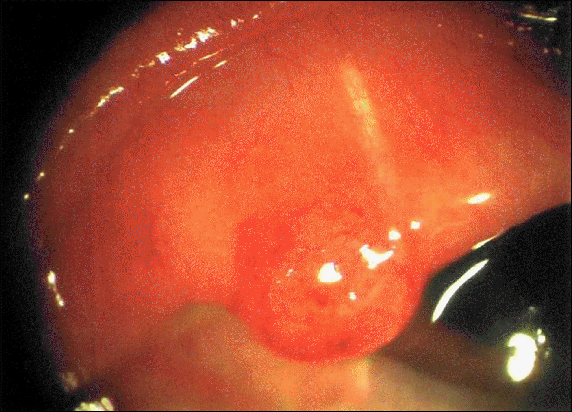
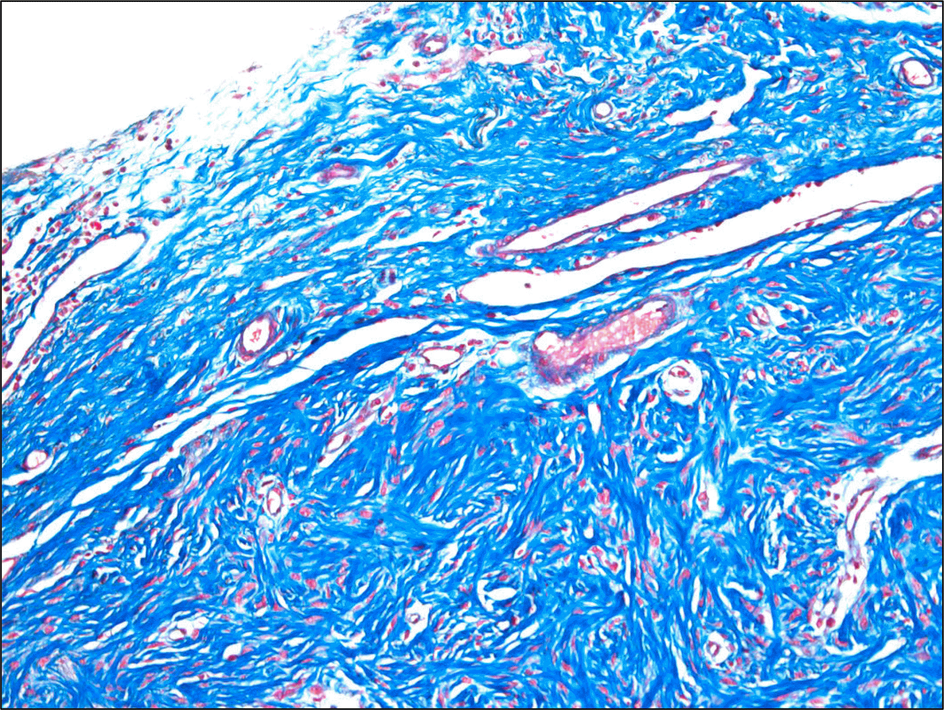
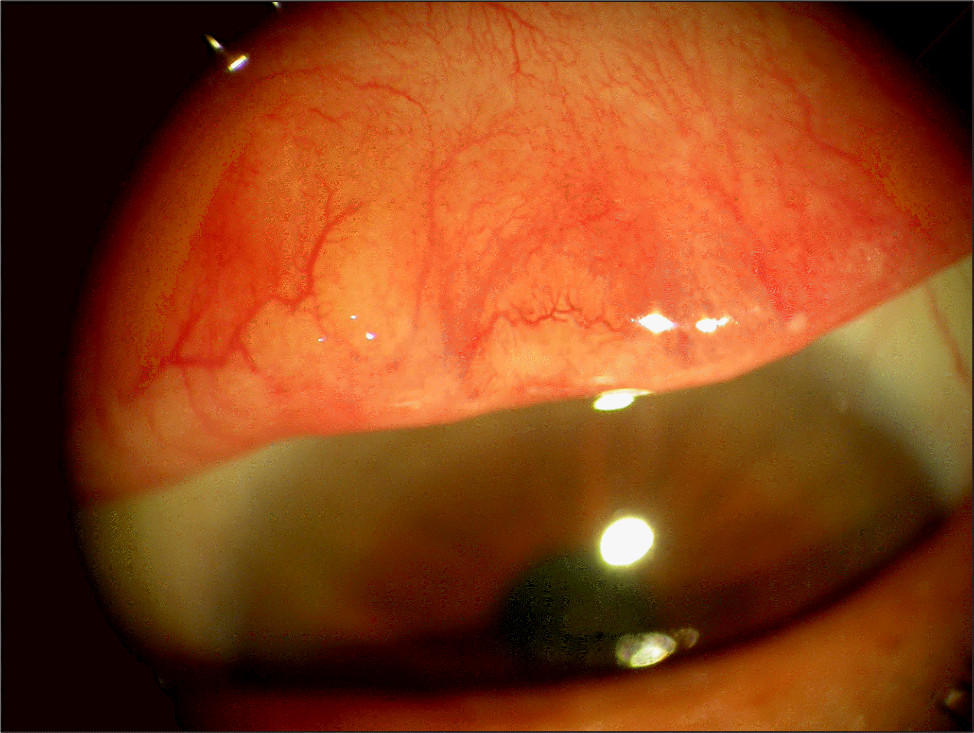
A 73-year-old male visited our clinic with sensations of irritation in his left upper eyelid that occurred one week prior. The patient did not have any evidence of external injuries, systemic inflammations, or any other specific findings except a history of hypertension. The best corrected visual acuity was 0.7 in the right and 0.8 in the left eye with normal IOP. On the slit-lamp biomicroscopic examinations, anterior segment showed no specific findings and the shapes and positions of the upper and lower eyelids were normal. Upon eversion of the upper eyelid, a definite solid 5 × 5-mm sized tumor with clear boundaries was observed in the upper tarsal conjunctiva. An excision biopsy was performed under local anesthesia. Gross examinations of the tumor revealed a 5 × 5 × 2-mm, gray, oval-shaped mass. On microscopic examinations, the tumor had minimal number of cells and was composed of dense collagens and scattered fibroblasts. Based on these findings, the patient was diagnosed with tarsal fibroma. The patient experienced no discomfort after the excision biopsy. At a one-year followup, there were no signs of recurrence.
References
1. Herschorn BJ, Jakobiec FA, Hornblass A, et al. Epibulbar subconjunctival fibroma. A tumor possibly arising from Tenon's capsule. Ophthalmology. 1983; 90:1490–4.
2. Clinch TJ, Kostick DA, Menke DM. Tarsal fibroma. Am J Ophthalmol. 2000; 129:691–3.

3. Mortada A. Fibroma of the orbit. Br J Ophthalmol. 1971; 55:350–2.
4. Schutz JS, Rabkin MD, Schutz S. Fibromatous tumor (desmoid type) of the orbit. Arch Ophthalmol. 1979; 97:703–4.
5. Jakobiec FA, Sacks E, Lisman RL, Krebs W. Epibulbar fibroma of the conjunctival substantia propria. Arch Ophthalmol. 1988; 106:661–4.
6. Jakobiec FA, DeVoe AG, Boyd J. Fibrous histiocytoma of the tarsus. Am J Ophthalmol. 1977; 84:794–7.
7. Font RL, Zimmerman LE. Nodular fasciitis of the eye and adnexa. A report of ten cases. Arch Ophthalmol. 1966; 75:475–81.
8. Hidayat AA, Font RL. Juvenile fibromatosis of the periorbital re-gion and eyelid. A clinicopathologic study of six cases. Arch Ophthalmol. 1980; 98:280–5.
9. Craig RD, Studd D. Extra-abdominal desmoid of the face involving the orbit. Br J Surg. 1978; 65:131–4.

 XML Download