

 PDF
PDF ePub
ePub Citation
 Print
Print


Abstract
Purpose
To report a case of fibrin pupillary block diagnosed by ultrasonic biomicroscopy (UBM) and treated by argon neodymium:YAG (Nd:YAG) laser in a patient with uveitis.
Case summary
A 56-year-old man visited the hospital for decreasing visual acuity and a sudden onset of pain in the right eye. Best corrected visual acuity was 0.02 in the right eye and 1.0 in the left eye. Intraocular pressure (IOP) was 48 mm Hg in the right eye and 18 mm Hg in the left eye. Slit-lamp examination revealed diffuse corneal stromal edema with Descemet's folds and uniform shallowing of the anterior chamber, with 360 degrees of peripheral iridocorneal touch in the right eye. A fibrin membrane was present across the pupil. UBM showed a fibrin membrane across the pupil, uniform shallowing of the anterior chamber, and peripheral angle closure. The lens was displaced posteriorly. A sequential Nd:YAG laser membranectomy was performed that same day, with immediate deepening of the anterior chamber and normalization of the IOP.
Conclusions
UBM can play an invaluable role in the diagnosis of fibrin pupillary block by showing the presence of a fibrin pupillary membrane, accumulation of aqueous in the posterior chamber, and a clear separation between the iris and the lens. Nd:YAG laser membranectomy can be performed effectively in a fibrin pupillary block.
References
1. Ritch R, Shields MB, Krupin T. The glaucomas. 2nd ed.2. St. Louis: Mosby-Year Book Inc.;1996. p. 1225–58.
2. Van Buskirk EM. Pupillary block after intraocular lens aberrations. Am J Ophthalmol. 1983; 95:55–9.
3. Samples JR, Bellows AR, Rosenquist RC, et al. Pupillary block with posterior chamber intraocular lenses. Arch Ophthalmol. 1987; 105:335–7.

4. Forrester JV, Mc Menamin PG. Immunopathogenic mechanisms in intraocular inflammation. Chem Immunol. 1999; 73:159–85.
5. Moorthy RS, Mermoud A, Baerveldt G, et al. Glaucoma associated with uveitis. Surv Ophthalmol. 1997; 41:361–94.
6. Jaffe GJ, Lewis H, Han DP, et al. Treatment of postvitrectomy fibrin pupillary block with tissue plasminogen activator. Am J Ophthalmol. 1989; 108:170–5.
7. Khor WB, Perera S, Jap A, et al. Anterior segment imaging in the management of postoperative fibrin pupillary-block glaucoma. J Cataract Refract Surg. 2009; 35:1307–12.
8. Sathish S, MacKinnon JR, Atta HR. Role of ultrasound biomicroscopy in managing pseudophakic pupillary block glaucoma. J Cataract Refract Surg. 2000; 26:1836–8.
9. Kim EA, Bae MC, Cho YW. Neodymium YAG laser and surgical synechiolysis of iridocapsular adhesions. Korean J Ophthalmol. 2008; 22:159–63.
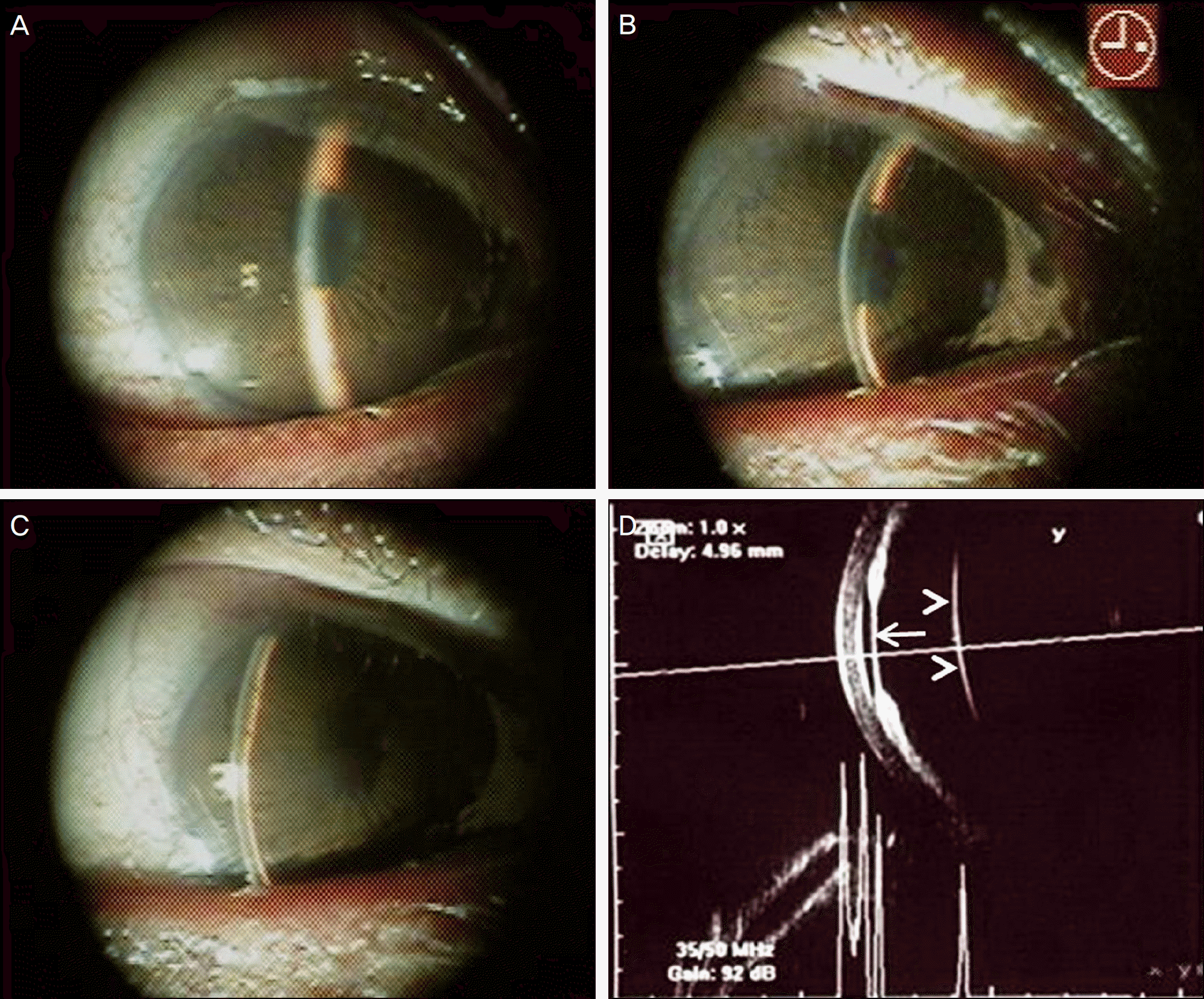
Figure 1.
Photographs of anterior segment and ultrasound biomicroscopic findings at the first visit. Anterior chamber shallowing with peripheral iridocorneal touch, and a fibrin membrane are noted across the pupil (A, B, C). Ultrasound biomicroscopy shows a fibrin membrane across the pupil (arrow), uniform shallowing of the anterior chamber, and peripheral angle closure (D). Lens is displaced posteriorly (arrow head) and separated from the iris and fibrin membrane by a large clear space (D).
 XML Download