Abstract
Purpose
To identify the impact of the presence of peripheral anterior synechia (PAS) on the depth of the anterior segment in patients with a shallow anterior chamber after laser iridotomy (LI) by analyzing changes in the anterior segment biometry using ultrasound biomicroscopy (UBM).
Methods
Twenty eyes of 20 patients with PAS and shallow anterior chamber, and another 20 eyes of 20 patients with shallow anterior chamber without PAS were studied. The changes in the anterior segment biometry for each group of patients were examined using gonioscopy and UBM before and after the LI.
Results
The central corneal thicknesses and scleral thicknesses of the two groups did not show significant differences (p > 0.05). The anterior chamber depths, anterior chamber angles, trabecular meshwork-iris distances, and angle-opening distances 500 increased significantly after the peripheral LI (p < 0.05) in both groups. However, the difference in the increases in the anterior segment biometries between the two groups was not statistically significant.
References
1. Quigley HA, Miller NR, George T. Clinical evaluation of nerve fiber layer atrophy as an indicator of glaucomatous optic nerve damage. Arch Ophthalmol. 1980; 98:1564–71.

2. Levene RZ. Low tension glaucoma: a critical review and new material. Surv Ophthalmol. 1980; 24:621–64.

3. Salmon JF. Predisposing factors for chronic angle-closure glaucoma. Prog Retin Eye Res. 1999; 18:121–32.

4. Ritch R, Lowe RF. Angle-closure glaucoma: mechanisms and epidermiology. Ritch R, Shields MB, Krupin T, editors. The Glaucomas. 2nd ed.St. Louis: CV Mosby;1996. 2:chap.p. 37.
5. Lowe RF. Aetilogy of the anatomical basis for primary angle-clo-sure glaucoma. Biometrical comparasions between normal eyes and eyes with primary angle-closure glaucoma. Br J Ophthalmol. 1970; 54:161–9.
6. Foster PJ, Aung T, Nolan WP, et al. Defining “occludable” angles in population surveys: drainage angle width, peripheral anterior synechia, and glaucomatous optic neuropathy in east Asian people. Br J Ophthalmol. 2004; 88:486–90.
7. Inoue T. Distribution and morphology of peripheral anterior synechia in primary angle-closure glaucoma. Nippon Ganka Gakkai Zasshi. 1993; 97:78–82.
8. Rivera AH, Brown RH, Anderson DR. Laser iridotomy vs surgical iridectomy. Have the indications changed? Arch Ophthalmol. 1985; 103:1350–4.

9. Robin AL, Pollack IP. Argon laser peripheral iridotomies in the treatment of primary angle closure glaucoma: long-term follow-up. Arch Ophthalmol. 1982; 100:919–23.
10. Hong C, Hahn SH, Sohn YH. Argon laser iridotomy for angle-clo-sure glaucoma. J Korean Ophthalmol Soc. 1983; 24:807–11.
11. Han CW, Kim JW. Effect of YAG laser iridotomy on IOP in chronic angle-closure glaucoma. J Korean Ophthalmol Soc. 1995; 36:1730–6.
12. Lee HB, Hwang WS, You JM, et al. Sequential Argon and Nd:YAG laser iridotomies in angle closure glaucoma. J Korean ophthalmol Soc. 1999; 40:2245–51.
13. Gazzard G, Friedman DS, Devereux FG, et al. A prospective ultrasound biomicroscopy evaluation of changes in anterior segment morphology after laser iridotomy in Asian eyes. Ophthalmology. 2003; 110:630–8.

15. Caronia RM, Liebmann JM, Stegman Z, et al. Increase in iris-lens contact after laser iridotomy for papillary block angle closure. Am J Ophthalmol. 1996; 122:53–7.
16. Kashiwagi K, Abe K, Tsukahara S. Quantitative evaluation of changes in anterior segment biometry by peripheral laser iridotomy using newly developed scanning peripheral anterior chamber depth analyzer. Br J Ophthalmol. 2004; 88:1036–41.
17. Pavlin CJ, Sherar MD, Foster FS. Subsurface ultrasound imaging of the intact eye. Ophthalmology. 1990; 97:244–50.
18. Pavlin CJ, Harasiewicz K, Sherar MD, Foster FS. Clinical use of ultrasound biomicroscopy. Ophthalmology. 1991; 98:287–95.

19. Marchini G, Pagliarusco A, Toscano A, et al. Ultrasound biomicroscopic and conventional ultrasonographic study of ocular dimensions in primary angle-closure glaucoma. Ophthalmology. 1998; 105:2091–8.
20. Ishikawa H, Liebmann J, Ritch R. Quantitiative assessment of the anterior segment using ultrasound biomicroscopy. Curr Opin Ophthalmol. 2000; 11:133–9.
21. Mandell MA, Palvin CJ, Weisbrod DJ, et al. Anterior chamber depth in plateau iris syndrome and papillary block as measured by ultrasound biomicroscopy. Am J Ophthalmol. 2003; 136:900–3.
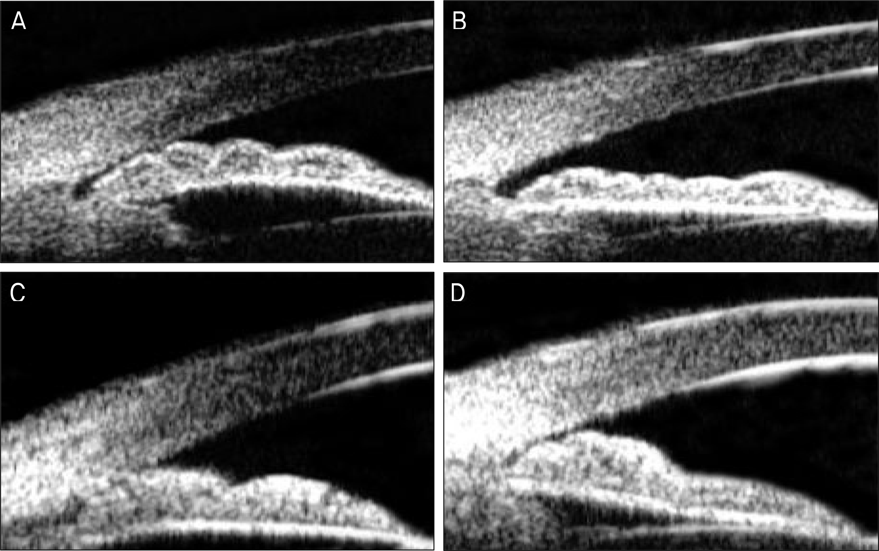
Figure 1.
UBM images of the temporal angle of eyes before and after LI in PAS-positive eye and PAS-negative eye that show flattening of the iris and widening of the angle in detail. (A) Before LI in PAS-negative eye. (B) After LI in PAS-negative eye. (C) Before LI in PAS-positive eye. (D) After LI in PAS-positive eye.

Table 1.
Patients' data base
| |
Study subjects (n=40) |
|
|---|---|---|
| PAS-positive (n=20) | PAS-negative (n=20) | |
| Gender | | |
| Male | 5 | 1 |
| Female | 15 | 19 |
| Age (years) | | |
| Mean ± SD* | 65.5 ± 11.0 | 66.2 ± 6.7 |
| Range | 49∼79 | 57∼82 |
| Laterality of affected eye | | |
| Right | 10 | 10 |
| Left | 10 | 10 |
| Refractive errors († S.E.: ‡ D) | | |
| Mean ± SD* | 0.900 ± 0.609 | 0.500 ± 0.846 |
| Range | −0.25∼+2.00 | −0.75∼+2.75 |
| Intraocular pressure (mmHg) | | |
| Mean ± SD* | 17.4 ± 2.5 | 16.6 ± 4.4 |
| Range | 13∼23 | 8∼24 |
Table 2.
UBM parameters in PAS-negative and PAS-positive eyes before and after LI
| UBM parameters |
PAS-positive s |
PAS-negative |
||||
|---|---|---|---|---|---|---|
| Before LI | After LI | p value | Before LI | After LI | p value | |
| CCT* (μ m) | 497.7 ± 14.6 | 498.5 ± 10.2 | 0.666 | 493.7 ± 13.5 | 494.7 ± 11.4 | 0.428 |
| ACD† (mm) | 1.96 ± 0.11 | 1.97 ± 0.09 | 0.049 | 2.15 ± 0.11 | 2.17 ± 0.09 | 0.021 |
| ACA‡ (°) | 11.71 ± 0.98 | 14.08 ± 0.99 | 0.006 | 14.57 ± 0.66 | 18.42 ± 0.68 | <0.001 |
| ST§ (μ m) | 866.1 ± 2.56 | 866.3 ± 2.38 | 0.444 | 861.5 ± 2.33 | 862.3 ± 3.00 | 0.238 |
| TID∏ (μ m) | 90.43 ± 0.77 | 110.93 ± 1.14 | <0.001 | 103.68 ± 0.65 | 125.87 ± 0.59 | <0.001 |
| AOD500# (μ m) | 120.37 ± 2.96 | 150.43 ± 2.12 | <0.001 | 131.75 ± 2.37 | 162.43 ± 1.35 | <0.001 |
Table 3.
UBM parameters depending on the numbers of quadrants eyes in PAS-positive eyes before and after LI
| UBM parameters |
1 quadrant |
2 quadrants |
3 quadrants |
4 quadrants |
||||||||
|---|---|---|---|---|---|---|---|---|---|---|---|---|
| Before LI | After LI | p value | Before LI | After LI | p value | Before LI | After LI | p value | Before LI | After LI | p value | |
| CCT* (μ m) | 503.75 | 502.50 | 0.668 | 485.00 | 48.00 | <0.001 | 507.50 | 502.50 | 0.092 | 487.14 | 494.28 | 0.008 |
| | ± 10.60 | ± 5.97 | | ± 0.00 | ± 0.00 | | ± 6.45 | ± 8.66 | | ± 16.03 | ± 12.05 | |
| ACD† (mm) | 2.05 | 2.04 | 0.598 | 2.15 | 2.10 | <0.001 | 1.86 | 1.91 | 0.375 | 1.88 | 1.92 | 0.008 |
| | ± 0.92 | ± 0.72 | | ± 0.00 | ± 0.00 | | ± 0.75 | ± 0.75 | | ± 0.55 | ± 0.63 | |
| ACA‡ (°) | 12.09 | 14.53 | <0.001 | 10.50 | 13.00 | 0.030 | 12.00 | 15.00 | <0.001 | 11.28 | 13.25 | <0.001 |
| | ± 1.22 | ± 1.48 | | ± 0.57 | ± 0.81 | | ± 1.36 | ± 1.71 | | ± 1.18 | ± 1.43 | |
| ST§ (μ m) | 860.9 | 862.8 | 0.050 | 862.5 | 863.7 | 0.391 | 882.5 | 882.1 | 0.791 | 863.3 | 861.7 | 0.142 |
| | ± 19.44 | ± 17.86 | | ± 6.45 | ± 6.29 | | ± 4.83 | ± 7.29 | | ± 15.98 | ± 16.67 | |
| TID∏ (μ m) | 94.68 | 113.12 | <0.001 | 95.00 | 110.00 | 0.024 | 89.68 | 112.81 | <0.001 | 85.35 | 107.67 | <0.001 |
| | ± 6.60 | ± 9.22 | | ± 4.08 | ± 5.77 | | ± 4.98 | ± 5.46 | | ± 2.69 | ± 6.15 | |
| AOD500# (μ m) | 121.71 | 151.40 | <0.001 | 118.75 | 150.00 | 0.002 | 124.68 | 156.87 | <0.001 | 116.60 | 148.57 | <0.001 |
| | ± 8.38 | ± 7.74 | | ± 4.78 | ± 4.08 | | ± 10.71 | ± 4.03 | | ± 4.31 | ± 12.16 | |
Table 4.
UBM parameters in PAS-positive and PAS-negative eyes in the Superior, Inferior, Temporal, and Nasal quadrants before and after LI
| UBM parameters |
Superior |
Inferior |
||||||||||
|---|---|---|---|---|---|---|---|---|---|---|---|---|
|
PAS-positive |
PAS-negative |
PAS-positive |
PAS-negative |
|||||||||
| Before LI | After LI | p value | Before LI | After LI | p value | Before LI | After LI | p value | Before LI | After LI | p value | |
| ACA* (°) | 10.90 | 14.25 | <0.001 | 14.45 | 18.00 | 0.034 | 11.85 | 15.25 | 0.004 | 15.25 | 19.10 | <0.001 |
| | ± 0.64 | ± 1.25 | | ± 0.68 | ± 1.45 | | ± 1.22 | ± 1.11 | | ± 1.11 | ± 1.07 | |
| TID† (μ m) | 89.75 | 109.50 | <0.001 | 102.75 | 125.50 | 0.001 | 90.50 | 104.00 | <0.001 | 104.00 | 125.50 | <0.001 |
| | ± 5.72 | ± 8.25 | | ± 4.72 | ± 7.93 | | ± 6.46 | ± 5.52 | | ± 5.52 | ± 9.44 | |
| AOD500‡ (μ m) | 116.50 | 149.25 | <0.001 | 132.00 | 160.00 | 0.025 | 119.75 | 131.00 | <0.001 | 131.00 | 161.00 | 0.040 |
| | ± 8.59 | ± 7.82 | | ± 9.65 | ± 8.73 | | ± 6.58 | ± 7.18 | | ± 7.18 | ± 8.04 | |
| |
Temporal |
Nasal |
||||||||||
|---|---|---|---|---|---|---|---|---|---|---|---|---|
|
PAS-positive |
PAS-negative |
PAS-positive |
PAS-negative |
|||||||||
| Before LI | After LI | p value | Before LI | After LI | p value | Before LI | After LI | p value | Before LI | After LI | p value | |
| ACA* (°) | 13.05 | 15.40 | <0.001 | 14.90 | 18.90 | <0.001 | 11.05 | 13.10 | 0.041 | 13.70 | 17.70 | 0.020 |
| | ± 1.19 | ± 1.53 | | ± 0.64 | ± 0.91 | | ± 0.68 | ± 1.48 | | ± 0.73 | ± 1.45 | |
| TID† (μ m) | 90.00 | 112.25 | <0.001 | 103.75 | 125.75 | 0.030 | 91.50 | 111.25 | 0.040 | 104.25 | 126.75 | <0.001 |
| | ± 7.25 | ± 6.97 | | ± 6.04 | ± 9.90 | | ± 6.09 | ± 7.92 | | ± 5.68 | ± 10.10 | |
| AOD500‡ (μ m) | 123.25 | 153.00 | <0.001 | 131.75 | 161.50 | 0.009 | 122.00 | 153.50 | 0.029 | 136.25 | 163.25 | 0.037 |
| | ± 6.93 | ± 9.23 | | ± 7.48 | ± 8.12 | | ± 9.09 | ± 10.52 | | ± 9.30 | ± 7.48 | |


 PDF
PDF ePub
ePub Citation
 Print
Print


 XML Download