

 PDF
PDF ePub
ePub Citation
 Print
Print


Abstract
Purpose
To investigate the clinical features of low vision patients due to macular degeneration and to evaluate the efficacy of low vision aids in patients with the disease.
Methods
Out of 283 patients who visited the vision clinic of Seoul National University Hospitalfrom March 2004 to January 2007, the number of patients with macular degeneration was 38. Their medical records were reviewed retrospectively.
Results
The study group consisted of 23 male and 15 female patients. The average age was 69.7±11.2 years and 34 patients (89.5%) were over 50 years of age. From the visits to the low vision clinic, results showed thenear vision improved in 63%, distant vision in 5.3%, and both in 7.9% of the patients. With the help of low vision aids, near visual acuity of 0.4 or better was achieved in 80% of the patients. Low vision aids were prescribed for near vision in 30 patients and for distant vision in 2 patients.
References
1. Kwon JW, Kim HG, Kim SJ, et al. Development of an electronic low vision aid using a computer mouse. J Korean Ophthalmol Soc. 2006; 47:455–8.
2. Hwang JS, Han YK, Kwon JW. A case of low vision device application in a patient with visual acuity counting fingers. J Korean Ophthalmol Soc. 2007; 48:1012–6.
3. Park JH, Moon NJ. Clinical analysis of 500 low vision patients. J Korean Ophthalmol Soc. 2005; 46:345–52.
4. Lee HI, Song KS, Moon NJ. Clinical analysis of 350 low vision patients. J Korean Ophthalmol Soc. 2000; 41:391–400.
5. An GJ, Park JH, Paik HJ. Clinical analysis of low vision patients. J Korean Ophthalmol Soc. 1998; 39:1523–7.
6. Whang CH, Moon NJ. Low vision care for elderly patients over 60. J Korean Ophthalmol Soc. 1999; 40:2884–92.
7. Faye EE. Clinical low vision. 2nd ed.Boston: Little, Brown and Company;1984. p. 4–5.
8. Shuttleworth GN, Dunlop A, Collins JK, James CR. How effective is an integrated approach to low vision rehabilitation? Two year follow up results from South Devon. Br J Ophthalmol. 1995; 79:719–23.

9. Kim YD, Park SC, Kim DH. Epidemiological analysis and study of social welfare of low vision patients. J Korean Ophthalmol Soc. 2007; 48:111–6.
10. Bonastre J, Le Pen C, Soubrane G, Quentel G. The burden of age-related macular degeneration: results of a cohort study in two French referral centres. Pharmacoeconomics. 2003; 21:181–90.
11. Schmier JK, Halpern MT, Covert D, et al. Impact of visual impairment on use of caregiving by individuals with age-related macular degeneration. Retina. 2006; 26:1056–62.
12. Brown MM, Brown GC, Stein JD, et al. Age-related macular degeneration: economic burden and value-based medicine analysis. Can J Ophthalmol. 2005; 40:277–87.
13. Virtanen P, Laatikainen L. Primary success with low vision aids in age-related macular degeneration. Acta Ophthalmol. 1991; 69:484–90.
14. Oh SY, Ham DI, Ji YH. Clinical Effect of Low Vision Aids. J Korean Ophthalmol Soc. 1997; 38:281–5.
15. Korean Council for the Low Vision, Korean Foundation for the Prevention of Blindness. Understanding of Low Vision. 4th ed.Seoul, Korea: Newest Medicine Company;2007. p. 43.
Figure 1.
Goldmann perimetry in a patient with age-related macular degeneration shows bilateral large central scotoma. (R) right, (L) left.
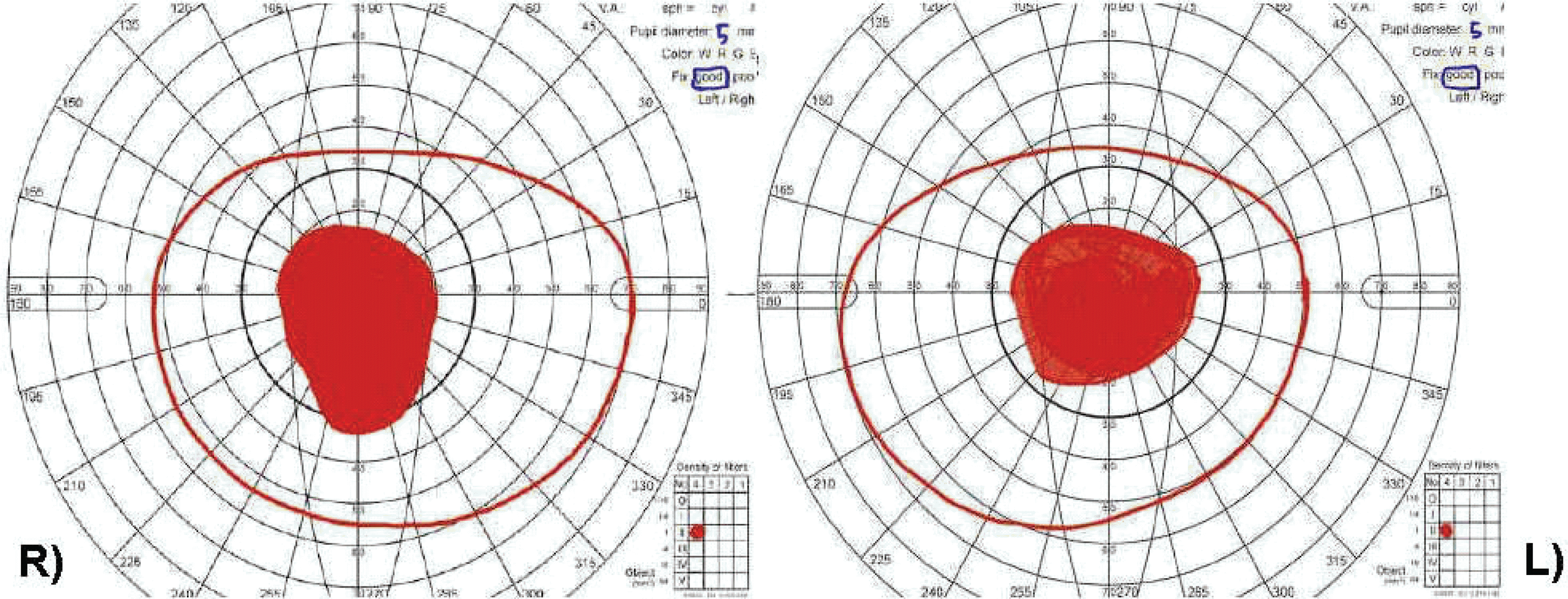
Table 1.
Age distribution of the total patients group and that of the macular degeneration group.
| Age (yr) | Total | Macular degeneration |
|---|---|---|
| 0∼10 | 33 | 0 |
| 11∼20 | 34 | 0 |
| 21∼30 | 24 | 0 |
| 31∼40 | 23 | 0 |
| 41∼50 | 40 | 4 |
| 51∼60 | 44 | 2 |
| 61∼70 | 48 | 12 |
| 71∼80 | 29 | 15 |
| 81∼ | 8 | 5 |
| Total | 283 | 38 |
Table 2.
Distribution of binocular visual acuity of patients (far, near visual acuity before, and after the correction with low vision aids)
Table 3.
Binocular near visual acuity of the patients before and after the correction with the low vision aids
| Cases |
V/A* before correction |
V/A after correction† |
||
|---|---|---|---|---|
| Decimal | LogMAR | Decimal | LogMAR | |
| 1 | 0.63 | 0.2 | 0.63 | 0.2 |
| 2 | 0.1 | 1.0 | 0.5 | 0.3 |
| 3 | 0.1 | 1.0 | 0.5 | 0.3 |
| 4 | 0.08 | 1.1 | 0.63 | 0.2 |
| 5 | 0.2 | 0.7 | 0.4 | 0.4 |
| 6 | 0.12 | 0.9 | 0.5 | 0.3 |
| 7 | 0.063 | 1.2 | 0.5 | 0.3 |
| 8 | 0.12 | 0.9 | 0.5 | 0.3 |
| 9 | 0.063 | 1.2 | 0.5 | 0.3 |
| 10 | 0.1 | 1.0 | 0.2 | 0.7 |
| 11 | 0.05 | 1.3 | 0.4 | 0.4 |
| 12 | 0.1 | 1.0 | 0.63 | 0.2 |
| 13 | 0.1 | 1.0 | 0.32 | 0.5 |
| 14 | 0.08 | 1.1 | 0.25 | 0.6 |
| 15 | 0.1 | 1.0 | 0.4 | 0.4 |
| 16 | 0.1 | 1.0 | 0.4 | 0.4 |
| 17 | 0.16 | 0.8 | 0.5 | 0.3 |
| 18 | 0.12 | 0.9 | 0.5 | 0.3 |
| 19 | 0.16 | 0.8 | 0.32 | 0.5 |
| 20 | 0.32 | 0.5 | 0.4 | 0.4 |
| 21 | 0.05 | 1.3 | 0.12 | 0.9 |
| 22 | 0.2 | 0.7 | 0.4 | 0.4 |
| 23 | 0.25 | 0.6 | 0.8 | 0.1 |
| 24 | 0.12 | 0.9 | 0.63 | 0.2 |
| 25 | 0.12 | 0.9 | 0.5 | 0.3 |
| 26 | 0.16 | 0.8 | 0.63 | 0.2 |
| 27 | 0.2 | 0.7 | 0.63 | 0.2 |
| 28 | 0.16 | 0.8 | 0.2 | 0.7 |
| 29 | 0.3 | 0.5 | 0.4 | 0.4 |
| 30 | 0.08 | 1.1 | 0.5 | 0.3 |
| Mean V/A‡ | 0.90±0.25 | 0.37±0.17 | ||
 XML Download