

 PDF
PDF ePub
ePub Citation
 Print
Print


Abstract
Purpose
To investigate the long-term results and complications of orbital wall fracture reconstruction using the bioabsorbable orbital implant, Macropore® orbital floor liner.
Methods
This retrospective study included patients who underwent the reconstruction of an orbital wall fracture using Macropore®
orbital floor liner and completed a postoperative follow-up longer than 6 months. The enophthalmic values as well as the data of ocular movement and diplopia was collected from the medical records of each patient and analyzed.
Results
A total of 35 patients were evaluated with an average follow-up period of 14.0 months. The average enophthalmic value of 29 patients, whose reconstruction was primarily indicated from enophthalmic tissue, was 0.90 mm at the last follow-up. No significant progression of enophthalmos was observed at postoperative 12, 18 and 24 months when compared with the enophthalmic value at postoperative 6 months. All 15 patients who have had the limitation of ocular movement or diplopia preoperatively resolved completely or improved to the degree that no clinically significant limitation or diplopia further existed. No complications such as dislocation of implant, infection, and aggravation of ocular limitation were observed during the follow-up period.
References
1. Holmes RE, Cohen SR, Cornwall GB, et al. MacroPore resorbable devices in craniofacial surgery. Clin Plast Surg. 2004; 31:393–406.

2. Park YJ, Chung IY, Seo SW. An analysis of orbital reconstruction with bioresorbable plate though orbital volume assessment. J Korean Ophthalmol Soc. 2008; 49:1046–53.
3. Chi MJ, Jeung JW, Lee JH. Reconstruction of orbital wall fracture with resorbable copolymer mesh. J Korean Ophthalmol Soc. 2006; 47:1021–30.
4. Kontio RK, Laine P, Salo A, et al. Reconstruction of internal orbital wall fracture with iliac crest free bone graft: clinical, computed tomography, and magnetic resonance imaging follow-up study. Plast Reconstr Surg. 2006; 118:1365–74.
5. Krishnan V, Johnson JV. Orbital floor reconstruction with autogenous mandibular symphyseal bone grafts. J Oral Maxillofac Surg. 1997; 55:327–30.
6. Kraus M, Gatot A, Fliss DM. Repair of traumatic inferior orbital wall defects with nasoseptal cartilage. J Oral Maxillofac Surg. 2001; 59:1397–400.
7. Guerra MF, Perez JS, Rodriguez-Campo FJ, Gias LN. Reconstruction of orbital fractures with dehydrated human dura mater. J Oral Maxillofac Surg. 2000; 58:1361–6.
8. Kim HK, Lim HS, Chung WS. Surgical Effect of Medpor in the Reconstruction of Orbital Wall Fracture. J Korean Ophthalmol Soc. 1998; 39:623–30.
9. Villarreal PM, Monje F, Morillo AJ, et al. Porous polyethylene implants in orbital floor reconstruction. Plast Reconstr Surg. 2002; 109:877–85.
10. Mauriello JA Jr., Wasserman B, Kraut R. Use of Vicryl (polyglactin-910) mesh implant for repair of orbital floor fracture causing diplopia: a study of 28 patients over 5 years. Ophthal Plast Reconstr Surg. 1993; 9:191–5.
11. Burres SA, Cohn AM, Mathog RH. Repair of orbital blowout fractures with Marlex mesh and Gelfilm. Laryngoscope. 1981; 91:1881–6.
12. Iizuka T, Mikkonen P, Paukku P, Lindqvist C. Reconstruction of orbital floor with polydioxanone plate. Int J Oral Maxillofac Surg. 1991; 20:83–7.
13. Tuncer S, Yavuzer R, Kandal S, et al. Reconstruction of traumatic orbital floor fractures with resorbable mesh plate. J Craniofac Surg. 2007; 18:598–605.
14. Hwang JH, Kwak MS. Residual Diplopia and Enophthalmos after Reconstruction of Orbital Wall Fractures. J Korean Ophthalmol Soc. 2003; 44:1959–65.
15. Hu J, Liu X, Ma PX. Induction of osteoblast differentiation phenotype on poly (L-lactic acid) nanofibrous matrix. Biomaterials. 2008; 29:3815–21.
16. Rozema FR, Bos RR, Pennings AJ, Jansen HW. Poly (L-lactide) implants in repair of defects of the orbital floor: an animal study. J Oral Maxillofac Surg. 1990; 48:1305–9.
Figure 1.
Classification of orbital wall fracture according to the percent of bony defect in coronal CT scan in which the maximum defect is observed. (A) Small size fracture: less than 25% of bony defect, (B) Medium size fracture: between 25 and 75% of defect. (C) Large size fracture: more than 75% of defect.
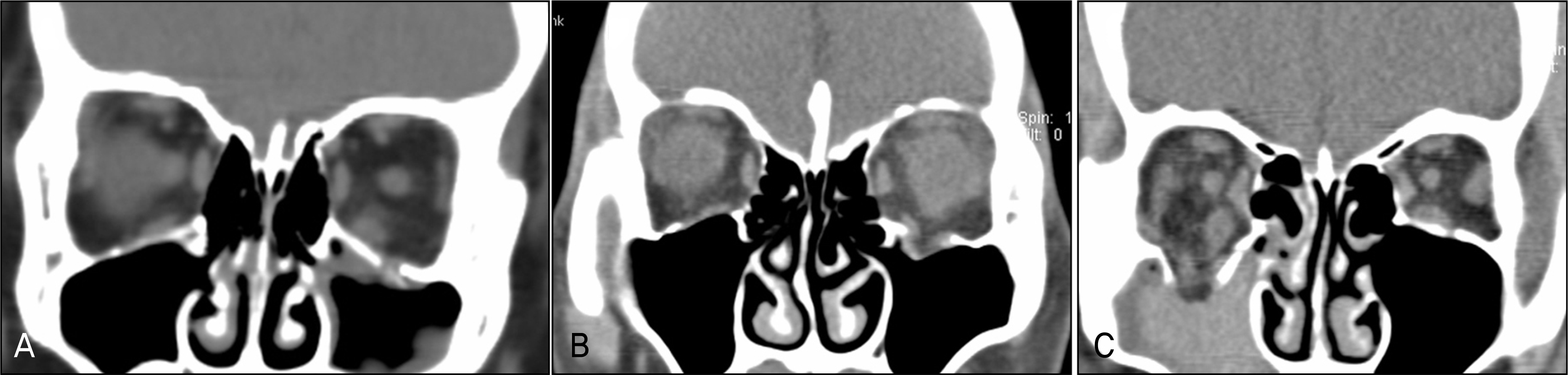
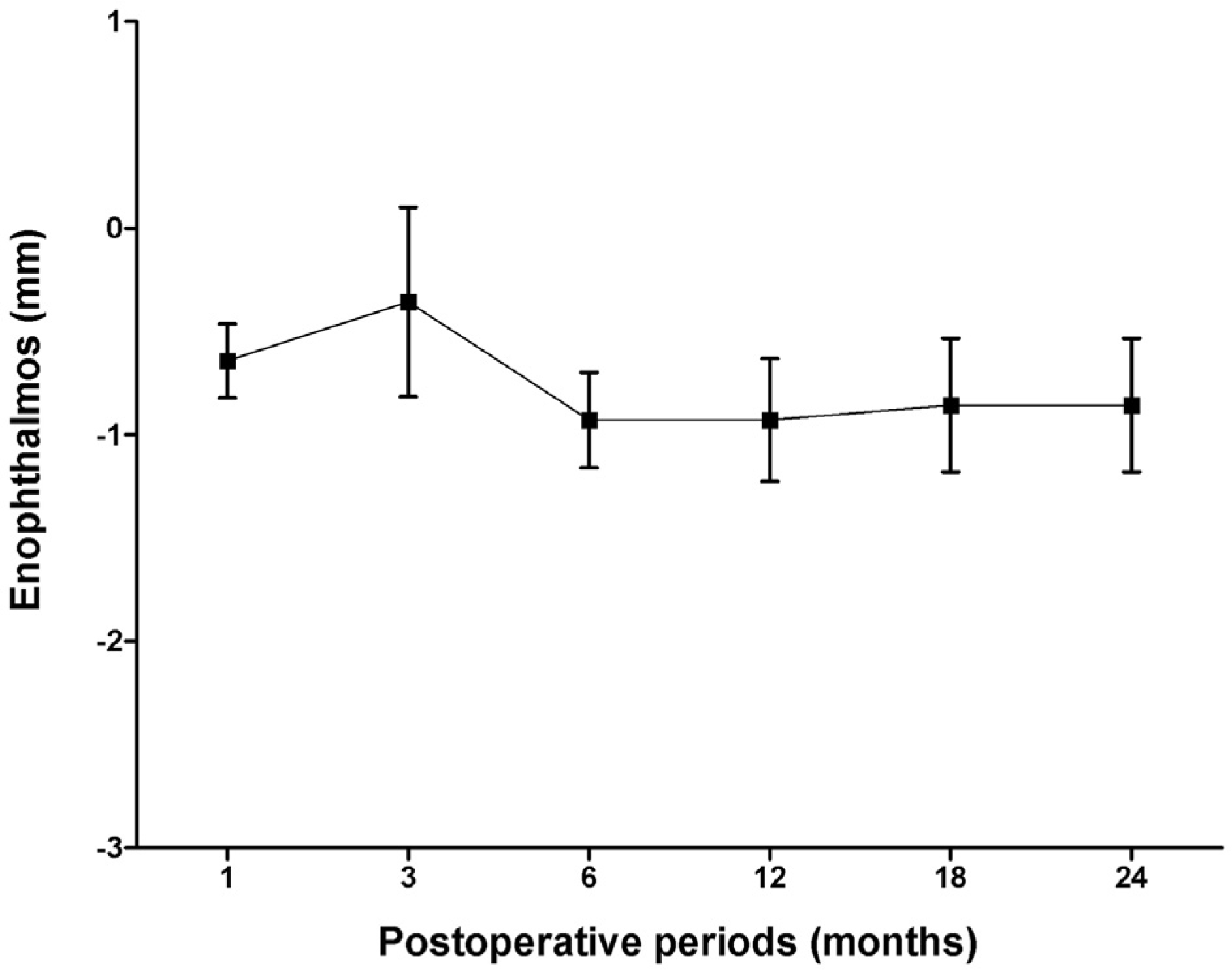
Figure 3.
Preoperative (A) and immediate postoperative (B) CT of the patient who showed 3 mm of enophthalmos at postoperative 6 months. (A) Large-size right inferior orbital wall fracture is noted preoperatively. (B) Well reconstructed inferior orbital wall with Macropore® orbital floor liner is observed (arrow).
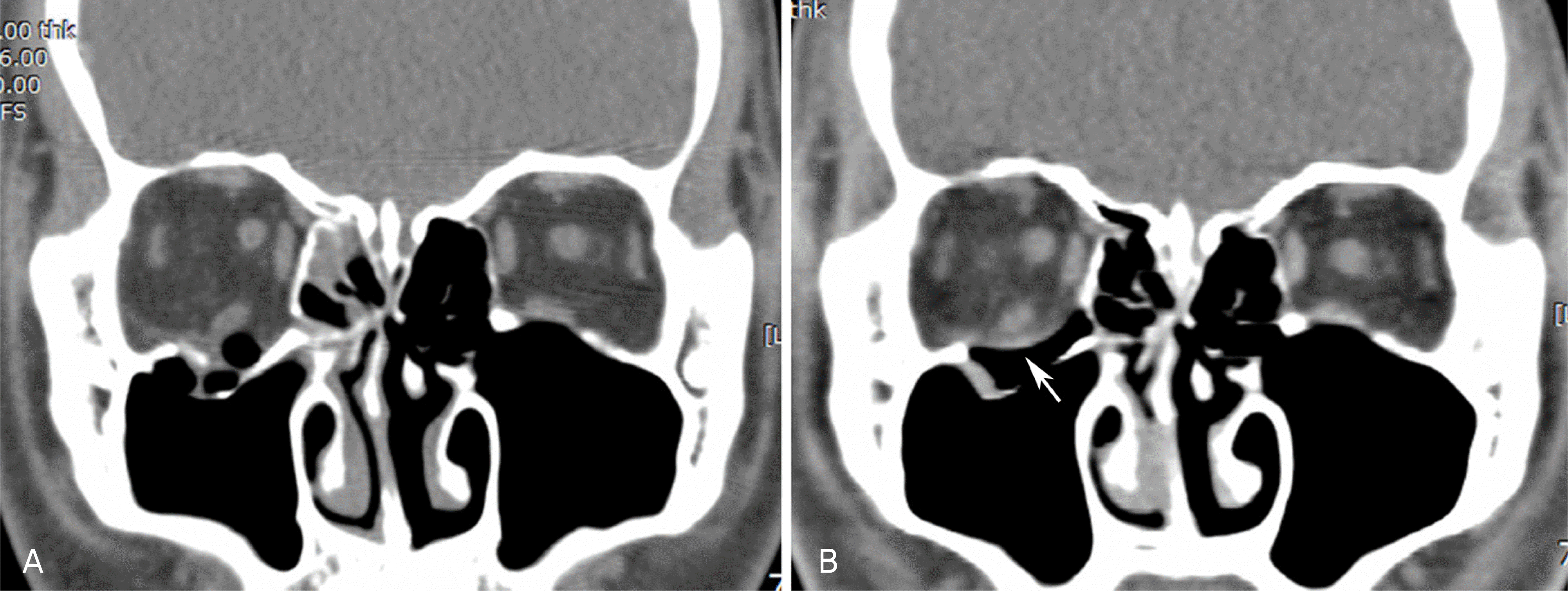
Figure 4.
Computerized tomography (CT) of patient who were followed up to postoperative 24 months. (A) Large size right inferior orbital wall fracture is noted preoperatively. (B) In immediate postoperative CT scan, orbital floor is well reconstructed with Macropore, which is seen as a radiolucent line. (C) The contour of orbital floor including inferior rectus muscle is well maintained at postoperative 24 months. The area where Macropore was located previously is replaced by soft tissue (probably thought as fibrotic tissue) (short arrow), and the line with bone density outlines the orbital floor, which suggests calcification (long arrow).
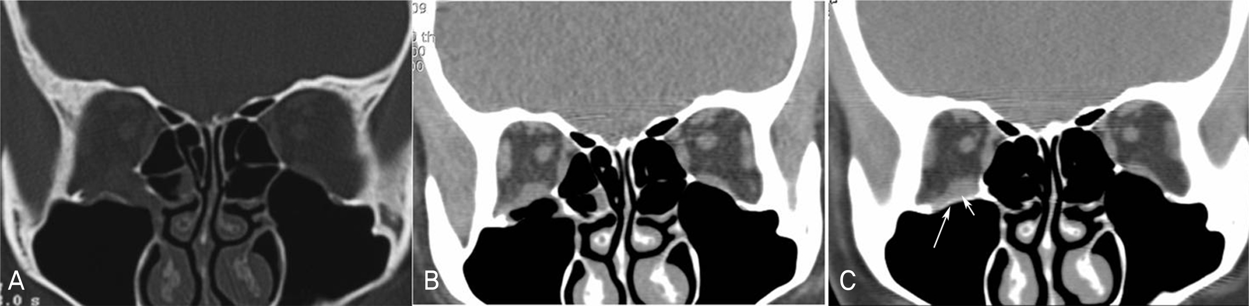
Table 1.
Demographic data of patients
| Characteristics | Number of patients (%) | |
|---|---|---|
| Sex | Male | 25 (71.4) |
| Female | 10 (28.6) | |
| Cause of trauma | Violence | 16 (45.7) |
| Fall down | 8 (22.9) | |
| Sports | 3 (8.6) | |
| Traffic accident | 6 (17.1) | |
| Others | 2 (5.7) | |
| Involved wall | Medial wall | 12 (34.3) |
| Inferior wall | 20 (57.1) | |
| Both | 3 (8.6) | |
| Size of fracture* | Small | 6 (17.1) |
| Medium | 9 (25.8) | |
| Large | 20 (57.1) |
Table 2.
The enophthalmic values of patients at postoperative follow-up periods (mm)
| Follow up | Postoperative 6 months | Postoperative 12 months | Postoperative 18 months | Postoperative 24 months |
|---|---|---|---|---|
| 24 months (n=7)† | 1.00±0.71 | 0.93±0.79* | 0.86±0.85* | 0.86±0.85* |
| 18 months (n=13)‡ | 1.04±0.80 | 1.08±0.86* | 1.00±0.91* | N/A |
| 12 months (n=20) | 1.00±0.87 | 0.83±0.92* | N/A | N/A |
Table 3.
The number of patients showing limitation of ocular movement and diplopia according to each grade at preoperative and last follow up
|
Limitation of motion* |
Diplopia† |
|||
|---|---|---|---|---|
| Preoperative | Postoperative | Preoperative | Postoperative | |
| Grade 0 | 0 (0.0 %) | 13 (86.7 %) | 0 (0.0 %) | 13 (86.7 %) |
| Grade 1 | 4 (26.7 %) | 2 (13.3 %) | 0 (0.0 %) | 2 (13.3 %) |
| Grade 2 | 7 (46.7 %) | 0 (0.0 %) | 5 (33.3 %) | 0 (0.0 %) |
| Grade 3 | 2 (13.3 %) | 0 (0.0 %) | 9 (60.0 %) | 0 (0.0 %) |
| Grade 4 | 2 (13.3 %) | 0 (0.0 %) | 1 (6.7 %) | 0 (0.0 %) |
Table 4.
The number of cases showing complications after reconstruction of orbital wall fracture with Macropore® orbital floor liner
| Complication | Case / Patients |
|---|---|
| Infection | 0 / 35 |
| Dislocation of implant | 0 / 35 |
| Loss of vision | 0 / 35 |
| Limitation of motion or diplopia | 0 / 20* |
 XML Download