Abstract
Purpose
To report a case of isolated posterior pole-penetrating ocular injury treated by nonsurgical methods such as argon laser photocoagulation and administration of antibiotics.
Case summary
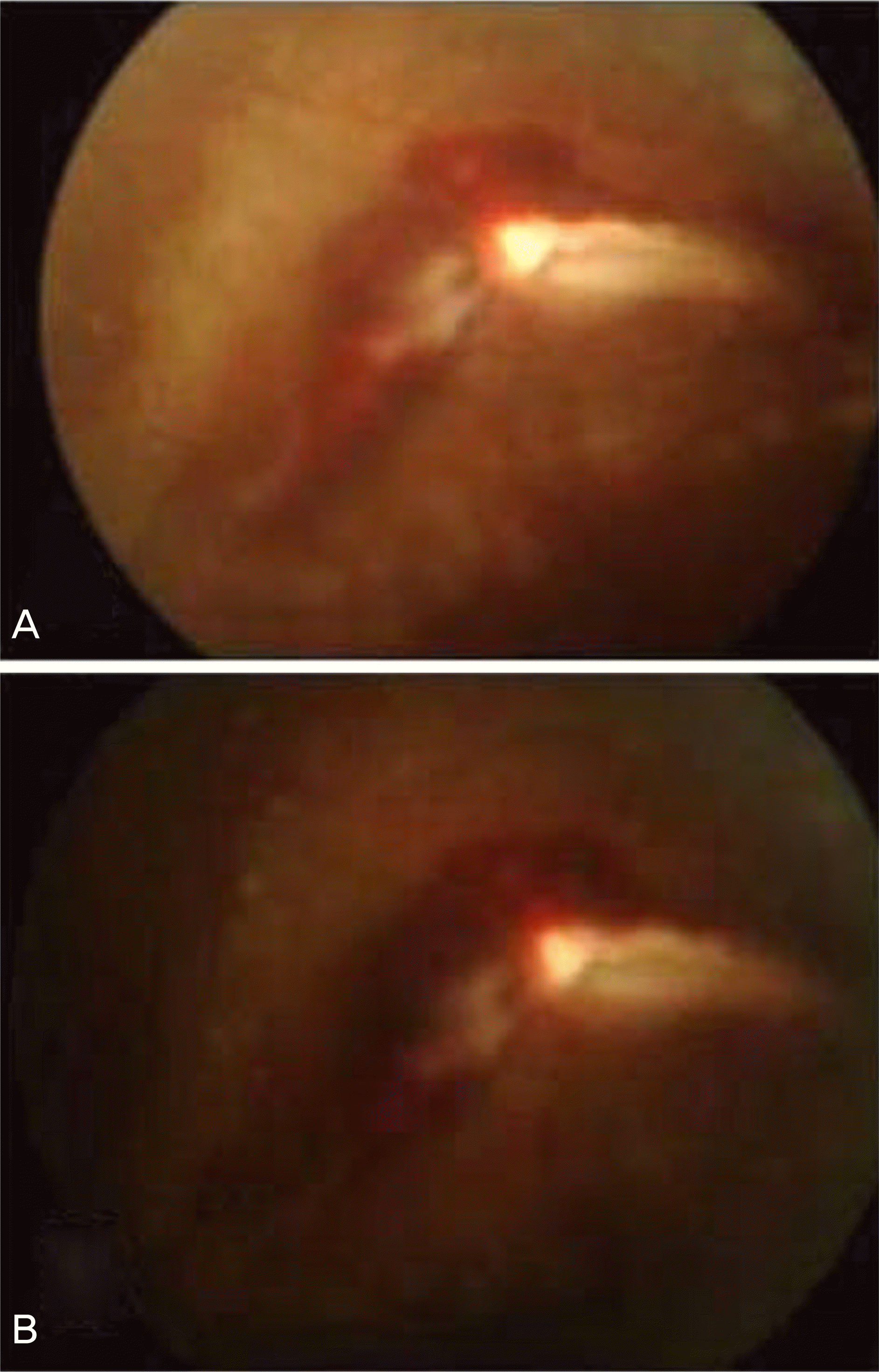
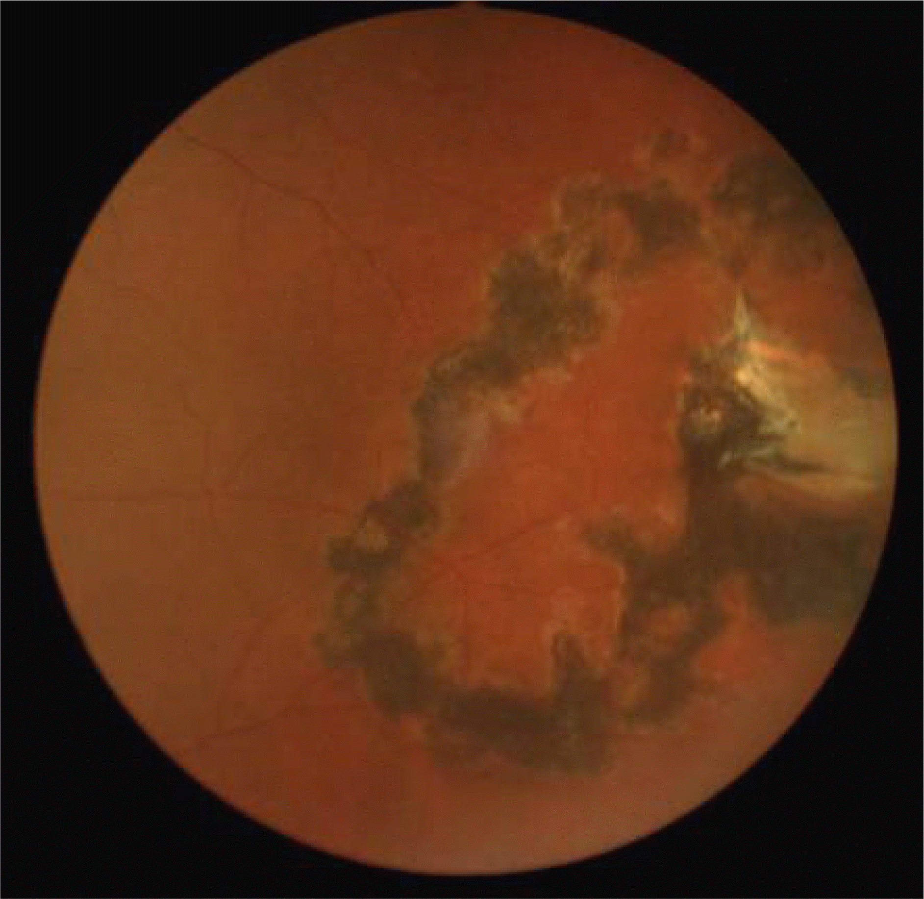
A 46-year-old male visited the hospital complaining of floaters in his left eye which had occurred when his cheek was penetrated by scissors from the inferior posterior part to the superior anterior part while working earlier that day. Upon initial examination, his best corrected visual acuity (BCVA) in the left eye was 0.8, and his intraocular pressure (IOP) was 10 mmHg. No cells or aqueous flares were observed in the anterior chamber. Fundus examination was performed, and three disc diameter-large breaks of the retina and choroid, scleral rupture and vitreous hemorrhage were observed at the posterior pole three disc diameters away from the fovea. It was difficult to make a surgical approach as the lesion was situated on the posterior pole, and there was the risk of prolapse of the eye contents. Therefore, we first performed argon laser photocoagulation around the lesion and administered topical as well as and systemic antibiotics. After admission the patient was observed carefully as the tractional retinal fold was located at the posterior pole. Additional argon laser photocoagulation was performed. After six months of treatment, BCVA in the left eye was 1.0, IOP was 16 mmHg, and no pathologic change was observed on fundus examination.
References
1. Chung SM. Choi JY. A clinical study of penetrating ocular injuries. J Korean Ophthalmol Soc. 1997; 38:491–8.
2. Kim JY, Kim JW, Lee J. Clinical evaluations of penetration ocular injuries. J Korean Ophthalmol Soc. 1992; 33:919–24.
3. Ahn JW, Moon SH, Lee DH, Lee CY. Factors influencing final visual acuity after Penetrating ocular injuries. J Korean Ophthalmol Soc. 1998; 39:2451–8.
4. Park YG, Lee Y, Cho SH. A Clinical Analysis of Inpatients in the Department of Ophthalmology. J Korean Ophthalmol Soc. 1972; 20:555–8.
5. Kim JT. Clinical Study of Perforating Eye Injuries. J Korean Ophthalmol Soc. 1982; 23:645–54.
7. Adhikary HP, Taylor P, Fitzmaurice DJ. Prognosis of perforating eye injury. Br J Ophthalmol. 1976; 60:737–9.

8. Jang Y, Oh S, Ji NC. A clinical observation of ocular injuries of inpatients. J Korean Ophthalmol Soc. 1993; 34:257–63.
9. Han YS, Shyn KH. A statistical observation of the ocular injuries. J Korean Ophthalmol Soc. 2005; 46:117–24.
10. Pieramici DJ, MacCumber MW, Humayun MU, et al. Open global injury, Update on types of injuries and visual results. Ophthalmology. 1996; 103:1798–803.
11. Ryan SJ, Liggett PE. Posterior penetrating ocular trauma. Ryan SJ, editor. Retina. 4th ed.St. Louis: CV Mosby;2006. v. 3. chap. 23.
12. Cleary PE, Ryan SJ. Histology of wound, vitreous, and retina in experimental posterior penetrating eye injury in the rhesus monkey. Am J Ophthalmol. 1979; 88:221–31.

13. Cleary PE, Minckler DS, Ryan SJ. Ultrastructure of traction retinal detachment in rhesus monkey eyes after posterior penetrating ocular injury. Am J Ophthalmol. 1980; 90:829–45.
14. Deutsch TA, Feller DB. Paton and Goldberg's management of ocular injuries. 2nd ed.Philadelphia: WB Saunders;1985. p. 148–9.
15. Kim MJ, Chang BH, Kim IC, et al. Posttraumatic bacillus cereus endophthalmitis. J Korean Ophthalmol Soc. 2005; 46:1597–604.
16. Thomson JT, Parver LM, Enger CL, et al. Infectious endophthalmitis after penetrating injuries with retained intraocular foreign bodies. Ophthalmology. 1993; 100:1468–74.

17. Punnonen E, Laatikainen L. Prognosis of perforating eye injuries with intraocular foreign bodies. Acta Ophthalmol. 1989; 67:483–91.

18. Yoo SJ, Cho SW, Kim JW. Clinical Analysis of Posttraumatic Endophthalmitis. J Korean Ophthalmol Soc. 2004; 45:69–78.
19. Baum JL. Current concepts in ophthalmology. Ocular infection. N Engl J Med. 1987; 299:28–31.
21. Kunimoto DY, Das T, Sharama S, et al. Microbiologic spectrum and susceptibility of isolates: Part II. Postoperative endophthalmitis. Am J Ophthalmol. 1999; 128:242–4.
22. Woodcock JM, Andrews JM, Boswell FJ, et al. In vitro activity of BAY 12-8039, a new fluoroquinolone. Antimicrob agents chemother. 1997; 41:101–6.

23. Donati M, Rodriguez Fermepin M, Olmo A, et al. Comparative in-vitro activity of moxifloxacin, minocycline, and azithromycin, against Chlamydia spp. J Antimicrobe Chemother. 1999; 43:825–37.

24. Aldridge KE, Ashcraft DS. Comparison of the in vitro activities of BAY 12-8039, a new quinolone, against clinically important anae-robe. Antimicrob Agents Chemother. 1997; 41:709–11.
Figure 1.
At the first visit. (A) Clinical photograph shows the scissors penetrating injury from the left lateral cheek to the middle of the lower eyelid. When he came to the clinic, primary repair had already been done at a local plastic clinic. (B) Fundus photograph of the left eye shows 3 disc diameter-large breaks of the retina and choroid, scleral rupture and vitreous hemorrhage were observed at the posterior pole 3 disc diameters away from the fovea. (C) Illustration shows mechanism of ocular isolated posterior pole injury by scissors.


 PDF
PDF ePub
ePub Citation
 Print
Print


 XML Download