

 PDF
PDF ePub
ePub Citation
 Print
Print


Abstract
Purpose
To evaluate the additional protective effect of the soft shell technique on corneal endothelial cells in torsional mode phacoemulsification.
Methods
Torsional mode phacoemulsification was performed on 60 eyes in 38 patients who were diagnosed with moderate cataracts. Thirty eyes were assigned to the soft shell technique group, and a control group with 30 eyes using only cohesive viscoelastics was created to evaluate ultrasound (US) time, mean US intensity, cumulative dissipated energy (CDE), and amount of used balanced salt solution. Corneal endothelium and corneal thickness were measured before and after the procedure.
Results
There were no statistically significant differences in US time, mean US intensity, cumulative dissipated energy, and dosage of the balanced salt solution between the two groups. There were no statistically significant differences between the two groups in postoperative central corneal thickness. Corneal endothelial cell densities decreased significantly after surgery in both groups, but there were no statistically significant differences between the two groups.
References
1. Jacobs PM, Cheng H, Price NC, et al. Endothelial cell loss after cataract surgery–the problem of interpretation. Trans Ophthalmol Soc U K. 1982; 102:291–3.
2. Kreisler KR, Mortenson SW, Mamalis N. Endothelial cell loss following “modern” phacoemulsification by a senior resident. Ophthalmic Surg. 1992; 23:158–60.

3. Werblin TP. Long-term endothelial cell loss following phacoemulsification: model for evaluating endothelial damage after intraocular surgery. Refract Corneal Surg. 1993; 9:29–35.
4. Hayashi K, Nakao F, Hayashi F. Corneal endothelial cell loss after phacoemulsification using nuclear cracking procedures. J Cataract Refract Surg. 1994; 20:44–7.
5. Diaz-Valle D, Benitez del Castillo Sanchez JM, Castillo A, et al. Endothelial damage with cataract surgery techniques. J Cataract Refract Surg. 1998; 24:951–5.
6. Storr-Paulsen A, Norregaard JC, Ahmed S, et al. Endothelial cell damage after cataract surgery: divide-and-conquer versus phaco- chop technique. J Cataract Refract Surg. 2008; 34:996–1000.
7. Storr-Paulsen A, Norregaard JC, Farik G, Tarnhoj J. The influence of viscoelastic substances on the corneal endothelial cell population during cataract surgery: a prospective study of cohesive and dispersive viscoelastics. Acta Ophthalmol Scand. 2007; 85:183–7.
8. Baykara M, Ercan I, Ozcetin H. Microincisional cataract surgery (MICS) with pulse and burst modes. Eur J Ophthalmol. 2006; 16:804–8.
9. Fishkind W, Bakewell B, Donnenfeld ED, et al. Comparative clinical trial of ultrasound phacoemulsification with and without the WhiteStar system. J Cataract Refract Surg. 2006; 32:45–9.
10. Hoffman RS, Fine IH, Packer M. New phacoemulsification technology. Curr Opin Ophthalmol. 2005; 16:38–43.
11. Mackool RJ, Brint SF. AquaLase: a new technology for cataract extraction. Curr Opin Ophthalmol. 2004; 15:40–3.
12. Gimbel HV, da Reitz Pereira C. Advances in phacoemulsification equipment. Curr Opin Ophthalmol. 2002; 13:30–2.
13. Vargas LG, Holzer MP, Solomon KD, et al. Endothelial cell integrity after phacoemulsification with 2 different handpieces. J Cataract Refract Surg. 2004; 30:478–82.
14. Behndig A, Lundberg B. Transient corneal edema after phacoemulsification: comparison of 3 viscoelastic regimens. J Cataract Refract Surg. 2002; 28:1551–6.
15. Hayashi K, Hayashi H, Nakao F, Hayashi F. Risk factors for corneal endothelial injury during phacoemulsification. J Cataract Refract Surg. 1996; 22:1079–84.
16. Sugar J, Mitchelson J, Kraff M. The effect of phacoemulsification on corneal endothelial cell density. Arch Ophthalmol. 1978; 96:446–8.
17. Liu Y, Zeng M, Liu X, et al. Torsional mode versus conventional ultrasound mode phacoemulsification: randomized comparative clinical study. J Cataract Refract Surg. 2007; 33:287–92.
18. Kim JH, Kang NY, Kwak NH, et al. Cataract. Seoul: Ilchokak;2008. p. 122–3.
19. Lane SS, Naylor DW, Kullerstrand LJ, et al. Prospective comparison of the effects of Occucoat, Viscoat, and Healon on intraocular pressure and endothelial cell loss. J Cataract Refract Surg. 1991; 17:21–6.
20. Hutz WW, Eckhardt HB, Kohnen T. Comparison of viscoelastic substances used in phacoemulsification. J Cataract Refract Surg. 1996; 22:955–9.
21. Bissen-Miyajima H. Ophthalmic viscosurgical devices. Curr Opin Ophthalmol. 2008; 19:50–4.
22. Arshinoff SA. Dispersive-cohesive viscoelastic soft shell technique. J Cataract Refract Surg. 1999; 25:167–73.
23. Lee DH, La TY, Oh JR. The Corneal Endothelial Protective Effect of Soft Shell Technique in Cataract Surgery. J Korean Ophthalmol Soc. 2003; 44:2523–8.
24. Kim H, Joo CK. Efficacy of the soft-shell technique using Viscoat and Hyal-2000. J Cataract Refract Surg. 2004; 30:2366–70.
25. Miyata K, Nagamoto T, Maruoka S, et al. Efficacy and safety of the soft-shell technique in cases with a hard lens nucleus. J Cataract Refract Surg. 2002; 28:1546–50.
26. Chylack LT Jr, Leske MC, McCarthy D, et al. Lens opacities classification system II (LOCS II). Arch Ophthalmol. 1989; 107:991–7.
27. Kelman CD. Phacoemulsification and aspiration. A new technique of cataract removal. A preliminary report. Am J Ophthalmol. 1967; 64:23–35.
28. Dick HB, Schwenn O. Viscoelastics in ophthalmic surgery. New York: Spinger;2000. p. 7–24.
29. Steinert RF. Cataract Surgery: Technique, Complications, and Management. 2nd ed.Philadelphia, Pa: Saunders;2004. p. 43.
30. Calciu-Rusu D, Rothfuss E, Eckelt J, et al. Rheology of sodium hyaluronate saline solutions for ophthalmic use. Biomacro-molecules. 2007; 8:1287–92.
31. Ray-Chaudhuri N, Voros GM, Sutherland S, Figueiredo FC. Comparison of the effect of sodium hyaluronate (Ophthalin) and hydroxypropylmethylcellulose (HPMC-Ophtal) on corneal endothelium, central corneal thickness, and intraocular pressure after phacoemulsification. Eur J Ophthalmol. 2006; 16:239–46.
32. Miyata K, Maruoka S, Nakahara M, et al. Corneal endothelial cell protection during phacoemulsification: low- versus high-molecular- weight sodium hyaluronate. J Cataract Refract Surg. 2002; 28:1557–60.
33. Davison JA. Ultrasonic power reduction during phacoemulsification using adjunctive NeoSoniX technology. J Cataract Refract Surg. 2005; 31:1015–9.
34. Zeng M, Liu X, Liu Y, et al. Torsional ultrasound modality for hard nucleus phacoemulsification cataract extraction. Br J Ophthalmol. 2008; 92:1092–6.
35. Alio J, Rodriguez-Prats JL, Galal A, Ramzy M. Outcomes of microincision cataract surgery versus coaxial phacoemulsification. Ophthalmology. 2005; 112:1997–2003.
36. Cameron MD, Poyer JF, Aust SD. Identification of free radicals produced during phacoemulsification. J Cataract Refract Surg. 2001; 27:463–70.
Figure 1.
Sequential changes of central cornea thickness (μm) in the soft shell technique and control group after cataract surgery.
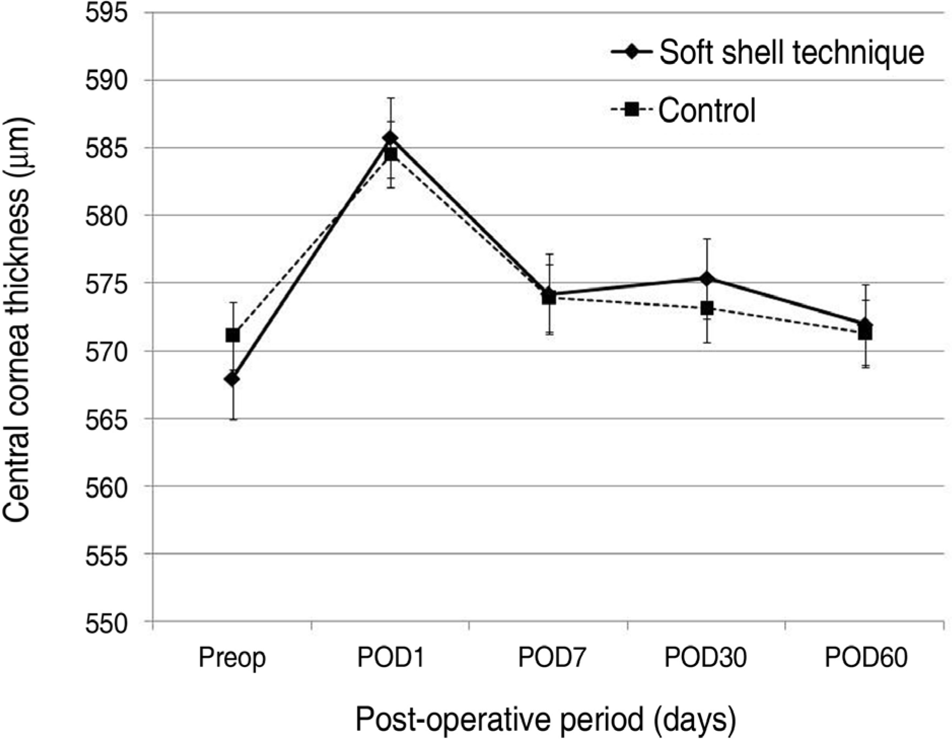
Figure 2.
Sequential changes of endothelial cell density (cells/mm2) in the soft shell technique and control group after cataract surgery.
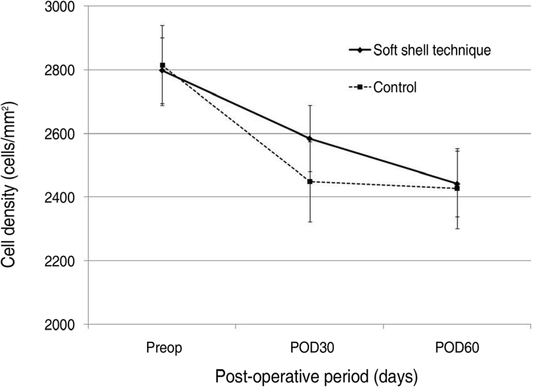
Table 1.
Intraoperative characteristics of the parameters
| Soft shell technique group | Conventional group | p value | |
|---|---|---|---|
| Mean BSS† volume (mL) | 61.4±16.72 | 55.0±19.35 | 0.17 |
| Mean Phaco time (sec) | 33.58±15.82 | 30.16±20.18 | 0.46 |
| Mean Phaco power (%) | 32.19±17.08 | 32.48±20.87 | 0.85 |
| CDE∗ | 3.97±2.13 | 3.68±2.98 | 0.67 |
Table 2.
Correlation between phacoemulsification parameter and decrement of endothelial cells
|
Correlation coefficient (p value) |
||
|---|---|---|
| Soft shell technique group | Conventional group | |
| Mean BSS volume(ml) | 0.150 (0.40) | 0.055 (0.77) |
| Mean Phaco time(sec) | 0.171 (0.36) | 0.027 (0.08) |
| Mean Phaco power(%) | 0.197 (0.29) | 0.020 (0.91) |
| CDE∗ | 0.088 (0.64) | 0.033 (0.86) |
 XML Download