

 PDF
PDF ePub
ePub Citation
 Print
Print


Abstract
Purpose
To investigate the effect of localized retinal detachment on both the detached and attached regions, and to determine the effect of triamcinolone on Müller cell gliosis.
Methods
Pars plana vitrectomy was performed in both eyes of 12 pigmented rabbits. A dome‐ shaped retinal detachment was made by injecting sodium hyaluronate into the subretinal space. Triamcinolone (5 mg) was applied intravitreally to one eye (12 eyes). The detached retinal area and the neighboring attached region were studied by light and electron microscopy 3, 7, and 28 days after surgery. Tissues were prepared in 5 um sections for hematoxylin-eosin staining and immunohistochemistry with antibody to glial fibrillary acidic protein (GFAP).
Results
In addition to the well-known degeneration of photoreceptor cells in the detached retina, an incomplete but severe loss of ganglion cell axons occurs in both the detached and the attached regions. The total retinal thickness gradually decreased in the detached areas, while the thickness of the inner retinal layers remained virtually unchanged over several weeks. Gliotic alterations were apparent in both the detached and non-detached retinal areas, and intravitreal triamcinolone did not alter these gliotic alterations of Müller cells.
References
1. Isashiki M, Ohba N. Recovery of differential light sensitivity following surgery for rhegmatogenous retinal detachment. Graefes Arch Clin Exp Ophthalmol. 1986; 224:184–90.

2. Erickson PA, Fisher SK, Anderson DH, et al. Retinal detachment in the cat: The outer nuclear and outer plexiform layers. Invest Ophthalmol Vis Sci. 1983; 24:927–42.
3. Anderson DH, Guerin CJ, Erickson PA, et al. Morphological recovery in the reattached retina. Invest Ophthalmol Vis Sci. 1986; 27:168–83.
4. Faude F, Francke M, Makarov F, et al. Experimental retinal detachment causes widespread and multilayered degeneration in rabbit retina. J Neurocytol. 2001; 30:379–90.
5. Francke M, Faude F, Pannicke T, et al. Glial cell‐ mediated spread of retinal degeneration during detachment: A hypothesis based upon studies in rabbits. Vision Res. 2005; 45:2256–67.
6. Fisher SK, Lewis GP. Cellular effects of detachment and reattachment on the neural retina and the retinal pigment epithelium. Ryan SJ, Wilkinson CP, editors. Retina. 4th ed.St. Louis: Mosby;2006. 3:chap.p. 115.
7. Jakson TL, Hillenkamp J, Williamson TH, et al. An experimental model of rhegmatogeneous retinal detachment: Surgical results and glial cell response. Invest Ophthalmol Vis Sci. 2003; 44:4026–34.
8. Uckermann O, Pannicke T, Wiedemann P, et al. Triamcinolone does not alter glial cell activation in the experimental detached rabbit retina. J Ocul Pharmacol Ther. 2005; 21:266–74.
9. Fisher SK, Lewis GP, Linberg KA, Verardo MR. Cellular remodeling in mammalian retina: results from studies of experimental retinal detachment. Prog Retin Eye Res. 2005; 24:395–431.
10. Massin P, Audren F, Haouchine B, et al. Intravitreal triamcinolone acetonide for diabetic diffuse macular edema. Ophthalmology. 2004; 111:218–25.
11. Jonas JB. Intravitreal triamcinolone acetonide as treatment for exudative retinal detachment. Br J Ophthalmol. 2004; 88:587–8.
12. Andrade RE, Muccioli C, Farah ME, et al. Intravitreal triamcinlone in the treatment of serous retinal detachment in Vogt‐ Koyanagi‐ Harada syndrome. Am J Ophthalmol. 2004; 137:572–4.
13. Jonas JB, Kreissig I, Degenring R. Intravitreal triamcinolone acetonide for treatment of intraocular proliferative, exudative, and neovascular diseases. Prog Retin Eye Res. 2005; 24:587–611.
14. Sakamoto T, Miyazaki M, Hisatomi T, et al. Triamcinolone-assisted pars plana vitrectomy improves the surgical procedures and decrease the postoperative blood‐ ocular barrier breakdown. Graefes Arch Clin Exp Ophthalmol. 2002; 240:423–9.
15. Sasoh M, Yoshida S, Kuze M, Uji Y. The multifocal electroretinogram in retinal detachment. Doc Ophthalmol. 1998; 94:239–52.
16. Yammana T, Kita M, Ozaki S, et al. The process of closure of experimental retinal holes in rabbit eyes. Graefes Arch Clin Exp Ophthalmol. 2000; 238:81–7.
Figure 1.
Fundus photographs of a rabbit eye after detachment surgery. (A) After a small retinal hole had been produced by a fluid stream tangential to the retinal surface, an injector needle was introduced through the hole and a solution of 0.25% sodium hyaluronate was infused between the retina and retinal pigment epithelium to create a localized retinal detachment. Note that a stable local retinal detachment is established at 3 days after surgery. (B) One week after surgery. (C) Four weeks after surgery, the surgically detached retinal area remained in the detached state over the entire period studied (up to four weeks).
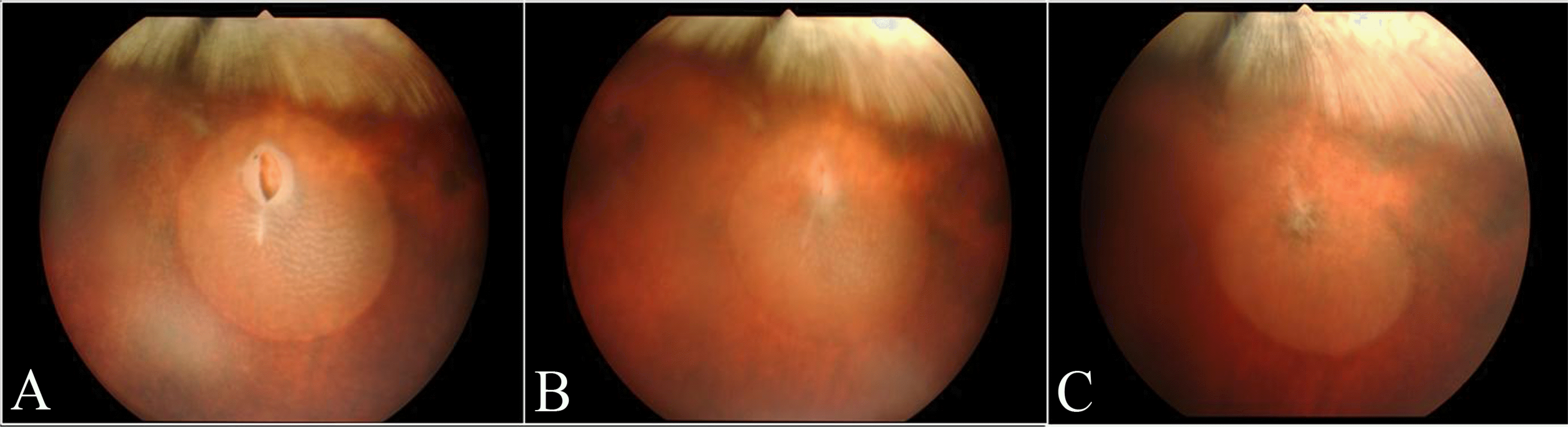
Figure 2.
Electron microscopic photographs of rabbit retina. (A) Ultrastructure of a non‐ detached area at the superior retina of medullary ray (normal retina). (B) Ultrastructure of the detached area four weeks after surgery, the detached retina underwent considerable cell loss and degeneration, and became rather thin. Note the cystic change of photoreceptor cell nuclei, abnormally swollen inner segments, and severely denatured outer segments. (C) Ultrastructure of a non‐ detached neighboring retina of (B), similar (though generally less severe) changes were observed. Note the degenerative changes of photoreceptor nuclei and inner segments.

Figure 3.
Ultrastructure of the detached rabbit retina four weeks after surgery. (A) The outer nuclear layer (ONL) is severe reduced in thickness. In the nerve fiber layer (NFL) and ganglion cell layer (GCL), empty spaces of various sizes are frequently observed. (B) Large empty spaces are found in the NFL just above a deformed ganglion cell. (C) Higher magnifications of the areas indicated in (B). A degenerating ganglion cell with nucleus consisting of disintegrated chromatin resides in close neighborhood to an intact ganglion cell axon (arrow) and empty spaces indicating sites of axon loss. IPL, inner plexiform layer; INL, inner nuclear layer; OPL, outer plexiform layer.

Figure 4.
Ultrastructure of the detached rabbit retina three days after surgery. (A) Within the vitreous body but in close approximity to the degenerating inner nuclear cell, a macrophage is located. (B) Note the degenerative changes of photoreceptor cell nuclei and denatured inner and outer segments.

Figure 5.
Light micrograph and graphs of the detached (regions A, B) and adhering non‐ detached (region C) rabbit retina after detachment surgery. (Upper) Hematoxylin‐ Eosin stained paraffin section of one rabbit four weeks after surgery, used for measurements of the thickness of individual retinal layers (ONL, outer nuclear layer; INL, inner nuclear layer), inner retina (IPL, inner plexiform layer; + GCL, ganglion cell layer; + NFL, nerve fiber layer), and total retinal thickness (between ILM, inner limiting membrane, and OLM, outer limiting membrane). (Bottom) Graphs of two‐ way analysis of variance (retinal area and duration of detachment). The total retinal thickness gradually decreased in the detached areas, with the most severe degeneration occurring in the center of detachment (region A), while the thickness of the inner retina and INL remained virtually unchanged over several weeks, and ONL was substantially reduced.
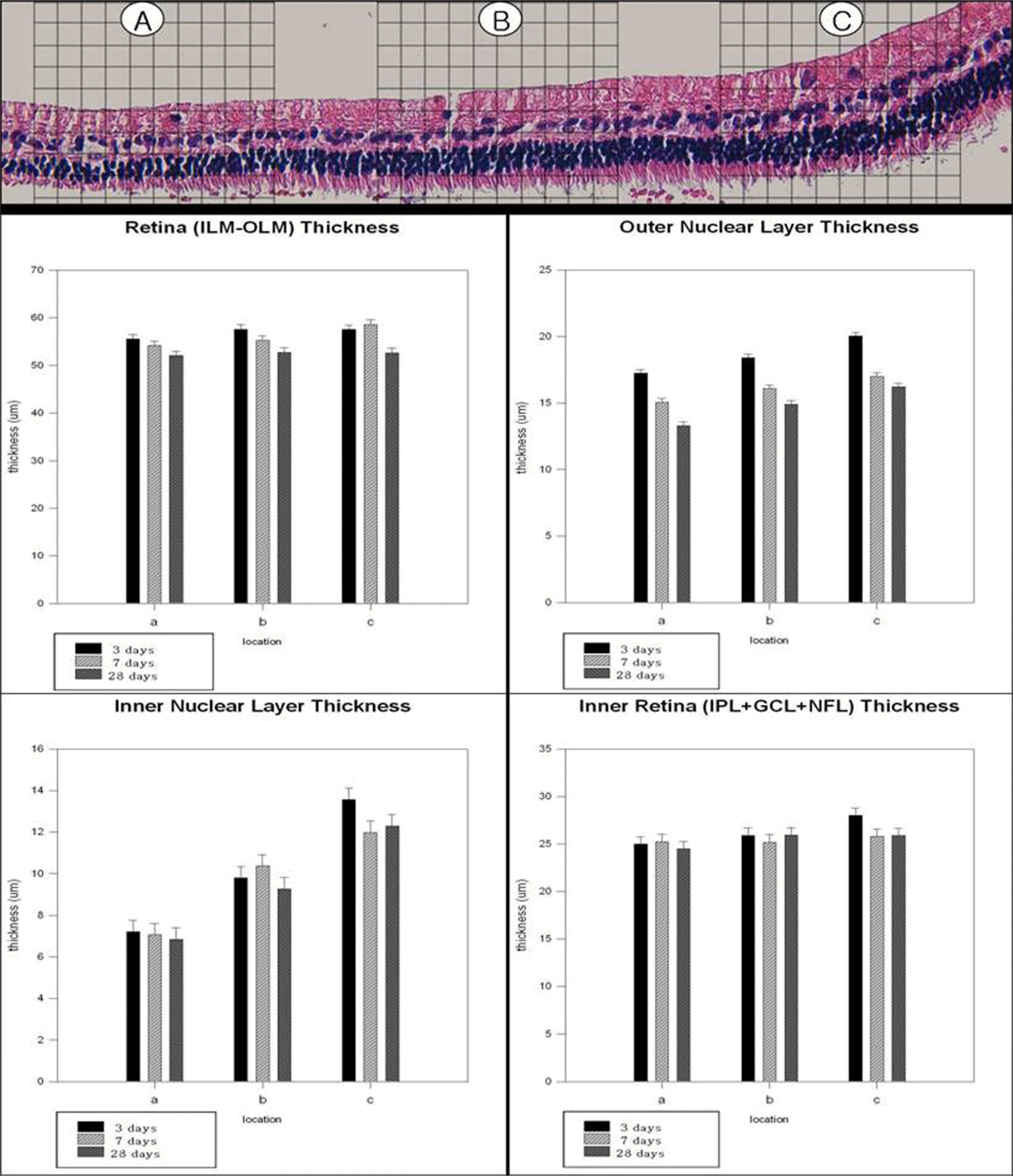
Figure 6.
Light micrograph and graph of the detached (regions A, B) and adhering non‐ detached (region C) rabbit retina after detachment surgery and intravitreal injection of triamcinolone. (Upper) Hematoxylin‐ Eosin stained paraffin section of one rabbit four weeks after retinal detachement surgery and intravitreal injection of triamcinolone (IVTA), used for measurements of the thickness of individual retinal layers (ONL, outer nuclear layer; INL, inner nuclear layer), inner retina (IPL, inner plexiform layer; + GCL, ganglion cell layer; + NFL, nerve fiber layer), and total retinal thickness (between ILM, inner limiting membrane, and OLM, outer limiting membrane). (Bottom) Multiple grouped‐ bar graph of Wilcoxon rank sum test (non‐ IVTA group and IVTA group). The total retinal thickness, ONL thickness, INL thickness, and inner retinal thickness were not significantly different between non‐ IVTA groups and IVTA groups.
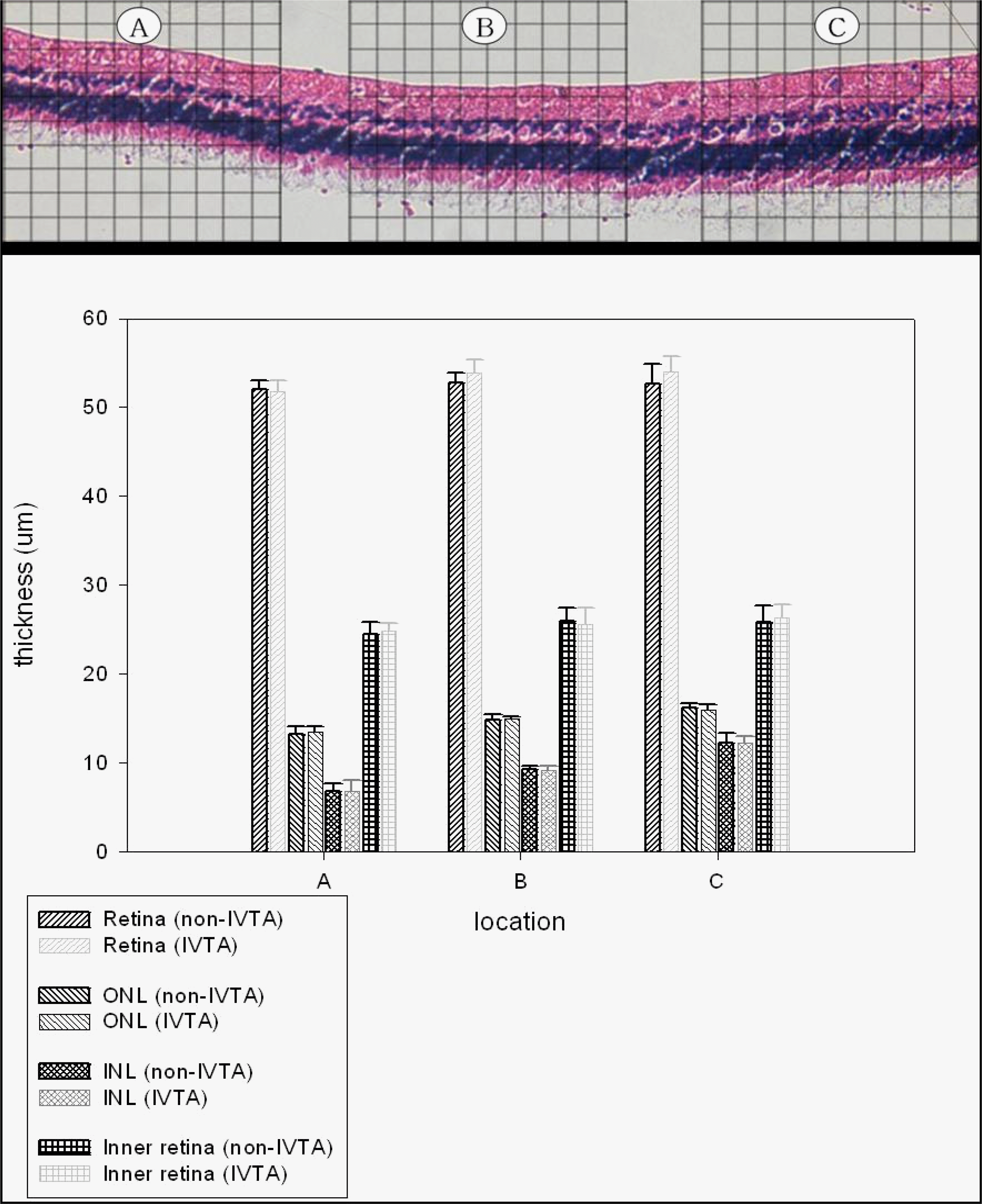
Figure 7.
Light micrographs (×200) of the detached and adhering non‐ detached rabbit retina. (A, B) Normal rabbit retinas (control eyes) show no glial fibrillary acidic protein (GFAP) immunoreacitivity. (C) Relatively strong GFAP immunoreactivity in adhering non‐ detached retina. (D) Relatively strong GFAP immunoreactivity in slice of a rabbit retina which was focally detached for four weeks and was treated with intravitreal triamcinolone injection (IVTA). (E) Strong GFAP immunoreactivity in slice of a rabbit retina which was focally detached for four weeks. (F) Strong GFAP immunoreactivity in slice of a rabbit retina which was focally detached for four weeks and was treated with IVTA. Note that IVTA did not alter these gliotic alterations (GFAP immunoreactivity).

 XML Download