

 PDF
PDF ePub
ePub Citation
 Print
Print


Abstract
Purpose
Angiogenesis is an integral part of wound healing, which is an unwanted process in the postoperative period after trabeculectomy. It was the aim of this study to report on the subconjunctival use of bevacizumab (Avastin®) as an antiproliferative agent to augment trabeculectomy.
Case summary
This clinical interventional case study included 2 patients with secondary glaucoma associated with uveitis who underwent antiglaucomatous filtering surgery combined with a subconjunctival injection of bevacizumab. Limbal-based trabeculectomy was performed, and subconjunctival injections (1.25 mg/0.05 ml) were given at the end of the surgery adjacent to the bleb, which was raised using a single-use 26 gauge needle. At 1 and 2 weeks and 1, 3, and 6 months after surgery, intraocular pressure was reduced in both patients to 11 mmHg with functioning filtering blebs. No complications were observed.
References
1. Skuta GL, Parrish RK 2nd. Wound healing in glaucoma filtering surgery. Surv Ophthalmol. 1987; 32:149–70.

2. Lama PJ, Fechtner RD. Antifibrotics and wound healing in glaucoma surgery. Surv Ophthalmol. 2003; 48:314–46.
3. Ferrara N, Hillan KJ, Gerber HP, Novotny W. Discovery and development of bevacizumab, an anti-VEGF antibody for treating cancer. Nat Rev Drug Discov. 2004; 3:391–400.
4. Cairns JE. Trabeculectomy: Preliminary report of a new method. Am J Ophthamol. 1968; 66:673–9.
5. Michaelson IC. Vascular morphogenesis in the retina of the cat. J Anat. 1948; 82:167–74.
6. Boulton M, Foreman D, Williams G, McLeod D. VEGF localisation in diabetic retinopathy. Br J Ophthalmol. 1998; 82:561–8.
7. Adamis AP, Miller JW, Bernal MT. . Increase vascular endothelia growth factor levels in the vitreous of eyes with proliferative diabetic retinopathy. Am J Ophthalmol. 1994; 118:445–50.
8. Patz A. Studies on retinal neovascularization. Invest Ophthalmol Vis Sci. 1980; 19:1133–8.
9. Spaide RF, Fisher YL. Intravitreal bevacizumab (Avastin) treatment of proliferative diabetic retinopathy complicated by vitreous hemorrhage. Retina. 2006; 26:275–8.
10. Iturralde D, Spaide RF, Meyerle CB. . Intravitreal bevacizumab (Avastin) treatment of macular edema in central retinal vein occlusion: a short-term study. Retina. 2006; 26:279–84.
11. Bahar I, Kaiserman I, McAllum P. . Subconjunctival bevacizumab injection for corenal neovascularization. Cornea. 2008; 27:142–7.
12. Oshima Y, Sakaguchi H, Gomi F, Tano Y. Regression of iris neovascularization after intravitreal injection of bevacizumab in patients with proliferative diabetic retinopathy. Am J Ophthalmol. 2006; 142:155–8.
13. Yazdani S, Hendi K, Pakravan M. Intravitreal bevacizumab (Avastin) injection for neovascular glaucoma. J Glaucoma. 2007; 16:437–9.
14. Jonas JB, Spandau UH, Schlichtenbrede F. Intravitreal Bevacizumab for filtering surgery. Ophthalmic Res. 2007; 39:121–2.
15. Kahook MY, Schuman JS, Noecker RJ. Needle bleb revision of encapsulated filtering bleb with bevacizumab. Ophthalmic Surg Lasers Imaging. 2006; 37:148–50.
16. Bakri SJ, Snyder MR, Reid JM. . Pharmacokinetics of Intravitreal Bevacizumab (Avastin). Ophthalmology. 2007; 114:855–9.
17. Michels S, Rosenfeld PJ, Puliafito CA. . Systemic bevacizumab (Avastin) therapy for neovascular age-related macular degeneration: twelve-week results of an uncontrolled open-label clinical study. Ophthalmology. 2005; 112:1035–47.
Figure 1.
Case. (1a) Conjuntival filtering bleb at one day after bevacizumab injection; (1b) Anterior segment photograph at 1 month after surgery.

Figure 2.
Case 2. (2a) Anterior segment photograph at 1 month after surgery; (2b) Well functioning filtering bleb three months after trabeculectomy with subconjunctival bevacizumab injection.

Figure 3.
Intraocular pressure (IOP) changes following trabeculectomy with subconjunctival bevacizumab injection.
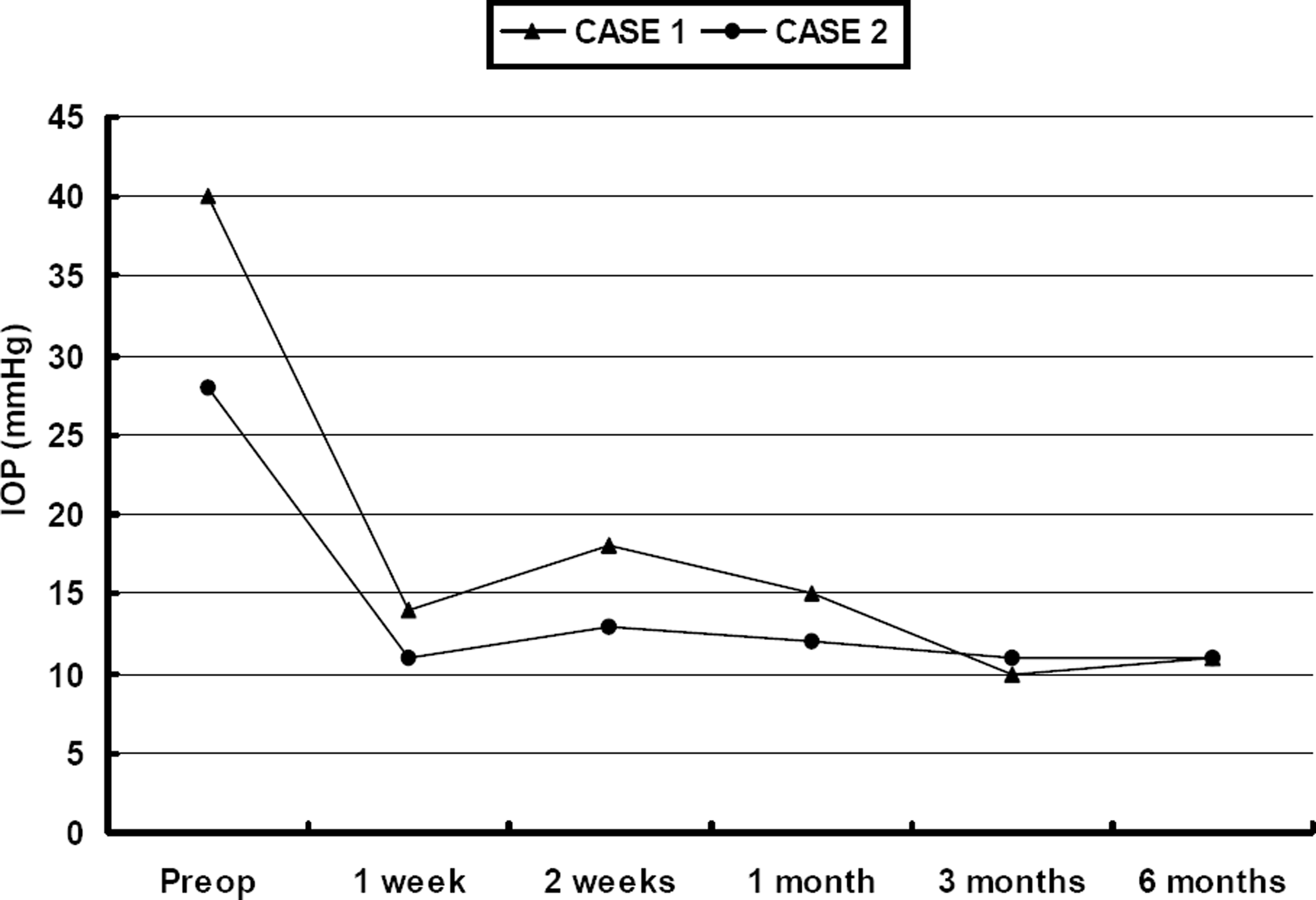
Table 1.
Patients’ characteristics and preoperative and postoperative intraocular pressure and best corrected visual acuity*
| Pre-operative | Post-operative | Post-operative | ||||
|---|---|---|---|---|---|---|
| Case | Age/sex | Diagnosis | Pre-operative IOP | BCVA* | 6 months IOP | 6 months BCVA* |
| 1 | 37/M | Posner-Schlossman syndrome | 40 mmHg | 0.1 | 11 mmHg | 0.15 |
| 2 | 28/M | Behcet disease | 28 mmHg | 0.1 | 11 mmHg | 0.1 |
 XML Download