

 PDF
PDF ePub
ePub Citation
 Print
Print


Abstract
Purpose
To study the clinical characteristics and visual prognosis of Leber's congenital amaurosis in Korea.
Methods
Children who were diagnosed with Leber's congenital amaurosis at Seoul Natioanl University Children's Hospital between 1992 and 2004, were included in this study. The medical records pertaining to the clinical characteristics and visual outcomes of the patients were retrospectively reviewed.
Results
The mean age of the patients at presentation and during the subsequent follow-up period was 15.5 and 62.7 months, respectively. The principal symptoms included lack of fixation (69.0%) and nystagmus (23.8%). At first observation, nystagmus was found in 40 patients, and the appearance of the fundi were variable, including ‘normal’ (23.8%), pigmentary degeneration (54.8%), vascular attenuation (35.7%) and macular coloboma (19.0%). At the age of three to five years, 44.4% of patients had hyperopia greater than +5D. At the time of last follow-up, the visual acuities of the patients who were examined after the age of four were ‘hand motion’ in 68.7%, >20/400 in 9.4%. No patient had visual acuity better than 20/200. Eight (25%) patients could read with glasses or low-vision aids.
Conclusions
The visual prognosis of patients with Leber's congenital amaurosis was poor in most cases, but a majority of our patients displayed a stable clinical course. Progression was rare, and one fourth of the patients were able to read with appropriate aides. In conclusion, regular follow-up care to assess visual function is necessary for optimal outcomes.
References
1. Perrault I, Rozet JM, Gerber S, et al. Leber congenital amaurosis. Mol Genet Metab. 1999; 68:200–20.

2. Heckenlively JR, Foxman SG, Parelhoff ES. Retinal dystrophy and macular coloboma. Doc Ophthalmol. 1988; 68:257–71.
3. Koenekoop RK. An overview of Leber congenital amaurosis: a model to understand human retinal development. Surv Ophthalmol. 2004; 49:379–98.
4. Schuil J, Meire FM, Delleman JW. Mental retardation in amaurosis congenital of Leber. Neuropediatrics. 1998; 29:294–7.
5. Casteels I, Spileers W, Demaerel P, et al. Leber congenital amaurosis—differential diagnosis, ophthalmological and neuroradiological report of 18 patients. Neuropediatrics. 1996; 27:189–93.
6. Schappert-Kimmijser J, Henkes HE, van den Bosch J. Amaurosis congenita (Leber). Arch Ophthalmol. 1959; 61:211–8.
7. Fazzi E, Signorini SG, Uggetti C, et al. Towards improved characterization of Leber congenital amaurosis: neurological and systemic findings. Am J Med Genet A. 2005; 132:13–9.
8. Heher KL, Traboulsi EI, Maumenee IH. The natural history of Leber's congenital amaurosis. Age-related findings in 35 patients. Ophthalmology. 1992; 99:241–5.
9. Lambert SR, Kriss A, Taylor D. Follow-up and diagnostic reappraisal of 75 patients with Leber's congenital amaurosis. Am J Ophthalmol. 1989; 107:624–31.
10. De Laey JJ. Leber's congenital amaurosis. Bull Soc Belge Ophtalmol. 1991; 241:41–50.
11. Fazzi E, Signorini SG, Scelsa B, et al. Leber's congenital amaurosis: An update. Eur J Paediatr Neurol. 2003; 7:2–22.
12. Weiss AH, Biersdorf WR. Visual sensory disorders in congenital nystagmus. Ophthalmology. 1989; 96:517–23.
13. Senior B, Friedmann AI, Braudo JL. Juvenile familial nephropathy with tapetoretinal degeneration. A new oculorenal dystrophy. Am J Ophthalmol. 1961; 52:625–33.
14. Saldino RM, Mainzer F. Cone-shaped epiphyses (CSE) in siblings with hereditary renal disease and retinitis pigmentosa. Radiology. 1971; 98:39–45.
15. Boltshauser E, Isler W. Joubert syndrome: episodic hyperpnea, abnormal eye movements, retardation and ataxia, associated with dysplasia of the cerebellar vermis. Neuropadiatrie. 1977; 8:57–66.
16. Harris EW. Leber's congenital amaurosis and RPE65. Int Ophthalmol Clin. 2001; 41:73–82.
17. Dagi LR, Leys MJ, Hansen RM, et al. Hyperopia in complicated Leber's congenital amaurosis. Arch Ophthalmol. 1990; 108:709–12.
18. Wagner RS, Caputo AR, Nelson LB, et al. High hyperopia in Leber's congenital amaurosis. Arch Ophthalmol. 1985; 103:1507–9.
19. Chew E, Deutman A, Pinckers A, et al. Yellowish flecks in Leber's congenital amaurosis. Br J Ophthalmol. 1984; 68:727–31.
20. Edwards WC, Macdonald R Jr, Price WD. Congenital amaurosis of retinal origin (Leber). Am J Ophthalmol. 1971; 72:724–8.
21. Schroeder R, Mets MB, Maumenee IH. Leber's congenital amaurosis. Retrospective review of 43 cases and a new fundus finding in two cases. Arch Ophthalmol. 1987; 105:356–9.
22. Leighton DA, Harris R. Retinal aplasia in association with macular coloboma, keratoconus and cataract. Clin Genet. 1973; 4:270–4.
23. Margolis S, Scher BM, Carr RE. Macular colobomas in Leber's congenital amaurosis. Am J Ophthalmol. 1977; 83:27–31.
24. Fulton AB, Hansen RM, Mayer DL. Vision in Leber congenital amaurosis. Arch Ophthalmol. 1996; 114:698–703.
25. Dharmaraj SR, Silva ER, Pina AL, et al. Mutational analysis and clinical correlation in Leber congenital amaurosis. Ophthalmic Genet. 2000; 21:135–50.
26. van der Spuy J, Kim JH, Yu YS, et al. The expression of the Leber congenital amaurosis protein AIPL1 coincides with rod and cone photoreceptor development. Invest Ophthalmol Vis Sci. 2003; 44:5396–403.
Figure 1.
Fundus photographs of patients with Leber's congenital amaurosis. (Left) Left eye of a\ 40-month-old girl is notable for pigmentary retinal degenration, retinal vessel attenuation, and macular coloboma. (Right) The right eye of a 6-year-old girl is notable for mild arterial attenuation. When she was 5 months old, fundus finding was normal without vessel attenuation.
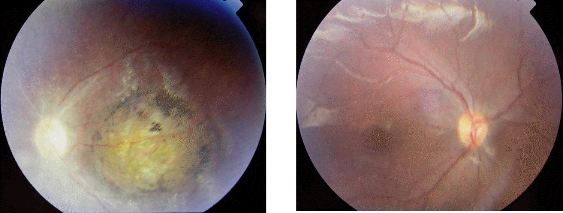
Figure 2.
Typical electroretinogram and visual evoked potential of a patient with Leber's congenital amaurosis. 5 year-old boy. (Left) Electroretinogram. (Right) Visual evoked potential. When he was 9 months old, the findings of both electroretinogram and visual evoked potential were same as above.
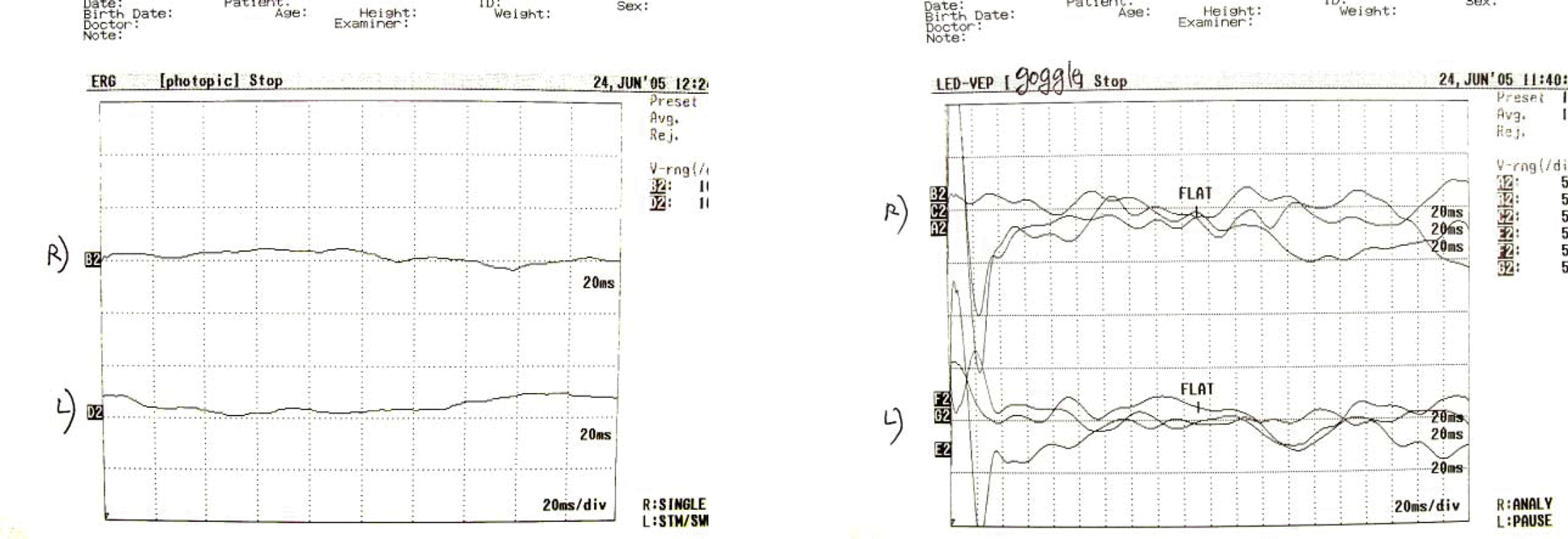
Figure 3.
Visual acuities of patients with Leber's congenital amaurosis at the time of the last follow-up visit. (N=32)
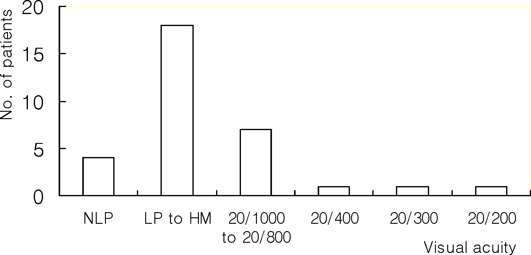
The patients who were examined only before 4 years old, were excluded in this graph.
NLP, no light perception; LP, light perception; HM, hand motion.
Table 1.
Demographics of patients with Leber's congenital amaurosis (N=42)
Table 2.
Principal symptoms of patients with Leber's congenital amaurosis (N=42)
| No fixation | 28 patients (66.7%) |
| Nystagmus | 10 (23.8%) |
| Photophobia | 2 (4.7%) |
| Esodeviation | 1 (2.4%) |
| Family history of poor visual acuity | 1 (2.4%) |
Table 3.
Refractive findings (spherical equivalent) in patients with Leber's congenital amaurosis at the age of 3 to 5 years (N=18)
| Myopia | 2 eyes (11.1%) |
| 0 to +3D | 5 (27.8%) |
| +3 to 5D | 3 (16.7%) |
| ≥+5D | 8 (44.4%) |
Table 4.
Fundus findings in patients with Leber's congenital amaurosis at presentation (N=42)∗
 XML Download