

 PDF
PDF ePub
ePub Citation
 Print
Print


Abstract
Purpose
To compare graft rejection of the first and second eyes after bilateral penetrating keratoplasty.
Methods
We performed a retrospective review of the cases of 16 patients who underwent bilateral penetrating keratoplasty. Nonparametric Kaplan-Meier survival analysis was used to account for variable follow-up among patients.
Results
The mean age of the 16 patients (12 M, 4 F) was 39.1 years. Mean follow-up was 89.8 months after surgery in the first eye and 42.8 months after surgery in the second eye. The average time interval between surgery on the first and second eyes was 47.0 months. The indications for surgery were keratoconus (37.5%), corneal dystrophy (25.0%), pseudophakic bullous keratopathy (12.5%), band keratopathy (12.5%), and inflammatory corneal opacity (12.5%). Endothelial rejection occurred in five of the first eyes but was treated successfully, remaining clear until the last follow-up. Endothelial rejections were seen in five of the second eyes, two of which failed. Survival analysis of endothelial rejection showed no significant difference between the first and second eyes; however, survival analysis of the graft failure showed a decreased survival rate of the second eyes compared with that of the first eyes. The best corrected visual acuity at the last follow-up of the second eyes seemed to be worse than that of the first eyes.
Conclusions
Survival analysis of the endothelial rejections showed no significant difference between the first and second eyes. Survival analysis of the graft failure showed decreased survival rate of the second eyes. At the last follow-up the best corrected visual acuity of the second eyes appeared to be worse than that of the first eyes.
References
1. Tuft SJ, Greory WM, Davison CR. Bilateral Penetrating Keratoplasty for Keratoconus. Ophthalmology. 1995; 102:462–8.

2. Choi SH, Lee YW, Kim HM, et al. Epidemiologic Studies of Keratoplasty in Korea. J Korean Ophthalmol Soc. 2006; 47:538–47.
3. Musch DC, Myer RF. Risk of Endothelial Rejection after Bilateral Penetrating Keratoplasty. Ophthalmology. 1989; 96:1139–43.
4. Khodadoust AA, Karnema Y. Corneal Grafts in the Second Eye. Cornea. 1984; 3:17–20.
5. Park SH, Kim JH, Joo CK. The Clinical Evaluations of the Penetrating Keratoplasty with Imported Donor Corneas. J Korean Ophthalmol Soc. 2005; 46:28–34.
6. Kim EC, Kim MS. Graft Rejection in Penetrating Keratoplasty. J Korean Ophthalmol Soc. 2003; 44:289–95.
7. Rao SK, Sudhir RR, Fogla R, et al. Bilateral Penetrating Keratoplasty- Indications, Results and Review of Literature. Int Ophthalmol. 2001; 23:161–6.
8. Arentsen JJ. Corneal Transplant Allograft Reaction: Possible Predisposing Factors. Trans Am Ophthalmol Soc. 1983; 81:361–402.
9. Chandler JM, Kaufman HE. Graft Reactions after Keratoplasty for Keratoconus. Am J Ophthalmol. 1974; 77:543–7.
10. Pouliquen Y, Rocher C. Keratoplasties successives et maladie du greffon. Arch Ophthalmol. 1975; 35:847–64.
11. Malbran ES, Fernandez-Meijide RE. Bilateral versus Unilateral Penetrating Graft in Keratoconus. Ophthalmology. 1982; 89:38–40.
12. Buxton JN, Schuman M, Pecego J. Graft Reactions after Unilateral and Bilateral Keratoplasty for Keratoconus. Ophthalmology. 1981; 88:771–3.
13. Ruedemann AD Jr.Clinical Course of Keratoconus. Trans Am Acad Ophthal Otolaryng. 1970; 74:384–98.
14. Donshik PC, Cavanagh HD, Boruchoff SA, Dohlman CH. Effect of Bilateral and Unilateral Grafts on the Incidence of Rejections in Keratoconus. Am J Ophthalmol. 1979; 87:823–6.
Figure 1.
Kaplan-Meier survival analysis of the time from penetrating keratoplasty to the first endothelial rejection episode after bilateral penetrating keratoplasty. There was no significant difference between survival rate of the first eyes and that of the second eyes.
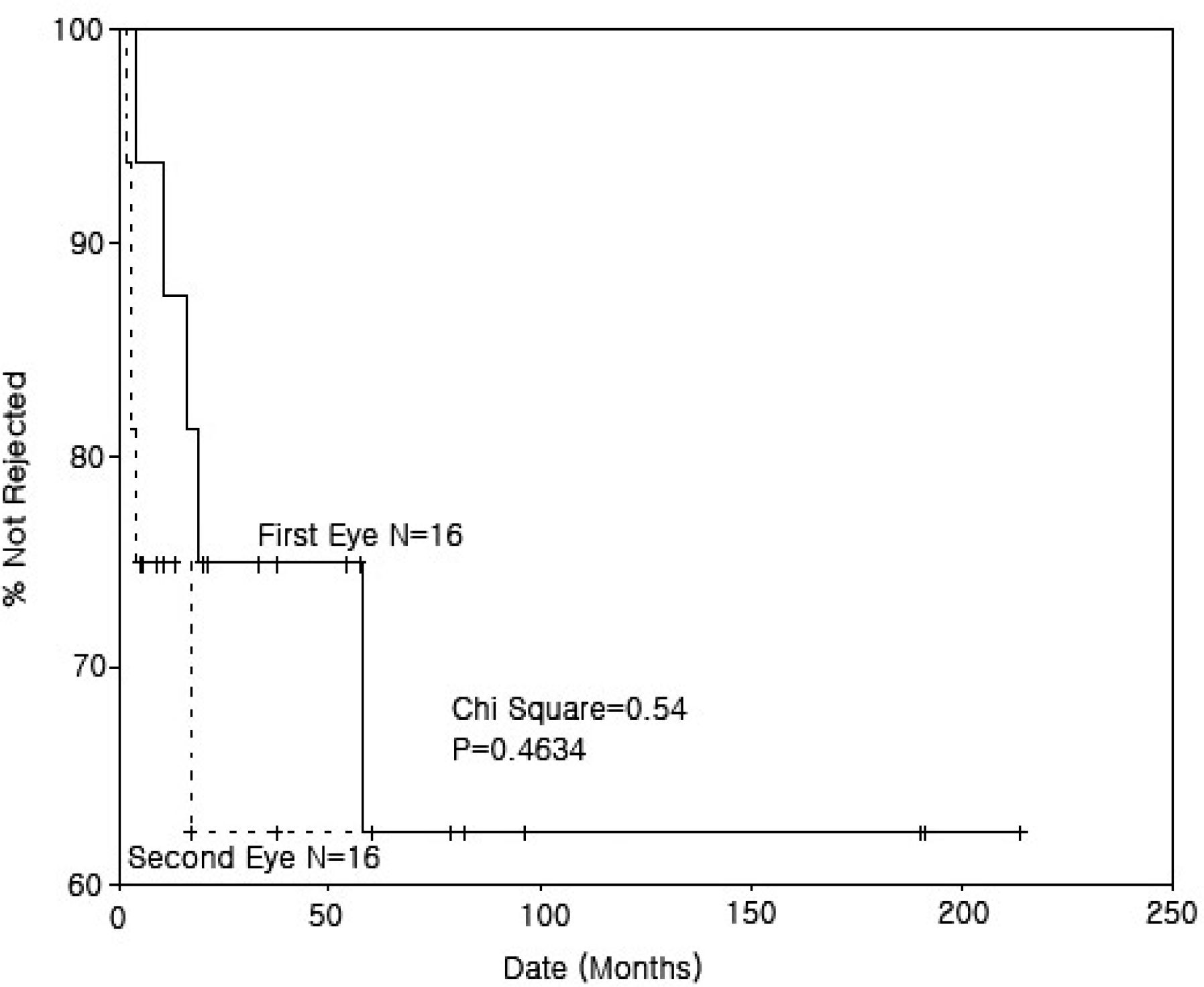
Figure 2.
Kaplan-Meier survival analysis of the time from penetrating keratoplasty to the first endothelial rejection episode in patients who underwent second surgery earlier than 2 years after first eye surgery. There was no significant difference between survival rate of the first eyes and that of the second eyes.
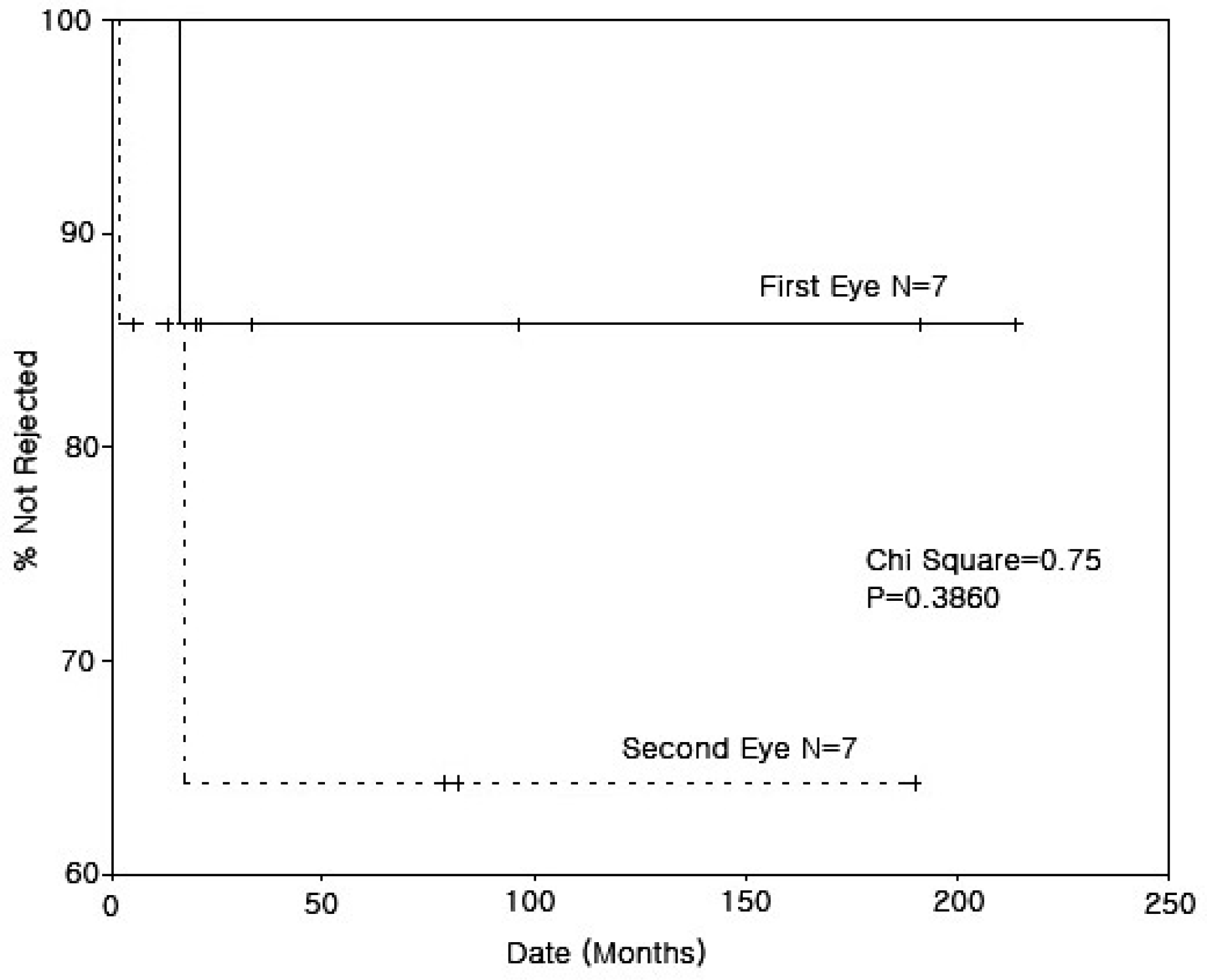
Figure 3.
Kaplan-Meier survival analysis of the time from penetrating keratoplasty to the first endothelial rejection episode in patients who underwent second surgery later than 2 years after first eye surgery. There was no significant difference between survival rate of the first eyes and that of the second eyes.
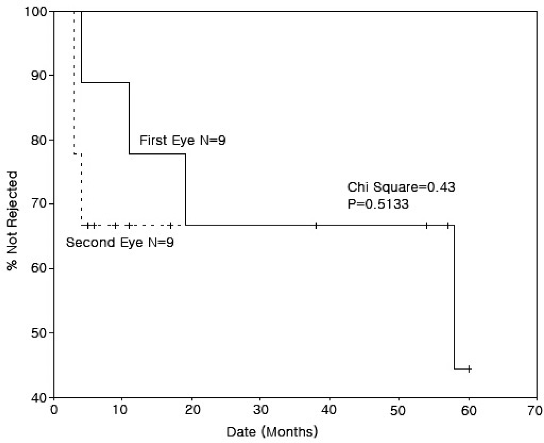
Figure 4.
Kaplan-Meier survival analysis curves of grafts in both eyes after bilateral penetrating keratoplasty. The survival rate of the second eyes decrease more rapidly than that of the first eyes.
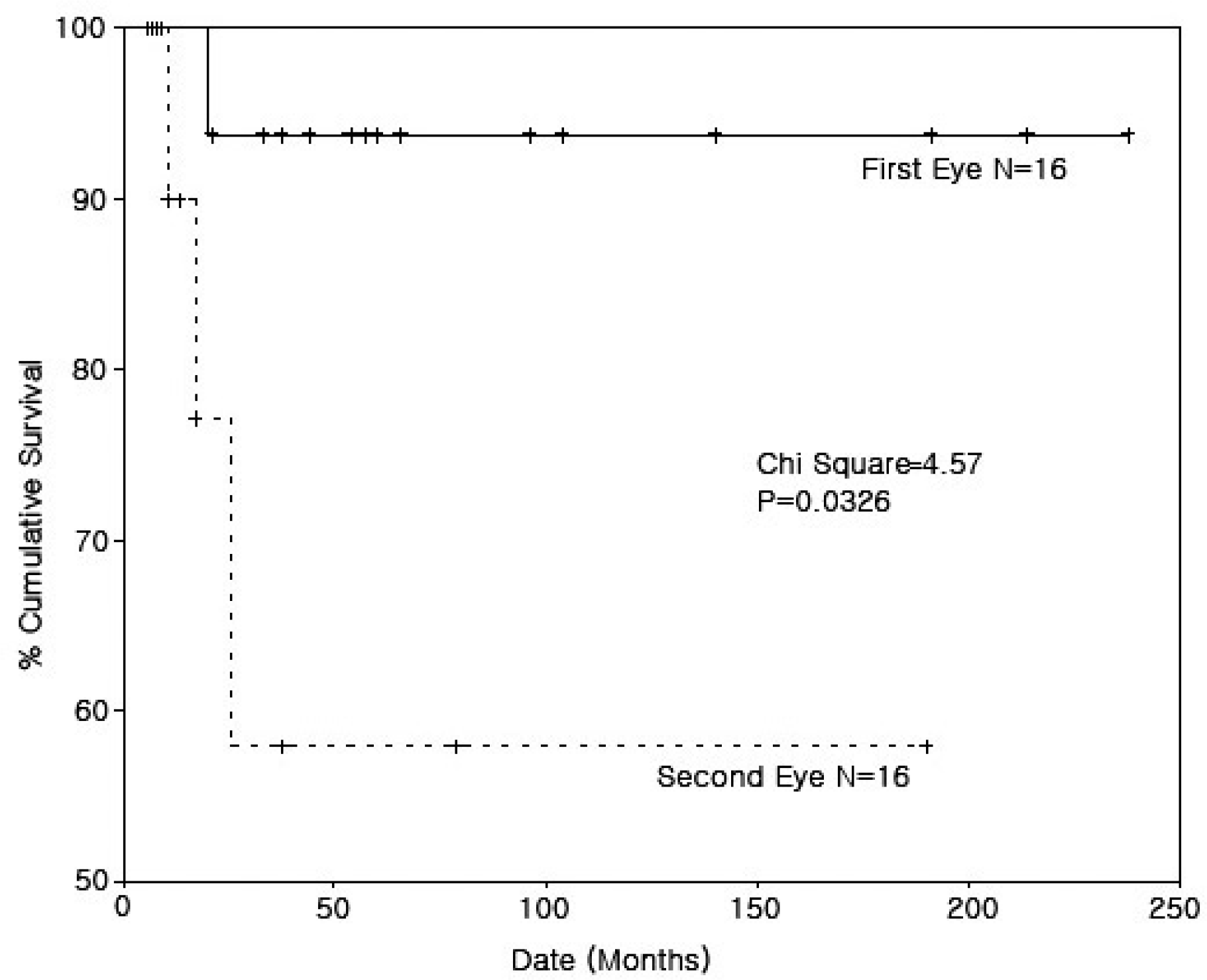
Table 1.
Indications for bilateral penetrating keratoplasty
Table 2.
Pre-, intra-, and post-operative ocular status in patients undergoing bilateral penetrating keratoplasty
| First eye No. of eyes (%) | Second eye No. of eyes (%) | |
|---|---|---|
| Ocular status | ||
| Aphakia | 0 (0) | 0 (0) |
| Pseudophakia | 2 (12.5) | 2 (12.5) |
| Prior antiglaucoma surgery | 1 (6.25) | 1 (6.25) |
| Preoperative best-corrected visual acuity | ||
| ≥0.5 | 0 (0) | 0 (0) |
| 0.1-0.5 | 4 (25.0) | 4 (25.0) |
| <0.1 | 11 (68.75) | 11 (68.75) |
| Could not be assessed | 1∗ (6.25) | 1∗ (6.25) |
| Preoperative corneal stromal vascularity† | 4 (25.0) | 3 (18.75) |
| Donor cornea | ||
| Dome stic/Im ported | 14/2 | 8/8 |
| Additional surgical procedures | ||
| IOL exchange | 1 (6.25) | 0 (0) |
Table 3.
Graft rejection or graft failure of the first eye and second eye after bilateral keratoplasty
| First eye No. of eyes (%) | Second eye No. of eyes (%) | |
|---|---|---|
| Graft rejection | 5 (31.25) | 5 (31.25) |
| Graft failure | 1 (6.25) | 3 (18.75) |
| Immunologic | 0 (0) | 2 (12.5) |
| Non immunologic | 1∗ (6.25) | 1† (6.25) |
Table 4.
Endothelial rejection during the first year alter surgery
| Second eye |
First eye |
Total | |
|---|---|---|---|
| Rejection: Yes | Rejection: No | ||
| Rejection: Yes | 1 | 3 | 4 |
| Rejection: No | 1 | 6 | 7 |
| Total | 2 | 9 | 11 |
Table 5.
Final visual outcomes in patients undergoing bilateral penetrating keratoplasty. The best-corrected visual acuity of the second eyes seemed to be worse than that of the first eyes
| Best-corrected visual acuity | First eye | Second eye |
|---|---|---|
| First eye No. of eyes (%) | Second eye No. of eyes (%) | |
| ≥0.5 | 7∗ (43.75) | 3∗ (18.75) |
| 0.1-0.5 | 7 (43.75) | 8 (50.0) |
| <0.1 | 1†(6.25) | 4† (25.0) |
| Could not be assessed | 1‡ (6.25) | 1‡ (6.25) |
| Total | 16 (100) | 16 (100) |
 XML Download