

 PDF
PDF ePub
ePub Citation
 Print
Print


Abstract
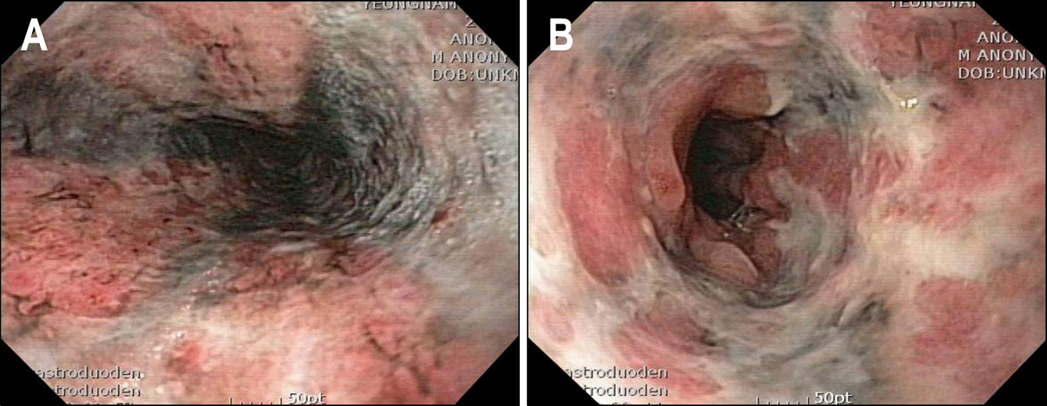
Acute necrotizing esophagitis, which presents as a black esophagus on endoscopy, is a rare disorder. We report a case of a 48-year-old male with acute necrotizing esophagitis, associated with alcoholic hepatitis. He was admitted to our hospital because of hematemesis. On initial upper esophagogastroduodenoscopy, diffuse friable black colored mucosa at whole length of the esophagus was observed. After conservative treatment with proton-pump inhibitor, esophageal mucosal lesion was healed without complication.
REFERENCES
1. Ben Soussan E, Savoye G, Hochain P, et al. Acute esophageal necrosis: a 1-year prospective study. Gastrointest Endosc. 2002; 56:213–217.
2. Gurvits GE, Shapsis A, Lau N, Gualtieri N, Robilotti JG. Acute esophageal necrosis: a rare syndrome. J Gastroenterol. 2007; 42:29–38.

3. Casella G, Perego D, Corti G, et al. Black esophagus: should it be considered an unfavorable prognostic factor? Dis Esophagus. 2001; 14:166–168.
4. Endo T, Sakamoto J, Sato K, et al. Acute esophageal necrosis caused by alcohol abuse. World J Gastroenterol. 2005; 11:5568–5570.
5. Augusto F, Fernandes V, Cremers MI, et al. Acute necrotizing esophagitis: a large retrospective case series. Endoscopy. 2004; 36:411–415.
6. Cattan P, Cuillerier E, Cellier C, et al. Black esophagus associated with herpes esophagitis. Gastrointest Endosc. 1999; 49:105–107.
7. Să ftoiu A, Cazacu S, Kruse A, Georgescu C, Comă nescu V, Ciurea T. Acute esophageal necrosis associated with alcoholic hepatitis: is it black or is it white? Endoscopy. 2005; 37:268–271.
8. Hong JW, Kim SU, Park HN, Seo JH, Lee YC, Kim H. Black esophagus associated with alcohol abuse. Gut Liver. 2008; 2:133–135.
9. Lacy BE, Toor A, Bensen SP, Rothstein RI, Maheshwari Y. Acute esophageal necrosis: report of two cases and a review of the literature. Gastrointest Endosc. 1999; 49:527–532.
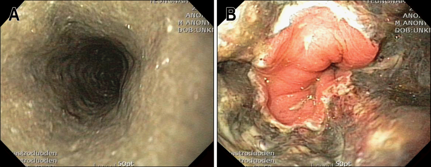
Fig. 1.
Endoscopic findings of the esophagus. (A) Friable and black colored mucosa was observed at whole length of the esophagus. (B) Black colored mucosa abrupt-ly disappeared at esophagogastric junction.
 XML Download