

 PDF
PDF ePub
ePub Citation
 Print
Print


INTRODUCTION
Vesicoureteral reflux (VUR) is present in about 1% of healthy children and in 30% to 50% of children with symptomatic urinary tract infections (UTIs) [1]. Treatments for VUR can be classified into four types: observation, antibiotic prophylaxis, open surgery, and endoscopic treatment. The endoscopic treatment of VUR was first introduced in 1981 by Matouschek with the use of polytetrafluoroethylene (Teflon) [2]. Recently, endoscopic treatment of VUR has gained widespread popularity and has become an alternative to ureteral reimplantation. Although ureteral reimplantation has a success rate in the range of 97% to 99% [34], relatively few complications, and minimal morbidity, it cannot compete with the advantages of endoscopic treatment, which leaves no scars, is fast to perform, and causes no or minimal pain. These features were recent arguments to accept endoscopic treatment, despite its lower success rate, which has been reported to range from 59.2% to 79.9% [567]. However, recent studies have reported success rates of up to 94% after a single injection and up to 98% after two injections [89]. Besides these high success rates, very low complication rates have been reported (about 1% of patients) [1011].
Ureteral obstruction is one potential complication after the endoscopic treatment of VUR. There have been isolated cases of ureteral obstruction or hydronephrosis following endoscopic treatment with dextranomer/hyaluronic acid copolymer (Dx/Ha, Deflux) [12]. Several studies have estimated the overall incidence of ureteral obstruction to be from 0.7% to 5.7% [111314]. In the present study, we examined the incidence, risk factors, and outcomes of postoperative ureteral obstruction after endoscopic treatment for VUR.
MATERIALS AND METHODS
We performed a retrospective chart review of all patients who underwent injection of Dx/Ha or polydimethylsiloxane (PDS, Macroplastique) to treat primary VUR at Pusan National University Yangsan Hospital between January 2008 and July 2013. Pusan National University Yangsan Hospital Institutional Review Board approval (12-046) was obtained.
Endoscopic treatment for VUR was performed in several instances. If conservative therapy failed owing to the occurrence of breakthrough UTI, or in the case of compliance problems with antibiotic prophylaxis in patients less than 1 year old, endoscopic treatment was given. Additionally, if the patient or parents elected to undergo endoscopic treatment, endoscopic treatment was performed. Finally, if the patient had persistent VUR, especially in those with renal scarring during a conservative follow-up period of at least 1 year, endoscopic treatment was performed.
Perioperative prophylaxis was given routinely. One operator (S.D.L.) performed the procedures. Endoscopic treatment was performed by using either the subureteral transurethral injection technique in cases with no distension of the ureteral orifice at the hydrodistention or the hydrodistentionimplantation technique in cases with distension of the ureteral orifice at the hydrodistention.
According to our follow-up protocol, we examined patients at 1 week, 1 month, and 3 months after endoscopic treatment. At 1 week after endoscopic treatment, we only checked for any unexpected symptoms or signs, such as high fever, nausea or vomiting, flank pain, and decreased urine output. Follow-up studies consisted of a postoperative ultrasound and urine analysis after 1 month and postoperative voiding cystourethrogram (VCUG) after 3 months. Additional follow-up studies including ultrasound and urine analysis were obtained in the case of unexpected symptoms or signs.
The diagnosis of ureteral obstruction was based on the finding of significant hydronephrosis by ultrasound during follow-up. Cases with new occurrences of hydronephrosis (society of fetal urology [SFU] grade 3 or more) or aggravation of previous hydronephrosis as shown on the postoperative ultrasound images were enrolled in the ureteral obstruction group and any accompanying symptoms were noted.
Charts of the patients with clinically relevant obstruction, which required intervention, were further analyzed, and data regarding the timing and symptoms of obstruction as well as the type of management were recorded. We analyzed the following factors: age, sex, injection material, laterality, voiding dysfunction, constipation, renal scarring, preoperative and postoperative ultrasound findings, endoscopic findings (such as mound shape, ureteral orifice shape and lateralization, bladder trabeculation, and others), injection number, and injection volume. We checked the postoperative findings of ultrasound including new occurrences of hydronephrosis (SFU grade 3 or more) or aggravation of previous hydronephrosis and mound diameter. Additionally, we reviewed the clinical manifestations, natural course, management, and outcome of ureteral obstruction after endoscopic treatment.
Statistical analysis was performed by using IBM SPSS Statistics ver. 19.0 (IBM Co., Armonk, NY, USA) with Mann-Whitney U test (age, mound diameter, and injection volume) or Kruskal-Wallis test (for the other variables). Data for the patients who underwent endoscopic treatment were analyzed by use of nonparametric tests. A p-value of <0.05 was considered statistically significant.
RESULTS
1. Patients
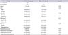
Ninety patients (132 ureters; 55 males and 35 females) underwent endoscopic treatment for VUR. These patients were classified into two groups according to ureteral obstruction: the nonobstruction group (83 cases, 122 ureters; mean age, 7.0±2.8 years) and the obstruction group (7 cases, 10 ureters; mean age, 6.2±8.1 years) (Table 1).
2. Outcome and complications
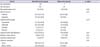
The incidence of postoperative ureteral obstruction after the endoscopic treatment was 7.6% (10/132 ureters). When we compared the two groups according to sex, age, injection material, ureter laterality, renal scarring, constipation, preand postoperative ultrasound findings, cystoscopic findings, injection numbers, and injection volume, there were no statistically significant differences in risk factors between the two groups (Table 2).
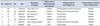
Three of the patients showed no symptoms or signs of a ureteral obstruction. Most patients' ureteral obstructions resolved spontaneously within 5 weeks with conservative therapy (cases 1, 3-5, and 7). However, two patients needed temporary ureteral stents to release the ureteral obstruction (cases 2 and 6). The first, a male, showed no symptoms; however, he had unilateral hydronephrosis of SFU grade 3-4 at his first follow-up ultrasound. Therefore, he was treated with a ureteral stent for 2 weeks, at which point he showed complete resolution of the ureteral obstruction. The second patient, a female, showed symptoms of pain, nausea, and oliguria on postoperative day 1. We immediately inserted a ureteral stent and kept it for 6 weeks. After removal of the ureteral stent, she showed complete resolution of the ureteral obstruction (Table 3). Finally, the two patients who needed temporary ureteral stents to release the ureteral obstruction showed no recurrence of VUR in postoperative VCUG after 3 months.
DISCUSSION
Renal function and renal scarring, clinical presentation, bladder-bowel dysfunction, age, and VUR grade are essential variables for choosing a conservative or a more invasive treatment for VUR. Endoscopic treatment offers the advantages of a minimally invasive method. The main advantages include decreased posttreatment pain, bladder spasm, and infection and the absence of a surgical scar [15]. The availability of this procedure in the outpatient setting, the short procedure time, the rapid time to discharge, and the minimal use of postoperative analgesics have been shown to be beneficial for both the patient and the physician [16]. The procedure can also be performed after an initial failure with either implantation or surgery [17]. Owing to the various advantages of endoscopic treatment for VUR, it has been commonly used as an alternative to open surgery.
The ideal bulking agent should be effective, stable over time, and safe (nonmigratory, nonantigenic, and biocompatible). Biological materials such as collagen, chondrocytes, PDS, and Dx/Ha were introduced for the purpose of endoscopic treatment [18]. PDS and Dx/Ha are currently the most popularly used injection materials. We also used either PDS or Dx/Ha in our study.
Besides the high success rates of endoscopic treatment, very low complication rates have recently been reported. Ureteral obstruction is one potential complication after endoscopic treatment. In 2004 Snodgrass reported the first case of ureteral obstruction occurring after Dx/Ha injection and attributed it to the dysmorphic nature of the injected ureter in an otherwise healthy female [12]. Studies on the use of PDS have reported ureteral obstruction in 1.8% and 0.65% of monitored cases, respectively [1920]. Serrano Durba et al. [20] observed some form of complications in 10 of the 516 patients included in their study. Of the 10 patients, 8 were treated with PDS. The complications described in the present four cases all appeared after an injection of PDS, raising the question of whether the complication rate is truly independent of the type of material used.
The most widely used material, Dx/Ha, still results in singular cases of ureteral obstruction after injection. For example, ureteral obstruction occurred in one case of a dysmorphic ureter [12], in another case leading to a filiform stenosis of a transplanted ureter after renal transplantation [21], and in two children mimicking distal ureteral stone formations after bilateral injection with apparent calcification [2223]. In a single retrospective study involving four institutions and a total of 745 patients (1,155 ureters), obstruction following Dx/Ha injection was reported in 5 children (<0.7%), 4 of whom had preexisting voiding dysfunction or neurogenic bladder, indicating an increased risk in this subgroup of pediatric patients [11]. In two recent retrospective studies with Dx/Ha, ureteral obstruction was reported in 5/475 ureters (1.05%) and in 5/87 ureters (5.7%), respectively [1314]. In our series, the incidence of ureteral obstruction (7.6%) was a little higher than in previous reports. This difference was due to how ureteral obstruction was defined. Garcia-Aparicio et al. [13] defined ureteral obstruction clinically in those patients with acute symptoms or by ultrasound findings and technetium-99m mercaptoacetyltriglycine (MAG3) diuretic renogram findings. Mazzone et al. [14] defined ureteral obstruction as clinically relevant obstruction that required intervention. We defined ureteral obstruction only on the basis of the finding of significant hydronephrosis in an ultrasound during follow-up. Therefore, asymptomatic ureteral obstruction may have occurred more than in other reports.
The following potential causes of obstruction were considered in our patients: (1) the bulking agent used, (2) injected volume, (3) injection number, and (4) surgical skill. Each is explained in further detail below.
1. Bulking agent
The bulking agent used does not appear to be significantly related to ureterovesical junction (UVJ) obstruction. Previous reports have suggested a very low incidence of ureteral obstruction after endoscopic treatment of VUR, regardless of the type of injection material used. Moreover, in our study, there was no significant difference between Dx/Ha and PDS. However, our personal feeling according to our data was that PDS may cause more obstruction than Dx/Ha because of 9 of 10 obstructive ureters in the PDS group.
2. Injection volume
In our cases, the injection volumes were 1.23±0.74 and 1.60±0.57 mL in the nonobstruction and obstruction groups, respectively. We initially considered injection volume as the most powerful risk factor before we analyzed the clinical data. According to our data, a greater injection volume tended to cause more obstruction; however, there was no significant difference between the two groups. Several authors used similar volumes in their studies. McMann et al. [24] reported using injected volumes ranging from 0.74 to 1.59 mL. A study by Sorensen et al. [25] showed a trend toward higher injection volumes of Dx/Ha. The mean amounts used per procedure increased from 1.67 to 2.22 mL from 2003 to 2008. Even though Mazzone et al. [14] reported a relatively high incidence rate (5.7%) compared with other studies, they used an injection volume of 0.7±1.2 mL. The volume injected seems to be irrelevant with regard to ureteral obstruction after endoscopic treatment of VUR, because all series showed a similar volume: from 0.7 to 2.2 mL [25].
3. Injection number
It is known that a giant cell inflammatory reaction occurs and fibrotic pseudoencapsulation is formed as a foreign body reaction in response to injections. This can account for decreased compliance [14]. On the other hand, in a case series including 149 reinjections and 19 third injections, no obstruction was reported by Puri et al. [26]. In our study, all obstruction patients were first-time injection patients, so this factor cannot be considered relevant.
4. Surgical skill
In our study, only one operator (S.D.L.) performed the endoscopic treatments for more than 10 years in one hospital. Therefore, surgical skill and error were not valuable discussion points in our study.
5. Other risk factors
Mazzone et al. [14] discussed risk factors of obstruction including predisposing anatomical variants, injected volume of Dx/Ha, repeated injections, technical errors, and surgical case load. Those researchers concluded that, although patients with associated urological anomalies may be at a higher risk of obstruction after injection, others without the anomalies showed no associations of the risk factors with obstruction.
The main cause of distal ureteral obstruction has been suggested to be edema of the UVJ or the aperistaltic distal ureter. It is possible that the combination of postinjection trauma contributed to a transient edema of the UVJ or aperistaltic distal ureter, which, in combination with ureteral bulking, led to an obstruction.
Treatments for ureteral obstruction include a ureteral stent, percutaneous nephrostomy, ureteral reimplantation, and nephrectomy in the case of poorly differential renal function. Garcia-Aparicio et al. [13] described two cases of ureteral stenting in acute clinical settings in which UVJ obstructions were successfully treated; in the follow-up period, there was no VUR on the VCUG.
Theoretically, it is possible that stenting increased the submucosal dispersion of the injected material or caused the injection material to mold around the ureteral orifice, creating an incompetent valve mechanism. In our study, most cases improved with conservative treatment and only 2 of 7 patients were treated with ureteral stents. All ureteral stents were easily passed. After the ureteral stents were removed, there was no recurrence of VUR.
Injection material mounds have been shown to lose volume within the first several weeks after surgery. Kirsch et al. [27] observed an approximately 20% volume loss in the first 3 months after an injection on the basis of sonographic measurements. However, once the mounds were fixed by encapsulation, there was no change in the volume of Dx/Ha mounds for up to 3 years after implantation [24]. Most cases improved spontaneously. We suggest that the resolution resulted from the improvement of transient edema, and that the injection material was then absorbed or spread around the ureteral orifice.
The observations in this report suggest that complications can occur early after surgical procedures (cases 4, 6, and 7), but that they can also develop over weeks (cases 1, 3, and 5) or even months (case 2). This finding suggests that obstruction may occur early, but that diagnosis can be delayed when there are no symptoms present. Obstruction may be difficult to diagnose owing to the lack of typical symptoms in young patients, especially infants. For example, a ureteral obstruction was found in an apparently healthy child. Therefore, we strongly recommend ultrasound studies be undertaken 1 month after endoscopic treatment. Although discussion remains about the use of follow-up imaging and useful algorithms for controlled examinations [2829], the postoperative course in these patients indicates that regular follow-up visits are warranted.
6. Limitations
Several limitations of this study merit discussion. First, we did not examine renal scans, such as diethylene triamine pentaacetic acid or MAG3, to determine whether there was a true functional obstruction. In our study, the diagnosis of obstruction was based on the findings of hydronephrosis in ultrasound images during the follow-up period. The MAG3 diuretic renogram is known to have limitations in detecting UVJ obstruction, but when used in combination with hydronephrosis on ultrasound, the resulting information should be suf f icient to accurately suspect ureteral obstruction. Second, because obstruction is so scarce, analysis is very difficult because of low statistical power. In our retrospective study, obstruction occurred in only 10 ureters. Therefore, further prospective studies that last a long time are needed to overcome the limitations of retrospective study.
CONCLUSIONS
We found that the incidence of ureteral obstruction after endoscopic treatment was a little higher than reported in the literature. There were no predictive risk factors for developing ureteral obstruction after endoscopic treatment. Although most ureteral obstructions spontaneously resolved within 1 month, some cases needed drainage to relieve symptoms or to prevent renal function deterioration. Standardized follow-up examinations would help to monitor the success of the performed procedure and facilitate an earlier diagnosis of developing ureteral obstruction.
 XML Download