

 PDF
PDF ePub
ePub Citation
 Print
Print


INTRODUCTION
Renal artery pseudoaneurysms, whether traumatic or iatrogenic in origin, always seem to play hide-and-seek with urologists. Patients usually present with hematuria in the postoperative period that continues for days to weeks. Initial treatment is usually conservative with bed rest and monitoring of hemoglobin. Patients having continuous hematuria require intervention. Percutaneous embolization techniques have long been used in these cases and are at present the first-line treatment for renal artery pseudoaneurysm. Micro coils, Gelfoam particles, and cyanoacrylate glue have all been used with comparable efficacies. The overall success rate for these techniques is more than 90%. We report here a rare complication that occurred during angioembolization of an intrarenal pseudoaneurysm (IRP).
CASE REPORT
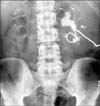
A 40-year-old man with a solitary functioning left kidney harboring staghorn renal stones and chronic renal failure presented to the Emergency Department with fever, anuria, and hypotension. He was evaluated and found to have left pyonephrosis, hyperkalemia, acidosis, and sepsis. Acute management included hemodialysis and left percutaneous nephrostomy (PCN) along with parenteral antibiotics (Fig. 1). After day 3, the patient began to have bleeding through the PCN catheter. Initially, the patient was managed conservatively with rest, hydration, and serial monitoring of hemoglobin, pulse rate, and blood pressure. Clamping of the nephrostomy was done, but the patient continued to bleed and was taken for digital subtraction angiography by the intervention radiology team.
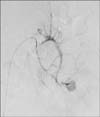
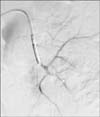
During the angiography, a pseudoaneurysm was found in the territory of the inferior segmental branch of the left renal artery (Fig. 2). Selective catheterization and angioembolization of the inferior segmental branch was done with a micro coil, but the pseudoaneurysm was still filling. Thus, the angiocatheter was withdrawn a little proximally and cyanoacrylate glue was injected. After the glue was deployed, the angiocatheter could not be pulled back and was found to be stuck inside the inferior segmental vessel (Fig. 3). Gentle attempts to pull out the catheter failed. The whole kidney was seen moving under fluoroscopy with movement of the angiocatheter. The catheter was left in situ and the patient was optimized (which included one session of hemodialysis) and prepared for emergency exploration.
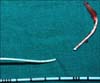
A laparotomy was done with a midline incision and the left kidney was mobilized. After taking hilar control, the inferior segmental branch was dissected out. The catheter and glue cast could be palpated. A longitudinal arteriotomy was done (Fig. 4), and the glue cast with the tip of the angiocatheter were removed (Fig. 5) and the inferior segmental artery was ligated. The patient was maintained on hemodialysis during the postoperative period and is now on maintenance hemodialysis while waiting for a renal donor. The left PCN is still in situ with an output of around 300 to 400 mL in 24 hours. The patient is scheduled to undergo a native nephrectomy before renal transplantation.
DISCUSSION
Patients with long-standing stones commonly present with pyonephrosis. Percutaneous drainage has emerged as the most appropriate primary intervention in these patients. Percutaneous renal procedures can cause several renovascular injuries, such as hematomas, arteriovenous fistulas, or pseudoaneurysms. The reported incidence of renal pseudoaneurysms following percutaneous nephrolithotomy is 1% and it is usually assessed by renal angiography. Selective renal embolization is currently considered the most appropriate technique in the treatment of these complications, with a success rate greater than 80% and a low complication rate [1]. Pseudoaneurysms must be occluded with a permanent agent at the fistulous point where the risk of hemorrhage is greater [2].
Endovascular management is minimally invasive and results in occlusion of the fistula itself as well as the proximal portion of venous drainage and helps to save the kidney in many patients. Coaxial catheter systems with small-caliber microcatheters are highly reliable for achieving superselective catheter positions in small-caliber arteries, thereby minimizing the volume of infarcted tissue and allowing maximal preservation of organic function [3].
Many substances can be used for embolization, such as ethanol, polyvinyl alcohol, Gelfoam particles, micro coils, detachable balloons, and N-butyl-2-cyanoacrylate (NBCA). Ethanol injected intra-arterially has been used to reduce the vascularity of tumors to facilitate their surgical resection. Selective arterial injection causes luminal thrombi with endothelial loss and varying degrees of medial necrosis, leading to occlusion. Polyvinyl alcohol has been used as a particle embolic agent because of its biocompatibility and inertness. Polyvinyl alcohol quickly occludes the arteries and its effect is thought to be permanent. According to histological examination, however, most large vessels containing polyvinyl alcohol are incompletely occluded, with particles becoming embedded in the walls. Incomplete occlusion in the kidneys causes excessive excretion of rennin, which in turn results in hypertension [4].
Onyx is a newer biocompatible liquid embolic agent that consists of an ethylene vinyl alcohol copolymer dissolved in an 8% concentration of dimethyl sulfoxide. Onyx has been used in the treatment of pelvic arteriovenous malformations (AVMs) and traumatic deep femoral artery pseudoaneurysms. Although not reported previously, onyx can be used as a liquid embolic agent in the treatment of IRPs. The advantages of this material are its easy handling and the ability to perform transembolization angiography (not possible with cyanoacrylates), which enables assessment of the need for additional embolic agents. Onyx can be delivered in a slower and more controlled manner than NBCA. This property is especially important in the prevention of distal embolization. However, onyx is much more expensive than NBCA [5].
Gelfoam (gelatin sponge) embolization has several shortcomings, such as the following: (1) reflux of embolic material into the normal arteries can occur, particularly if a small distal vessel has not been super-selectively cannulated; (2) a larger vessel may be difficult to occlude, as may a more generalized embolization of the arterial tree; (3) Gelfoam can undergo resorption and allow recannulation of the vessel. The results of the use of gelatin sponges include total occlusion with or without recanalization. In case of recanalization, the remaining AVMs were smaller than the lesion treated initially. Infarction of part of the renal parenchyma is reported to be about 14% [6]. Absorbable gelatin sponge embolization increases the risk of sudden rupture by increasing the pressure inside the cavity [7].
In moderate-sized vessels, steel coils or detachable balloons may also be used. Coils are the most commonly used embolic agent in visceral and renal pseudoaneurysms. The main disadvantage with coils is the cost of the embolic material. Usually, more than one coil is required for adequate occlusion. Because pseudoaneurysms lack a true wall, the endovascular approach should be done with care and with low-pressure delivery systems. Otherwise, rupture of the pseudoaneurysms may result in fatal complications [8].
Cyanoacrylate glue (NBCA) is a lasting and efficient occluding agent that has been successfully used for more than 20 years [2]. The polymerization time of NBCA can be delayed by adding iodized oil. As more iodized oil is added, polymerization is delayed and radiopacity is improved. However, the longer the polymerization time, the greater the risk of distal embolization, because the embolic material may be washed away before it polymerizes. The NBCA mixture (NBCA and Lipiodol) is difficult to handle because of the need to prepare the appropriate proportion of the mixture, the restricted injection time, and the necessity of rapid withdrawal of the microcatheter after injection. A ratio of 1:2 is ideal for peripheral pseudoaneurysms to prevent distal embolization. In AVMs and pseudoaneurysms, NBCA mixed with Lipiodol, if placed super-selectively, is safe, produces excellent results, and does not interfere with possible future retreatment. It has good results for both initial and repeat embolization provided the mixture can be delivered safely super-selectively inside the nidus. For best results in AVM, the glue should penetrate the immediate proximal draining vein to promote occlusion of outflow from other compartments of the renal AVM, which carries a small risk of causing bleeding, and to minimize the chance of a recurrent lesion due to recruitment of collateral vessels distal to the site of glue placement. With controlled injection of an adequately diluted amount of glue, ischemic complications of the kidney or pulmonary embolization should be avoided [9]. Extended contact of acrylics with the tip of the microcatheter during injection can glue the catheter tip to the arterial wall. Another major disadvantage of NBCA is that the microcatheter used for injection of the NBCA mixture can only be used once, and residual aneurysm filling requires re-catheterization. Therefore, considerable experience is required to achieve optimal results with NBCA [10].
In conclusion, endovascular management of intra-parenchymal renal artery pseudo-aneurysms is a reasonable and effective therapeutic technique. It preserves maximum renal tissue and reduces the potential risk of nephrectomy. NBCA as an embolizing agent provides accurate, definitive, and cost-effective embolization of post-traumatic IRP. NBCA embolization may be hazardous in inexperienced hands and requires familiarity with the use with this embolic agent, especially quick removal of the microcatheter after deploying NBCA, which otherwise can get stuck to the arterial wall. In such a situation, exploration and arteriotomy with removal of the angiocatheter tip seems to be the most appropriate treatment option.

 XML Download