

 PDF
PDF ePub
ePub Citation
 Print
Print


INTRODUCTION
Bladder cancer is the second-most-common malignancy of the genitourinary tract [1]. In our previously reported work [2], approximately 90% of bladder cancers are pure urothelial carcinoma (UC), and the remaining 5% to 10% consisted of UC with aberrant differentiation or non-UC. UC may exhibit a diverse array of morphologies. This propensity of divergent differentiation may manifest as squamous or glandular cancers. Conventional UC, particularly the high-grade and high-stage cancer, may exhibit full range of variant morphology. The term "variant" is used to describe these distinctively different histomorphologic phenotypes of a certain type of neoplasm [2,3]. In recent years, several variants have been described, along with their clinical significance at various levels, to avoid diagnostic misinterpretation, aid in risk prognostication, and present differences in therapeutic approach [4]. Multiple case series and case reports on the aggressive nature of these tumors are available in the literature, but there are few data comparing the outcomes of various types of these tumors with non-UC as well as conventional UC. A single large series has documented that histologic variants are common in high-grade UCs of the renal pelvis; they made up 40% of the cases in the series [5].
This study is focused on a comparison of the outcome of patients who underwent radical cystectomy (RC) for bladder cancers of different histopathologic types. Divergent histopathology has different clinical course and survival outcomes, as shown in several case series [4, 5, 6]. There is a dearth of data on the subject, and reported series have variable incidence; however, one factor common to all series is that variant histology increases the likelihood of locally advanced disease and metastasis. The aim of this study is to compare the clinical outcomes in different variants of bladder tumor that have not previously been reported in the literature.
MATERIALS AND METHODS
Between January 1988 and December 2010, 201 patients underwent RC for treatment of bladder cancer. These patients were identified through the hospital database by using the International Classification of Diseases, Ninth Revision, Clinical Modification (ICD-9CM). Thirty-one patients were excluded, of whom 18 were lost to follow-up, while 13 had missing records. Data were sealed in 2010 for follow-up and production of a final data set for analysis.
Bladder cancer was diagnosed by cystoscopy, which was followed by transurethral resection of the bladder tumor. The preoperative staging work-up included a complete history and a physical examination, followed by complete laboratory work-up, chest roentgenogram, computed tomography scan, and magnetic resonance imaging or ultrasound of abdomen and pelvis. A radio-isotopic bone scan was done only if indicated. Indications for RC were based on clinical and pathologic staging of the disease, including muscle-invasive bladder cancer, high-grade tumor, high-volume superficial cancer, invasive tumor refractory to radiotherapy and/or systemic chemotherapy, or highly recurrent superficial cancer refractory to endoscopic resection and intravesical chemotherapy. All patients underwent standard bilateral pelvic lymphadenectomy with RC and urinary diversion. The tumors were staged according to TNM 1997 classification. The retrospective nature of this current work precluded a standardized postoperative follow-up protocol in all patients. However, patients were initially seen at one week after RC, after a month, then after every 3 months for 1 year, and, finally, every 6 months until disease progression or death. Except for the initial follow-up, patients were followed with complete physical examination, complete renal profile and blood count and electrolytes, chest x-ray, computed tomography of the abdomen and pelvis, and bone scan if indicated.
All specimens were re-reviewed in consultation with pathologists, and the presence or absence of histologic differentiation was recorded. Patients were divided into four groups on the basis of their histopathologic type-that is, UC, squamous cell carcinoma (SCC), adenocarcinoma (ADC), and other, a type that includes small-cell carcinoma, large-cell carcinoma, and micropapillary variant; UC with squamoid, glandular, or sarcomatoid differentiation; and signet cell carcinoma.
The variables evaluated were age, gender, tumor cell type, pathologic stage, and nodal involvement. The chi-square test and independent sample t-test were used for statistical analysis. The association of histopathologic variance with categorical variables was assessed by using the Fisher exact test or the chi-square test, while the Mann-Whitney U test was used for continuous variables. Results were presented as mean±standard deviation for quantitative variables and as number (%) of patients for qualitative variables. Univariate and multivariate analyses were performed to determine the impact of different clinicopathologic variables on survival outcome. Survival estimates were constructed by using the Kaplan-Meier product limit methods, and the log rank test was applied to determine statistical significance. All data were analyzed by using SPSS ver. 16 (SPSS Inc., Chicago, IL, USA). For all tests, p<0.05 was considered to indicate a statistically significant difference.
RESULTS
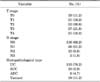
The mean age of the patients was 61±13.1 years (range, 27-87 years); males greatly outnumbered (143, 84%) females (27, 16%). Mean follow-up was 67 months (range, 6-132 months). Clinicopathologic features are described in Table 1. Nineteen patients had histopathologic variants: seven (37%) each with squamoid differentiation and large-cell variant, two (11%) with sarcomatoid differentiation, and one each with signet cell, glandular differentiation, or micropapillary variant.
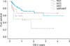
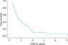
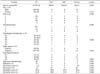
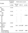
The majority of the patients had advanced disease at the time of presentation, with 18% of them having invasion of the surrounding pelvic structures. The overall survival (OS) was 55%, while cancer-specific survival (CSS) was 35% at a median follow-up of 58 months (range, 6-162 months). The OS for the pathologic stage pT0-pT1 was 83%, compared to 60% for the group with stage pT2-pT4 (p=0.03). The OS for node-positive patients was 16%, compared to 60% for node-negative patients (p<0.01). The subgroup analysis and univariate analysis showed no significant difference (p>0.05) in the overall mortality of the patients with variant pathology compared to UC, SCC, and ADC (Table 2). Univariate and multivariate analyses showed that age, gender distribution, T stage, and nodal status are not statistically significant in predicting survival, whereas variant pathology was an independent predictor of CSS (Table 3). On Cox regression analysis, variant pathology was also shown to have a significant impact on OS (hazard ratio, 2.81; 95% confidence interval, 1.32-5.97).
DISCUSSION
Bladder cancer is a heterogeneous disease with an array of histologic findings. High-grade and muscle-invasive bladder cancers treated with RC have varied prognoses [6,7]. In recent years, several "variant" morphologies have been described, and most were recognized in the 2004 World Health Organization Classification [8]. These histologic variants of UC have clinical significance for diagnosis, prognosis, and therapy. Awareness of the morphologic variant is essential to avoiding diagnostic misinterpretations. It has been shown that variant pathologic tumors are aggressive, a fact that has significant prognostic implications. In view of this factor, early recognition may require novel therapeutic interventions, such as the administration of a therapy distinctive from that used for conventional invasive UC [6].
Currently, clinical stage is the strongest predictor of 5-year progression-free survival. Patient risk stratification is essential for optimal management of patients with bladder cancer. Risk status determines the application and timing of adjuvant/neoadjuvant systemic chemotherapy and RC. One of the major factors in risk stratification is the presence of variant histologic patterns in the bladder tumor. These histologic variants of UC have clinical significance at various levels, including the diagnostic level; that is, awareness of the morphologic variant is essential to avoidance of diagnostic misinterpretations, to prognosis for patient risk stratification, and to therapy, in which a diagnostic assignment of a particular variant may be associated with the administration of a therapy distinctive from that used in conventional invasive UC [9]. The differential diagnosis of UC with variant pathology includes SCC and ADC. Pure SCC shows an exclusive squamous cell component. Similarly, ADC of the bladder is a term reserved for pure ADC.
The vast majority of tumors are conventional UCs, and the rest consist of UC with aberrant differentiation (i.e., squamous/glandular differentiation, small-cell carcinoma, sarcomatoid carcinoma, and micropapillary carcinoma) or non-UC (SCC and ADC). A review of contemporary literature suggests that all of the variant histologies portend a poor prognosis as compared to that for conventional UC. In our series, there was a trend toward poorer outcomes with variant histology; however, overall mortality was not statistically significantly different (p>0.05). This finding could potentially be due to the smaller numbers in our series.
RC remains the mainstay of treatment in high-grade and muscle-invasive bladder cancers, and the use of systemic chemotherapy may have a beneficial effect on OS. The timing of systemic chemotherapy has the potential to alter the ultimate outcome in patients at high risk of recurrence. The use of neoadjuvant chemotherapy in high-risk UC is recommended [10] and should be considered for variant histology as it also impart high risk. Scosyrev et al. [11] assessed the effect of neoadjuvant chemotherapy and noted that the presence of squamous or glandular differentiation in locally advanced UC of the bladder does not confer resistance to neoadjuvant methotrexate, vinblastine, adriamycin and cisplatin (MVAC) chemotherapy (chemotherapy combining methotrexate, vinblastine, doxorubicin, and cisplatin) and may, in fact, be an indication for the use of such chemotherapy before RC. For small-cell carcinoma, there is good evidence that MVAC chemotherapy is an appropriate strategy.
The established predictors of prognosis such as T stage and nodal status were predictors of survival in a univariate analysis but were not found to have a significant impact on survival in a multivariate analysis. This difference could perhaps be due to the smaller numbers of patients in the subgroups. Our study clearly indicates that, in a multivariate analysis, variant pathology is an independent predictor of survival.
 XML Download