Abstract
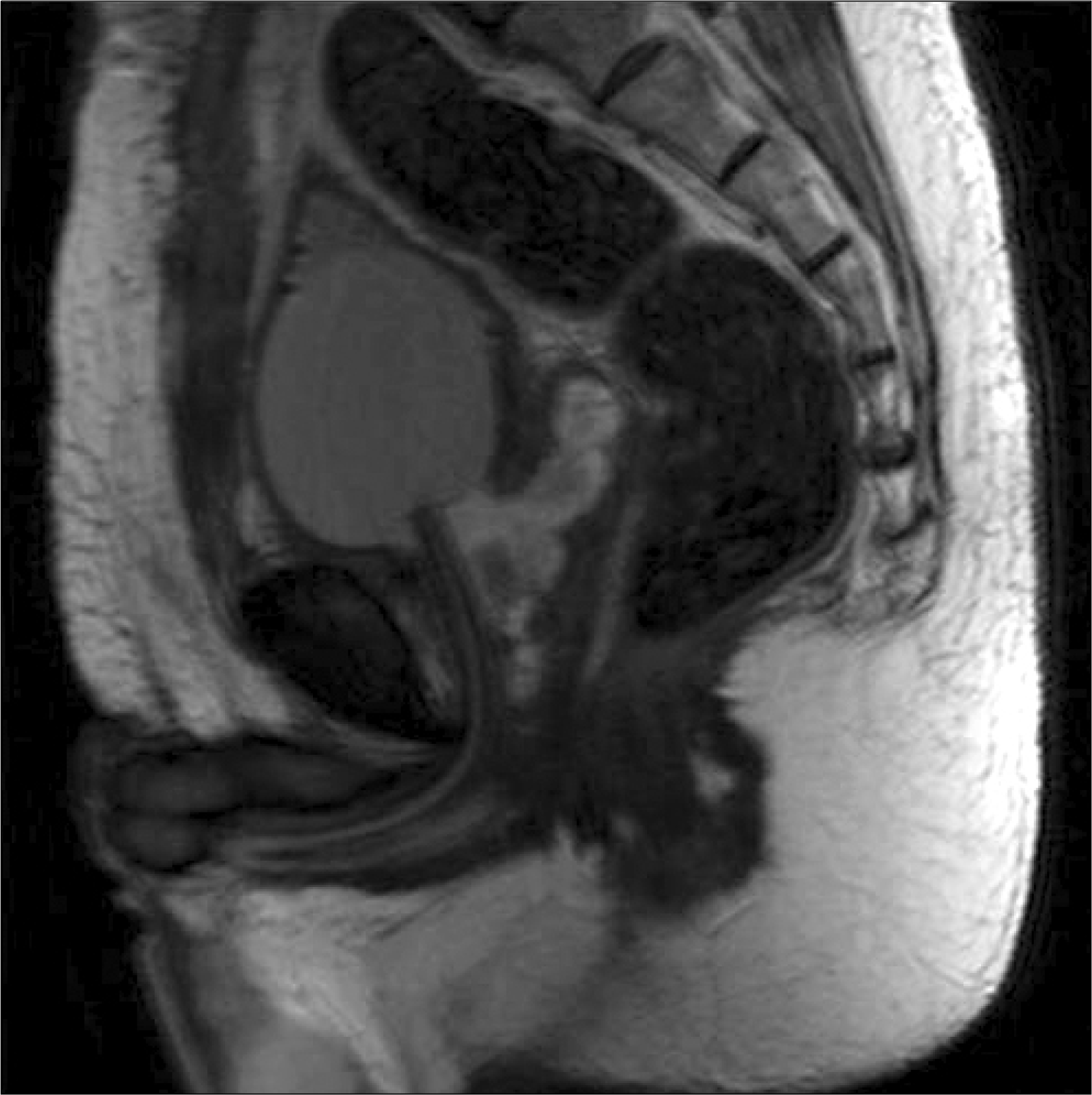
Gastrointestinal stromal tumor (GIST) are the most common mesenchymal malignancy of the gastrointestinal tract. They are diagnosed by combination of c-kit and CD34. We report a case of extragastrointestinal stromal tumor originating from the prostate diagnosed after retropubic open prostatectomy. The patient underwent additional retropubic radical prostatectomy again after 2 weeks. The possibility of secondary involvement by a rectal GIST was excluded by radiological, intraoperative and pathologic findings.
REFERENCES
1.Miettinen M., Sarlomo-Rikala M., Lasota J. Gastrointestinal stromal tumors: recent advances in understanding of their biology. Hum Pathol. 1999. 30:1213–20.

2.Hirota S., Isozaki K., Moriyama Y., Hashimoto K., Nishida T., Ishiguro S, et al. Gain-of-function mutations of c-kit in human gastrointestinal stromal tumors. Science. 1998. 279:577–80.
3.Fletcher CD., Berman JJ., Corless C., Gorstein F., Lasota J., Longley BJ, et al. Diagnosis of gastrointestinal stromal tumors: a consensus approach. Hum Pathol. 2002. 33:459–65.

4.Arce-Lara C., Shah MH., Jimenez RE., Patel VR., Benson DM Jr., Clinton SK, et al. Gastrointestinal stromal tumors involving the prostate: presentation, course, and therapeutic approach. Urology. 2007. 69:1209.

5.Lee CH., Lin YH., Lin HY., Lee CM., Chu JS. Gastrointestinal stromal tumor of the prostate: a case report and literature review. Hum Pathol. 2006. 37:1361–5.

6.Van der Aa F., Sciot R., Blyweert W., Ost D., Van Poppel H., Van Oosterom A, et al. Gastrointestinal stromal tumor of the prostate. Urology. 2005. 65:388.

7.Yinghao S., Bo Y., Xiaofeng G. Extragastrointestinal stromal tumor possibly originating from the prostate. Int J Urol. 2007. 14:869–71.

8.Dematteo RP., Heinrich MC., El-Rifai WM., Demetri G. Clinical management of gastrointestinal tumors: before and after STI-571. Hum Pathol. 2002. 33:466–77.
Fig. 1.
Preoperative transrectal and transabdominal ultrasonography. The large 65x75x70 mm sized prostate was well encapsulated.

Fig. 2.
(A) Prostate is replaced by cellular tumor mass. The tumor cells are spindle shaped with little atypia. They shows mainly fascicular arrangement. (B) Compressed prostatic glands are seen on upper left field. (C) Tumor cells are strongly positive for C-kit. (D) Tumor cells are strongly positive for CD34.


 PDF
PDF ePub
ePub Citation
 Print
Print


 XML Download