

 PDF
PDF ePub
ePub Citation
 Print
Print


Abstract
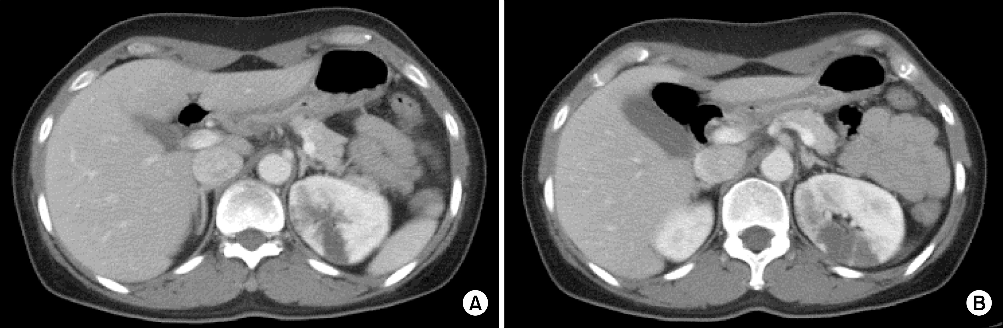
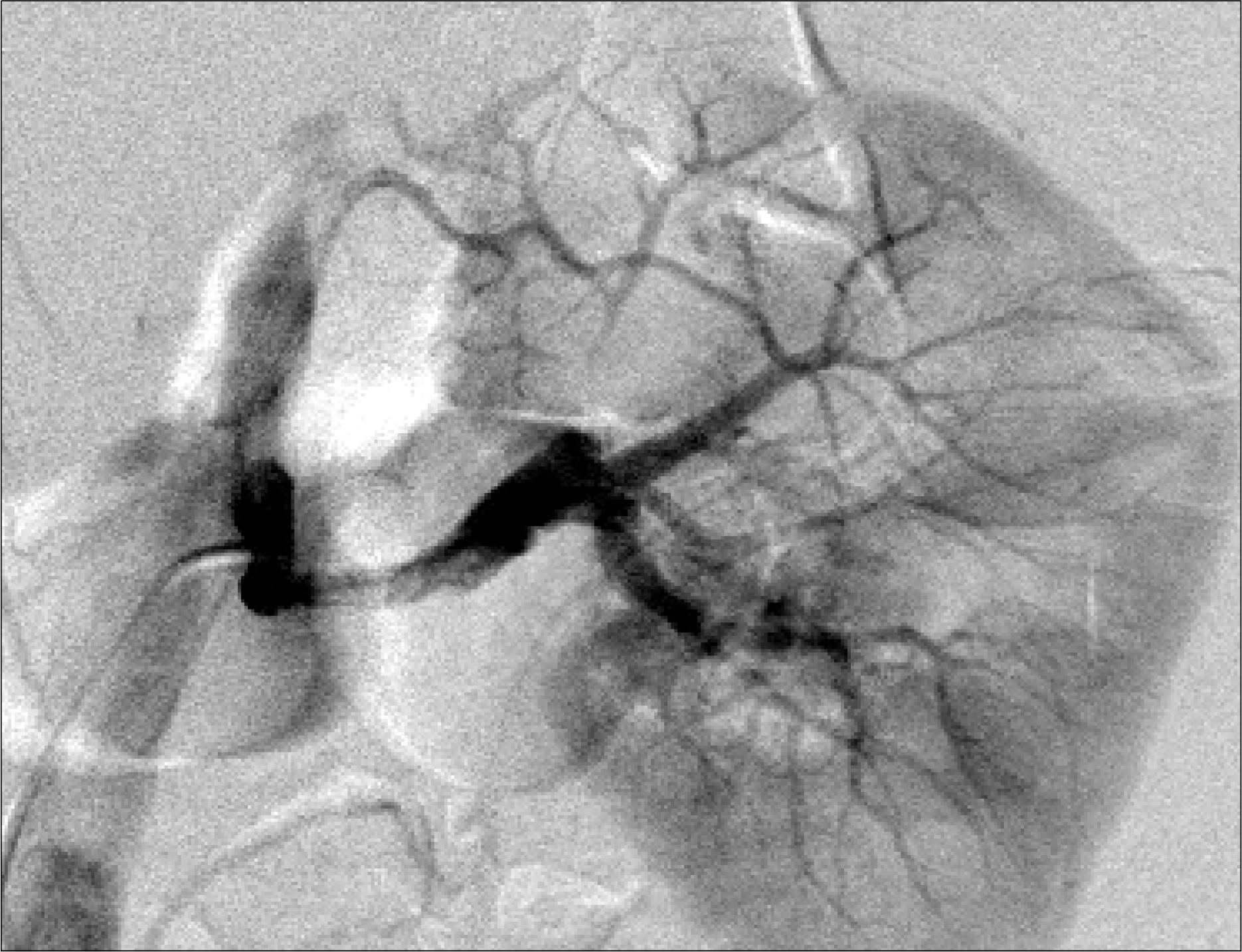
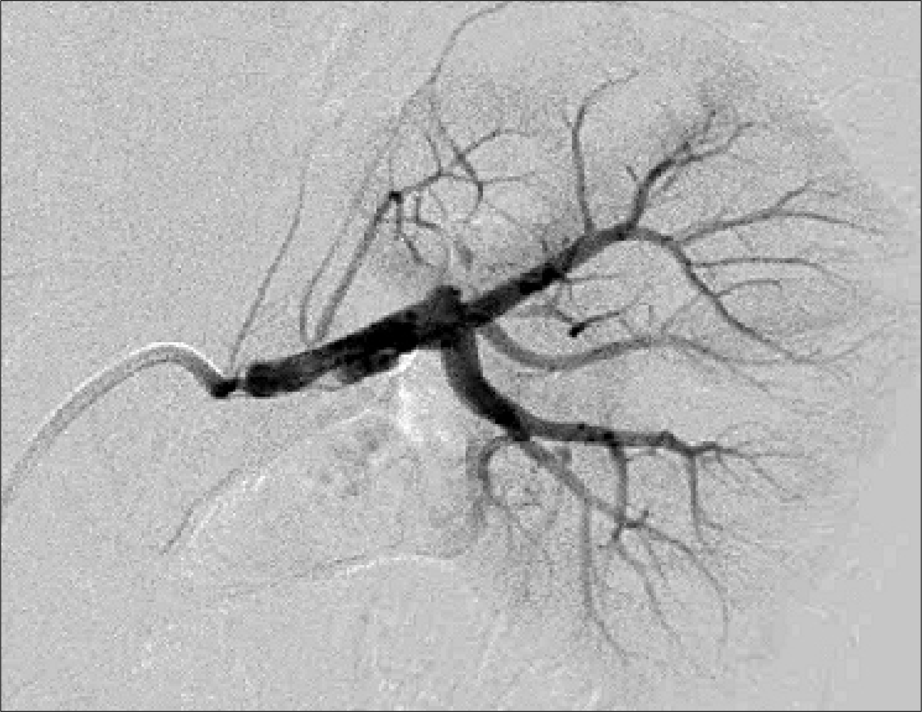
A previously healthy 44-year-old woman, with no notable medical history developed left flank pain. To rule out left renal infarction, enhanced abdominal computed tomography (CT) was done and a wedge shaped hypointense lesion was identified in the left posteromedial aspect of the interpolar region. Renal angiography revealed an isolated renal artery dissection that was causing renal infarction due to narrowing of the main stem of the left renal artery. The patient experienced pain with severe uncontrolled hypertension. The patient was successfully treated by percutaneous angioplasty and renal artery stenting.
REFERENCES
1.Wooley CF., Sparks EH., Hirata K., Boudoulas H. The aortopathy of heritable cardiovascular disease. Chap 19, in Functional Abnormalities of the Aorta. Armonk, NY: Futura Publishing;1996. 295-320.
2.Esayag-Tendler B., Yamase H., Ramsby G., White WB. Accelerated hypertension with encephalopathy due to an isolated dissection of a renal artery branch vessel. Am J Kidney Dis. 1994. 23:869–73.

3.Lee SH., Lee HC., Oh SJ., Park MC., Park KJ., Moon YS, et al. Percutaneous intervention of spontaneous renal artery dissection complicated with renal Infarction: a case report and literature review. Catheter Cardiovasc Interv. 2003. 60:335–8.
4.Reilly LM., Cunnungham CG., Maggisano R., Ehrenfeld WK., Stoney RJ. The role of arterial reconstruction in spontaneous renal artery dissection. J Vasc Surg. 1991. 14:468–77.
5.Edwards BS., Stanson AW., Holley KE., Sheps SG. Isolated renal artery dissection, presentation, evaluation, management and pathology. Mayo Clin Proc. 1982. 57:564–71.
6.Beroniade V., Roy P., Froment D., Pison C. Primary renal artery dissection, presentation of two cases and brief review of the literature. Am J Nephrol. 1987. 7:382–9.
7.Muller BT., Reiher L., Pfeiffer T., Muller W., Hort W., Voiculescu A, et al. Surgical treatment of renal artery dissection in 25 patients: indications and results. J Vasc Surg. 2003. 37:761–8.
8.Lawrie GM., Morris GC Jr., Debakey ME. Long-term result of treatment of the totally occluded renal artery in forty patients with renovascular hypertension. Surgery. 1980. 88:753–9.
 XML Download