

 PDF
PDF ePub
ePub Citation
 Print
Print


Abstract
Background
67Ga scintigraphy has been used for years in sarcoidosis for diagnosis and to determine the extent of the disease. The present report is a study of various findings of 67Ga scintigraphy in patients with sarcoidosis.
Methods
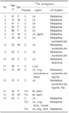
Between 1998 and 2007, 16 patients (male:female, 6:10; age, 35.9±15.3 years) with histologically proven sarcoidosis underwent clinical evaluation and 67Ga scintigraphy. According to the site of involvement, they were divided into subtypes and analyzed.
Results
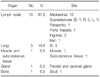
Sixteen patients with sarcoidosis had involvement of various organs, including lymph nodes (13/16, 81.3%), lung (3/16, 18.8%), muscle (1/16, 6.3%), subcutaneous tissue (1/16, 6.3%), glands (1/16, 6.3%), and bone (1/16, 6.3%). Sites of involved lymph nodes were thorax (12/13, 92.3%), supraclavicular area (5/13, 38.5%), inguinal area (2/13, 15.4%), abdomen (2/13, 15.4%), and pelvis (1/13, 7.7%).
Figures and Tables
Figure 1
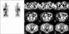
Sarcoidosis in a 32-year-old man with the lambda sign at 67Ga imaging. (A) A Chest radiograph shows both hilar enlargement. (B) Whole body 67Ga imaging shows increased tracer uptake in bilateral hilar and right paratracheal (lambda sign) and bilateral supraclavicular lymph nodes. (C) CT images (upper, preenhancement; lower, postenhancement) show multiple mediastinal lymphadenopathies with lower attenuation and mild enhancement. CT: computed tomography.
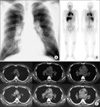
Figure 2
Sarcoidosis in a 27-year-old man with acute uveitis. Whole body 67Ga imaging shows lambda sign and focal uptake of tracer in the nasopharynx, parotid gland and lacrimal gland (panda sign).
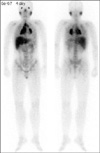
Figure 3
Sarcoidosis in a 59-year-old woman with involving lung, muscle and bones. (A) Whole body 67Ga imaging showed increased tracer uptake in multiple mediastinal lymph nodes. (B) Two years later, repeat 67Ga imaging (planar whole body and lower extremities imaging, SPECT scan for head) showed decreased uptake in multiple mediastinal lymph nodes compared with previous scan. However, new lesions were shown at both lung, lower extremities and skull. (C) The patient was treated with low-dose oral glucocorticoid. Six months later, repeat 67Ga imaging showed improvement.
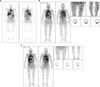
Figure 4
Sarcoidosis in a 28-year-old man with involving lung, subcutaneous tissue, and pelvic lymph nodes. Whole body 67Ga imaging shows increased tracer uptake in both lung and both inguinal lymph nodes. Multiple subcutaneous activities are also identified in upper extremities, abdomen and back.
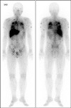
Figure 5
Sarcoidosis in a 37-year-old woman with involving multiple lymph nodes. Whole body 67Ga imaging shows increased tracer uptake in left supraclavicular, paraaortic, iliac, and inguinal areas corresponding to enlarge lymph nodes on enhanced CT images. CT: computed tomography.
References
1. Koyama T, Ueda H, Togashi K, Umeoka S, Kataoka M, Nagai S. Radiologic manifestations of sarcoidosis in various organs. Radiographics. 2004. 24:87–104.
2. Yi GW, Koh EM, Chung JK, Lee MC, Shim YS, Koh CS. Three cases of sarcoidosis evaluated by 67Ga scintigraphy. Korean J Nucl Med. 1988. 22:93–98.
3. Sulavik SB, Spencer RP, Weed DA, Shapiro HR, Shiue ST, Castriotta RJ. Recognition of distinctive patterns of gallium-67 distribution in sarcoidosis. J Nucl Med. 1990. 31:1909–1914.
4. Britt AR, Francis IR, Glazer GM, Ellis JH. Sarcoidosis: abdominal manifestations at CT. Radiology. 1991. 178:91–94.
5. Johnson DG, Johnson SM, Harris CC, Piantadosi CA, Blinder RA, Coleman RE. Ga-67 uptake in the lung in sarcoidosis. Radiology. 1984. 150:551–555.
6. Gupta RG, Bekerman C, Sicilian L, Oparil S, Pinsky SM, Szidon JP. Gallium 67 citrate scanning and serum angiotensin converting enzyme levels in sarcoidosis. Radiology. 1982. 144:895–899.
7. Sohn HS, Kim EN. A case of muscular sarcoidosis diagnosed by Gallium-67 scintigraphy and magnetic resonance imaging. Korean J Nucl Med. 1999. 33:543–548.
8. Moore SL, Teirstein AE. Musculoskeletal sarcoidosis: spectrum of appearances at MR imaging. Radiographics. 2003. 23:1389–1399.
9. Henry DA, Kiser PE, Scheer CE, Cho SR, Tisnado J. Multiple imaging evaluation of sarcoidosis. Radiographics. 1986. 6:75–95.
10. Kim MJ, Yoo HS, Lee JT, Suh JH, Park CY, Lee DY. 67Ga of primary hepatocellular carcinoma: correlation with angiography. Korean J Nucl Med. 1989. 23:27–34.
11. Tsan MF, Scheffel U. Mechanism of gallium-67 accumulation in tumors. J Nucl Med. 1986. 27:1215–1219.
12. Baughman RP, Shipley R, Eisentrout CE. Predictive value of gallium scan, angiotensin-converting enzyme level, and bronchoalveolar lavage in two-year follow-up of pulmonary sarcoidosis. Lung. 1987. 165:371–377.
13. Braun JJ, Kessler R, Constantinesco A, Imperiale A. 18F-FDG PET/CT in sarcoidosis management: review and report of 20 cases. Eur J Nucl Med Mol Imaging. 2008. 35:1537–1543.
14. Nishiyama Y, Yamamoto Y, Fukunaga K, Takinami H, Iwado Y, Satoh K, et al. Comparative evaluation of 18F-FDG PET and 67Ga scintigraphy in patients with sarcoidosis. J Nucl Med. 2006. 47:1571–1576.
 XML Download