

 PDF
PDF ePub
ePub Citation
 Print
Print


Abstract
Objectives
Endobronchial anthracofibrotic pigmentation, which presents as dark black or brown pigmentation mucosal changes of multiple bronchi combined with bronchial fibrosis and obstruction, is not a rare finding when performing diagnostic bronchoscopy for Koreans. This study was performed to define the clinical characteristics and to determine the association of these finding with the Korean life style and such other diseases as coal workers, pneumoconiosis or tuberculosis in the patients with anthracofibrotic pigmentation.
Methods
This retrospective analysis was conducted on 70 (5.2%) patients with endobronchial anthracofibrotic pigmentation, among a total of 1340 patients who underwent bronchoscopy. The distinctive clinical features, the personal life style, the past medical history, the histology and microbiology, the radiologic finding and the natures of the bronchoscopic lesions were analyzed.
Results
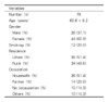
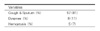
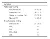
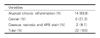
This mean age of the patients with anthracofibrotic pigmentation was 60.6 ± 9.2 year old and the male to female ratio was 1:1.7. The common respiratory symptoms of these patients were coughing and sputum (81%, 57/70), and this was followed in order by dyspnea and hemoptysisir. The symptoms were not related with smoking and an occupational history such as being a coal worker and so on. Pneumonia was most common finding on the radiologic studies. On bronchoscopy, the right middle lobe bronchus was most commonly involved. The most common associated disease was tuberculosis, and 40 cases (57.1%) were diagnosed by AFB staining, TB PCR, bronchoscopic guided tissue biopsy and a past history of tuberculosis. Other diseases related with anthracotic pigmentation were hypertension, diabetes, COPD, lung cancer, pneumoconiosis and asthma.
Figures and Tables
References
1. Vorwald AJ. Spencer H, editor. The Pneumoconiosis and other occupational lung disease. Pathology of the lung. 1985. Vol I:4th ed. Pergamon;456.
2. Woodruff CE, Moerke AG. Spencer H, editor. The pneumoconiosis and other occupational lung disease. Pathology of the lung. 1985. Vol I:4th ed. Pergamon;456.
3. Scheid KF. Ueber exogene und endogene eisenablagerungen in der lunge. Beitr Path Anat. 1931.
4. Dennis RJ, Maldonado D, Norman S, Baena E, Martinez G. Woodsmoke exposure and risk for obstructive airways disease among women. Chest. 1996. 109:115–119.
5. Park IW, Yoo CG, Kwon OJ, Kim YW, Han SK, Shim YS, et al. Clinical study of dark-blue pigmentation in the bronchial mucosa. Tuberc Respir Dis. 1991. 38:280–286.
6. Pandey MR. Domestic smoke pollution and chronic bronchitis in a rural community of the Hill Region of Nephal. Thorax. 1984. 39:337–339.
7. Kradine RL, Spirin PW, Mark EJ. Intrapulmonary lymph nodes. Chest. 1985. 87:662–667.
8. Brody AR, Craighead JE. Cytoplasmic inclusions in pulmonary macrophages of cigarette smokers. Lab Invest. 1975. 32:125–132.
9. Lee HS, Maeng JH, Park PG, Jang JG, Park W, Ryu DS, et al. Clinical features of simple bronchial anthracofibrosis which is not associated with tuberculosis. Tuberc Respir Dis. 2002. 53:510–518.
10. Choi DW, Park JN, Suh JE, Baik JH, Ma SD, Kim EB, et al. Chest CT finding of endobronchial tuberculosis patients. Tuberc Respir Dis. 1990. 37:195–202.
11. Kim HY, Im JG, Goo JM, Kim JY, Han SK, Lee JK, et al. Bronchial anthracofibrosis (inflammatory bronchial stenosis with anthracotic pigmentation): CT findings. AJR Am J Roentgenol. 2000. 174:523–527.
12. Chung MP, Lee KS, Han JH, Kim HJ, Rhee CH, Han YC, et al. Bronchial stenosis due to anthracofibrosis. Chest. 1998. 113:344–350.
13. Arbert RK, Petty TL. Endobronchial tuberculosis progressing to bronchial stensis. Chest. 1976. 70:537–539.
14. Pierson DJ, Lakshminarayan S, Petty TL. Endobronchial tuberculosis. Chest. 1973. 64:537–539.
15. Wilson NJ. Bronchoscopic observations in tuberculosis tracheobronchitis. Chest. 1945. 11:36.
16. Cohen AG. Atelectasis of the right middle lobe resulting from perforation of the tuberculosis lymph node into bronchi in adult. Ann Intern Med. 1951. 35:820–835.
17. Lee JH, Lee DH, Park SS. Endobronchial tuberculosis: clinical and bronchofiberscopic features. Korean J Intern Med. 1986. 1:229–232.
18. Han SW, Lee DH, Park SS, Lee JH. Clinical study of endobronchial tuberculosis. Tuberc Respir Dis. 1984. 31:57–61.
19. Song JH, Han SK, Heo IM. Clinical study on endobronchial tuberculosis. Tuberc Respir Dis. 1985. 32:276–283.
 XML Download