

 PDF
PDF ePub
ePub Citation
 Print
Print


Abstract
Purpose
To compare the changes in corneal topography between 23 gauge (G) pars plana vitrectomy (PPV) and 20 G PPV.
Methods
Forty-three eyes of 42 patients who underwent PPV were divided into 2 groups: 20G PPV (Group I, n=18) and 23 G PPV (Group II, n=25). Both groups were followed up over 3 months postoperatively. ORBSCAN II was performed to compare the corneal topographic change of 20 G PPV and 23 G PPV.
Results
In a 5 mm zone, significant postoperative change in the mean corneal surface cylinder was found between the 1st day and the 1st month in Group I, while no significant change was found in Group II. Postoperatively, the increase in the mean corneal surface cylinder in Group I was significantly higher than in Group II between the 1st day and the 1st month. Clinically significant astigmatism, induced by surgery, was found only in Group I one week after the operation. In a 3 mm zone, there was no significant change in the mean corneal surface cylinder in both groups postoperatively. The difference in the mean corneal surface cylinder between the 2 groups at the postoperative periods showed no significant change. Surgically induced astigmatism was significantly higher in Group I than in Group II between the 1st day and the 1st month postoperatively.
References
1. Jackson T. Modified sutureless sclerotomies in pars plana vitrectomy. Am J Ophthalmol. 2000; 129:116–7.

2. Assi AC, Scott RA, Charteris DG. Reversed self sealing pars plana vitrectomy. Retina. 2000; 20:689–92.
3. Rahman R, Rosen PH, Riddell C, Towler H. Self-sealing sclerotomies for sutureless pars plana vitrectomy. Ophthalmic Surg Lasers. 2000; 31:462–6.
4. Theelen T, Verbeek AM, Tilanus MA, van den Biesen PR. A novel technique for self sealing, wedge shaped pars plana sclerotomies and its features in ultrasound biomicroscopy and clinical outcome. Am J Ophthalmol. 2003; 136:1085–92.
5. Olsen T, Johansen MD, Bek T, Hjortdal JO. Corneal versus scleral tunnel incision in cataract surgery: A randomized study. J Cataract Refract Surg. 1997; 23:337–41.
6. Mendivile A. Frequency of induced astigmatism following phacoemulsification with suturing versus without suturing. Ophthalmic Surg Lasers. 1997; 28:377–81.
7. Yang HS, Ahn JH, Lew HM. The change of corneal shape after cataract operation analysed by Slit Scan Cornea tomography/ pachymetry system (ORBSCANTM). J Korean Ophthalmic Soc. 2000; 41:1544–55.
8. Joo CK, Han HK, Kim JH. Computer assisted video kera-tography to measure changes in astigmatism induced by sutureless cataract surgery. J Cataract Refract Surg. 1997; 23:555–61.
9. Koch DD, Haft EA, Gay C. Computerized videokeratographic analysis of corneal topographic changes induced by sutured and unsutured 4 mm scleral pocket incisions. J Cataract Refract Surg. 1993; 19:166–9.
10. Vass C, Menapace R, Rainer G. Corneal Topographic changes after frown and straight sclerocorneal incisions. J Cataract Refract Surg. 1997; 23:913–22.
11. Krishnamachary M, Basti S. Computerized topography of selective versus all-suture elease to manage high astigmatism after cataract surgery. J Cataract Refract Surg. 1997; 23:1380–3.
12. Jaffe NS, Clayman HM. The pathophysiology of corneal astigmatism after cataract extraction. Trans Am Acad Ophthalmol Otolaryngol. 1975; 79:615–30.
13. Naeser K. Conversion of keratometer readings to polar values. J Cataract Refract Surg. 1990; 16:741–5.
14. Naeser K, Behrens JK. Correlation between polar values and vector analysis. J Cataract Refract Surg. 1997; 23:76–81.
15. Alpins NA. A new method of analyzing vectors for changes in astigmatism. J Cataract Refract Surg. 1993; 19:524–31.
16. Park KJ, Hahn TW, Kim JH. Arcuate keratotomy for astigmatism after keratoplasty and advanced vector analysis. J Korean Ophthalmic Soc. 1998; 39:2558–68.
17. Kadonosono K, Yamakawa T, Uchio E, et al. Comparison of visual function after epiretinal membrane removal by 20 gauge and 25 gauge vitrectomy. Am J Ophthalmol. 2006; 142:513–5.
18. Weinberger D, Lichter H, Loya N, et al. Corneal topographic changes after retinal and vitreous surgery. Ophthalmology. 1999; 106:1521–4.
19. Wilson SE, Klyce SD. Advances in the analysis of corneal topography. Surv Ophthalmol. 1991; 35:269–77.
20. Dominez YY, Cahana M, Avni I. Corneal surface changes after pars plana vitrectomy and scleral buckling surgery. J Cataract Refract Surg. 2001; 27:868–72.
21. Wirbelauer C, Hoerauf H, Roider J, Laqua H. Corneal shape changes after pars plana vitrectomy. Graefes Arch Clin Exp Ophthalmol. 1998; 236:822–8.
22. Jampel HD, Thompson JC, Nunez M, Michels RG. Corneal astigmatic changes after pars plana vitrectomy. Retina. 1987; 7:223–6.
23. Yanyali A, Celik E, Horozoglu F, Nohutcu AF. Corneal topographic changes after transconjunctival (25 gauge) Sutureless vitrectomy. Am J Ophthalmol. 2005; 140:939–41.
Figure 1.
Changes in mean astigmatism (in 5-mm zone) after 20-gauge and 23-gauge pars plana vitrectomy.* Statistically significant change between preoperative and each postoperative periods; † Statistically significant change between Group I and Group II at each time interval.
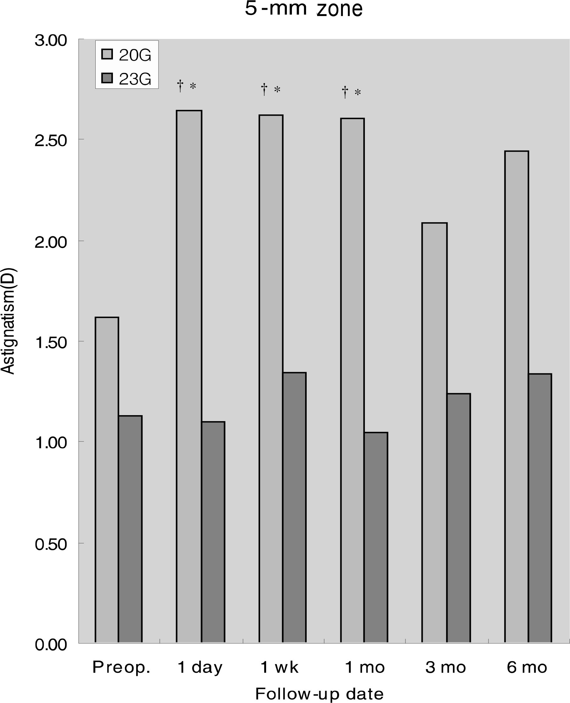
Figure 2.
Changes in mean astigmatism (in 3-mm zone) after 20-gauge and 23-gauge pars plana vitrectomy. Significant changes of mean astigmatism were not found in both groups postoperatively.
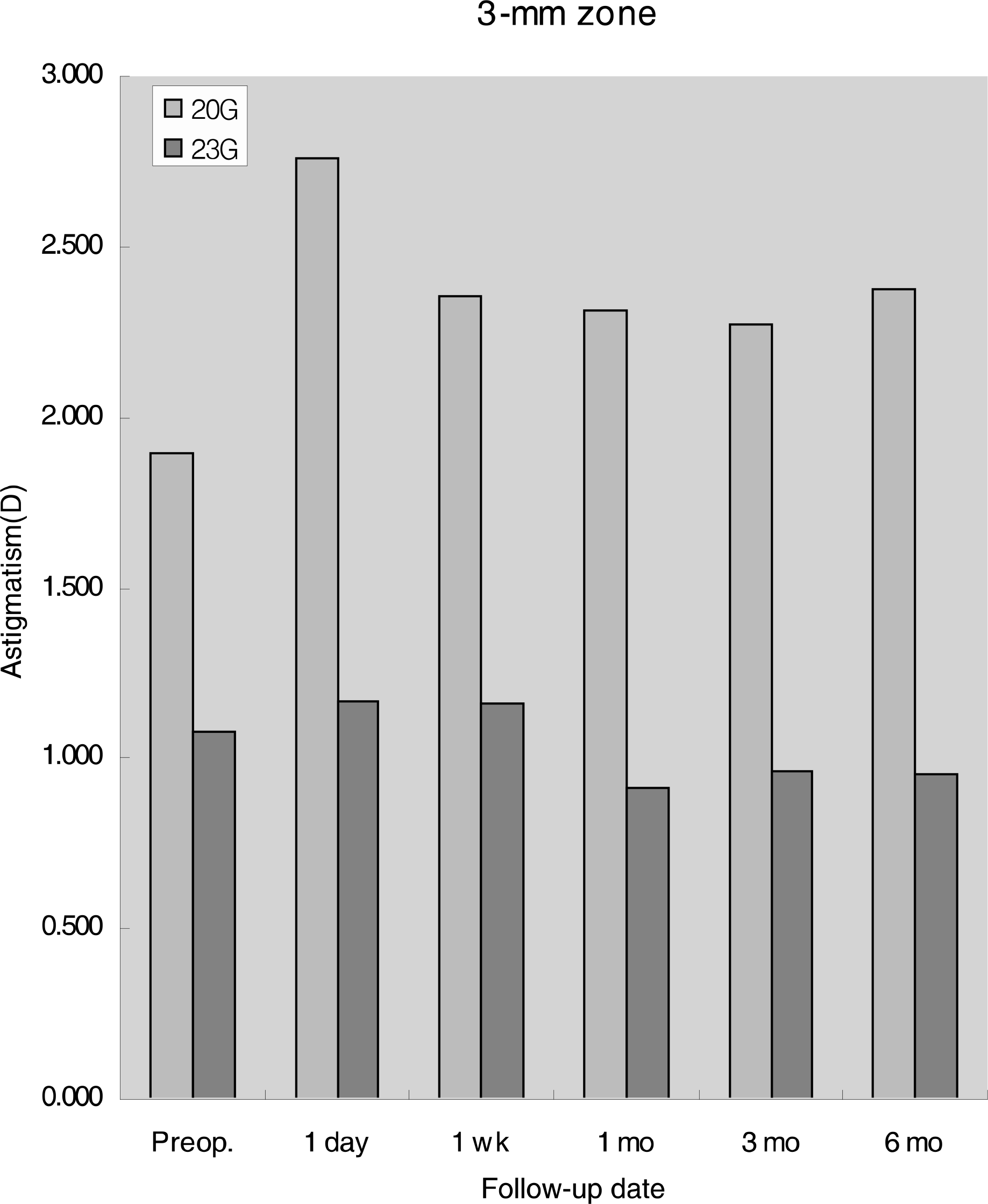
Table 1.
Methods of surgery
| No. of eyes | ||
|---|---|---|
| Group I | Group II | |
| PPV* | 0 | 14 |
| PPV+Silicone oil injection | 2 | 2 |
| PPV+Gas (C3 F8 or SF6) injection | 5 | 9 |
| Silicone oil removal | 11 | 0 |
| Total | 18 | 25 |
Table 2.
Changes of mean corneal power
| Preoperative (D*) | Postoperative (D) | |||||
|---|---|---|---|---|---|---|
| 1 day | 1 week | 1 month | 3 months | 6 months | ||
| Group I | 43.10±3.25 | 43.57±2.60 | 43.55±3.03 | 43.17±2.09 | 43.53±1.91 | 43.33±2.60 |
| Group II | 42.92±1.56 | 42.95±1.27 | 42.95±1.19 | 42.79±1.40 | 42.62±1.80 | 42.54±1.43 |
| p value | > 0.05 | > 0.05 | > 0.05 | > 0.05 | > 0.05 | > 0.05 |
Table 3.
Changes of anterior chamber depth
Table 4.
Surgically induced astigmatism after 20-gauge and 23-gauge pars plana vitrectomy in 5-mm zone
| Surgically induced astigmatism (D*) | |||||
|---|---|---|---|---|---|
| 1 day | 1 week | 1 month | 3 months | 6 months | |
| Group I | 1.56±1.58 | 1.48±1.73 | 1.30±1.07 | 0.33±1.07 | 0.45±2.41 |
| Group II | 1.25±1.16 | 0.02±1.38 | 0.29±1.45 | 0.00±0.79 | 0.21±0.89 |
| p value | < 0.05 | < 0.05 | < 0.05 | > 0.05 | > 0.05 |
 XML Download