

 PDF
PDF ePub
ePub Citation
 Print
Print


In 1923, the dentist Pierre Robin described a disorder characterized as a triad of micrognathia, glossoptosis, and airway obstruction.1 This disorder, now known as Pierre Robin sequence (PRS), has received much attention in dentistry due to its relationship with anomalies in the maxillofacial area. PRS does not always have a single pathogenesis, but rather is often associated with various syndromes.2 Izumi el al.3 found that 60% of cases of PRS were associated with another syndrome, while 40% were isolated.
Cerebro-costo-mandibular syndrome (CCMS) is a PRS-related syndrome associated with rib anomalies characterized by posterior rib gap defects.456 Since the first report of CCMS by Smith et al.4 in 1966, over 80 cases have been reported to date.5 In addition to PRS and multiple rib gaps, scoliosis is a common feature of CCMS. The heart, the kidneys, hearing ability, and intelligence are also affected in some patients with CCMS.7 Because the most consistent features of CCMS are associated with the hard tissues of the maxillofacial and thoracic vertebral area, radiographic evaluation, such as thoracic computed tomography, posteroanterior (PA) chest radiography, lateral cephalograms, and cone-beam computed tomography (CBCT), is required for the diagnosis and treatment planning of CCMS.
The patient in this report was suspected of having CCMS because she had PRS and severe scoliosis. However, no posterior rib gap defects were observed on PA chest radiography. Therefore, the authors could not make a definitive diagnosis of CCMS. Although many reports have been published on CCMS, few cases of PRS with scoliosis alone have been reported.8 This case report presents the clinical and radiological features of an adult patient with PRS and severe scoliosis, but without rib anomalies.
Case Report
A 35-year-old woman visited the Department of Orthodontics at Gangneung-Wonju National University Dental Hospital. Her chief complaints were a retruded mandible, a large overjet, and crowding on the upper incisors. She underwent soft palate surgery when she was an infant and spinal surgery for severe scoliosis at age 15. She had no familial history of any relevant syndromes.
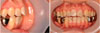
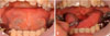
Clinical examination revealed an anterior open bite, crowding on the upper incisors, a narrow maxillary arch, and Class II canine relationship (Fig. 1). The bilateral lower second premolars and first molars were missing and replaced by fixed partial prostheses. Due to tongue-tie (Fig. 2A) and her anterior open bite, the patient had difficulty pronouncing sounds such as ‘s’ and ‘z’. Scar tissue was present on the soft palate, and mild rhinism was detected (Fig. 2B). There were no noteworthy symptoms of temporomandibular disorder except a non-reproducible click sound on the right temporomandibular joint when the mouth was opened. Additionally, the patient had no history of pain of either temporomandibular joint.
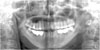
For the orthodontic diagnosis, a panoramic radiograph, a lateral cephalogram, and CBCT scans were taken. The panoramic radiograph showed bilateral absence of the lower second premolars and the lower first molars (Fig. 3). The bilateral lower second molars were affected by secondary dental caries, and the left lower third molar had a periapical lesion. Deep antegonial notches and posterior bowing of the bilateral condyles were observed.
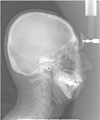
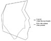
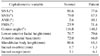
Cephalometric analysis showed a small and retruded mandible and a steep mandibular plane (Table 1). The A point-nasion-B point angle was 10.1°, indicating a severe skeletal Class II relationship. The mandibular plane angle was larger than normal, at 51.4°. Narrowing of the airway was also observed (Fig. 4). To compare the lateral cephalograms between the patient and normal control images,9 the profilograms were superimposed at the sella using the sella-nasion line as a horizontal reference line (Fig. 5). Severe bimaxillary retrusion, particularly of the mandible, as well as a relatively small mandible and a large inclination of the mandibular plane were found.
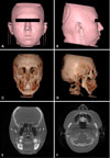
Mandibular asymmetry, with deviation of the chin to the left side and low-set ears (Figs. 6A–D), was exhibited on the CBCT images oriented according to the method described by Kim et al.10 A discontinuity of the nasal septum extending from the posterior hard palate to the superior nasal area was observed (Fig. 6E). A lack of soft tissue near the posterior nasal spine was also observed (Figs. 6E and F). The bilateral condyles were hypoplastic, but there were no signs of degenerative joint disease, such as erosive changes of the condyle or articular eminence.11
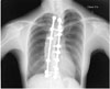
Because PRS and scoliosis were observed during the clinical and radiological examinations, this patient was initially suspected to have CCMS. A PA chest radiograph was taken to identify rib defects and scoliosis (Fig. 7). Severe scoliosis was identified, but no signs of posterior rib gap defects were detected.
Discussion
The patient in this report had PRS and severe scoliosis. PRS is known to be associated with various syndromes and genetic anomalies.2 Among the syndromes accompanied with PRS is CCMS, which is characterized by vertebral and rib anomalies. Campomelic dysplasia and diastrophic dysplasia also present with scoliosis,2 but no other symptoms of these syndromes - such as short stature, hearing loss, or short limbs - matched the patient's physical condition. Therefore, we suspected that this patient had CCMS.
Antenatally, patients with CCMS usually exhibit raised nuchal translucency, intrauterine growth retardation, polyhydramnios, and PRS,512 which are identified using ultrasonography or magnetic resonance imaging.12 After birth, various radiographic techniques are available to detect the rib anomalies characteristic of CCMS. Because the major radiological findings are a narrow thorax, posterior rib gap defects, and defects of costo-transverse articulation,5 thoracic computed tomography or PA chest radiography is required to identify CCMS. Posterior rib gap defects in CCMS may resemble the costal fractures of the newborn that can occur when resuscitation is performed.13 Several researchers have reported that these rib gaps may become ossified with increasing age.514 However, it is uncertain whether this ossification around the rib gaps in certain CCMS patients is true ossification or simply the calcification of fibroconnective tissue.14 Consequently, this indicates that the gaps may not be visible in adult CCMS patients. Other features of CCMS are severe scoliosis, a reduced number of ribs, the presence of rudimentary ribs or ribs with a total lack of ossification, and a lack of ossification at the center of the sternum.51516 Tooley et al.5 proposed that rudimentary ribs or ribs with a total lack of ossification are indicative of a severe phenotype with high mortality. Therefore, it is thought that these severe rib anomalies may be rare in adult patients with CCMS. Recently, the presence of an accessory ossicle arising from the lesser cornua of the hyoid bone in CCMS was reported, although the mechanism behind this is uncertain.5 In our patient, an accessory ossicle of the hyoid bone was not observed.
The dentofacial anomalies of PRS, along with the rib and vertebral anomalies, are major features of CCMS.57 Micrognathia and glossoptosis can be detected by a physical examination, but the skeletal anomalies in the maxillofacial area should be evaluated radiologically. The morphological features of the craniofacial area can be evaluated precisely by CBCT and lateral cephalograms (Figs. 4, 5, 6, 7, Table 1).
The dentofacial traits of this patient, including micrognathia, cleft soft palate, and tongue-tie, are typical features of PRS. The presence of nasal septum deviation aligns with an earlier study that found that the nasal septum deviation of the cleft group was more severe than that of the non-cleft group.17 On PA chest radiography, severe scoliosis was observed; however, there were no signs of posterior rib gap defects or other rib anomalies. A medical radiologist confirmed that posterior rib gap defects and traces of ossification were not observed upon further examination. Chest radiography at infancy or during the growth period could be useful to confirm the process of ossification, but the previous records of the patient could not be obtained.
Several researchers found that heterozygous mutations of the SNRPB gene are associated with CCMS.1819 Therefore, genetic tests, such as exome sequencing and Sanger sequencing, are recommended for confirmation of CCMS.19 However, our patient refused further genetic testing, so unfortunately, we could not confirm the presence of CCMS.
Maxillofacial radiologists, surgeons, and orthodontists play important roles in the skeletal and dental management of patients with PRS.20 The maxillofacial deformities associated with PRS are evaluated by an expert radiologist, and each evaluated deformity is managed by a maxillofacial surgeon. Generally, airway problems are the most critical. For those problems, noninvasive methods such as prone/lateral positioning, continuous positive airway pressure, or nasopharyngeal intubation are preferred. Invasive methods, including tracheostomy, tongue-lip adhesion, or mandibular distraction osteogenesis, may be also used. Palate surgery is usually performed approximately 1 year after birth.21 From an orthodontic perspective, problems associated with PRS include Class II malocclusion, crowding in the narrow maxillary arch, and hypodontia. A phase I orthodontic treatment plan can include maxillary expansion and induce the normal eruption of teeth. Although a lack of sufficient space for proper alignment of the teeth is observed, it is recommended that the decision whether to perform tooth extraction be delayed until phase II of treatment. After growth is complete, a large majority of cases require orthodontic treatment with orthognathic surgery to improve the patients' Angle Class II dentition and convex profiles due to anteroposterior discrepancies between the maxilla and the mandible. Only patients with mild discrepancies are typically treated using alternatives to orthognathic surgery.20
Because this patient showed a severe mandibular retrusion with a Class II relationship, an asymmetric mandible, orthognathic surgery was recommended for correction of the skeletal discrepancy. However, the patient did not want to undergo orthognathic surgery for financial and psychological reasons. Therefore, camouflage treatment for the correction of Class II malocclusion and prosthodontic rehabilitation for missing lower posterior teeth were planned. To advance the retruded chin, filler augmentation of the chin (or genioplasty) was also planned.
When treating a patient with PRS, clinicians should always suspect another underlying syndrome and, if necessary, identify the syndrome present. Radiological examinations are often useful to identify the syndrome, and additional genetic testing can be performed as well. This report provides a better understanding of the clinical, radiological, and cephalometric features of an adult presenting with PRS with scoliosis, but without rib anomalies.
 XML Download