

 PDF
PDF ePub
ePub Citation
 Print
Print


INTRODUCTION
The number of pediatric patients with inflammatory bowel disease (IBD) has gradually increased over the last several decades. Their clinical symptoms are so diverse or vague that many of them waste time for the proper diagnosis. Furthermore, others who do not have knowledge or experience with IBD, such as the patient's parents and even the primary physician may contribute to the delayed diagnosis of these diseases.
The clinical symptoms of mild IBD are similar to those of irritable bowel syndrome (IBS), infectious enterocolitis (IE), and allergic gastroenteritis, so systemic and aggressive diagnostic evaluations may not be undertaken in most primary clinics and/or larger institutions. This could be another reason why the proper diagnosis is delayed. Furthermore, the prevalence rate of IBD in Korea has been traditionally very low but has gradually increased during the last three decades. Thus most primary physicians in Korea may seldom initially consider the possibility of IBD, especially in the pediatric ages.
The symptoms of IBS are sometimes suggestive of those of initial state of IBD. Several clinical studies show that some patients with IBS develop to other diseases as well as IBD. Patients with IBS also show inflammatory and immunologic changes in the colon mucosa. Furthermore, IBS and IBD are aggravated after acute infectious gastroenteritis and psychological stimulations. Thus, many physicians suggest that IBS should never be overlooked as a simple transient disease [1,2].
Physicians also have problems to differentiating tuberculous enteritis (TE) from Crohn's disease (CD). When gastroenterologists make a diagnosis of CD, a differential diagnosis with TE is always performed and well executed in most cases. Alternatively, when physicians make a diagnosis of TE, the possibility of CD is rarely considered and CD is mostly ruled-out disease. However chronic infectious enteritis such as tuberculosis could mimic CD and mask early diagnosis.
One of the most confounding obstacles for reaching the timely and correct diagnosis of IBD is that patients sometimes do not seek a physician with the proper knowledge to solve the problems. Several patients in this study spent a significant amount of time under the care of unsuitable specialists.
This study was undertaken to know the typical bowel symptoms and clinical manifestations of IBD prior to diagnosis using the medical records of clinical pediatric case studies. Through these case studies, we investigated the time lag between clinical presentation and final diagnosis, and whether any useful laboratory predictors of CD or ulcerative colitis (UC) occurred among the routinely performed initial examinations.
MATERIALS AND METHODS
The medical records of all patients under 19 years of age who were ultimate diagnosed with IBD from 2000 to 2012 were reviewed. Indeterminate colitis was excluded for this study. CD was diagnosed in 14 children (10 male, 4 female) and UC in 17 children (15 male, 2 female). Medical histories prior to the IBD diagnosis were also reviewed. Their previous long-term symptoms and other, specific diagnosis made by former physicians were also reviewed. Among the routinely performed laboratory data, the results concerning white blood cell, hemoglobin, mean corpuscular volume (MCV), serum total protein, and serum albumin were evaluated to determine if they have any role as predictors of CD or UC. Other important laboratory data such as erythrocyte sedimentation rate (ESR), C-reactive protein (CRP), and anti-neutrophil cytoplasmic antibodies were not performed in all patients, so these data could not be evaluated. Two samples t-test's nonparametric test was applied to compare useful clinical indicators for CD and UC, and Fisher's exact test was applied for sexual difference in patients with either of the two diseases. SAS version 9.2 (SAS Institute Inc, Cary, NC, USA) was used for the statistical analysis. A p-value<0.05 was considered statistically significant when assessing the diagnostic time lag, and a p-value<0.1 was considered significant when comparing the clinical indicators of the two diseases.
RESULTS
The number of the male patients of CD and UC was more predominant than the female patients. The mean age at the time of final diagnosis was also also-similar in both diseases, but there were two preschool children (five and six years old) among the children with UC (Table 1).
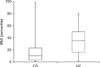
Body mass index (BMI) was used to as an anthropometric parameter to compare the nutritional status of patients with either of the two diseases. The mean BMI of patients with either of the two diseases was similar: 17.5 for children with CD, 18 for those with UC. Fig. 1 shows the detailed distribution of BMI among the children. Most children with CD showed a lower BMI than the children with UC. One child with CD showing very high BMI made the mean BMI similar with the children with UC (Fig. 1).
Of the 14 children with CD, nine (64%) had been experiencing diverse gastrointestinal (GI) symptoms, weight loss, and anemia. The same had been true for 10 (58%) of the 17 children with UC. Previously healthy but suddenly, severely ill children could rather get the correct final diagnosis on time, and the period until the final diagnosis was very short with statistical significances in both diseases (p<0.05) (Tables 2 and 3).
Physicians, mostly non-gastroenterologists, offered specific diagnoses that created time lags before other physicians could offer the correct final diagnosis of CD or UC in the children under study. The previous diagnoses were TE (2), duodenal ulcer (1), appendicitis (1), anal fistula (1), and irritable bowel disease (1) among the children with CD, and enterocolitis (3), hemorrhoids (2), rectal varix (1), and rheumatoid arthritis (1) among the children with UC. Table 4 shows that sometimes the final diagnosis was significantly delayed due to an incorrect initial diagnosis (six years after initial diagnosis of TE in a child and three years after the initial diagnosis of rheumatoid arthritis in another child).
To determine diagnostic predictors for differentiation between CD and UC, we compared initial laboratory and anthropometric data. Age, sexual distribution, and BMI were not significantly different. Children with CD showed lower hemoglobin, MCV, serum albumin, and protein levels than children with UC, but the differences were not statistically significant (Table 5).
DISCUSSION
The most common symptoms that presented prior to the prior the final diagnosis were abdominal pain, diarrhea, and bloody stool. Abdominal pain and diarrhea are also the classic, typical symptoms of IBS or IE. Some patients had suffered anal problems or joint diseases. The patients surely had visited so-called specialists such as rheumatologists or ano-rectal surgeon. However we found that many patients had to wait a significant amount of time time before a subsequent physician offered the correct diagnosis. It is not clear whether the former physicians let the patients cling to the doctors or there were no typical symptoms of IBD while the former physicians were taking care of them. It is strongly suggested that even though a patient may present with localized symptoms or clinical problems the responsible physician should evaluate the patient with an overall systemic inspection avoiding a simple decision based on a localized problem.
It is already known that IBD could develop among some patients who have suffered from longstanding IBS. Two large population studies show that the risk of IBD significantly increased (5-10 fold) among patients with IBS or IE when compared to those who did not have clinical histories of prior IBS, especially in CD [3,4]. The children in this study who had presented with diverse GI symptoms could have been the patients with IBS or IE, even though there were no specific clues or microbiologic evidences to support this. Infectious enteritis, chronic TE, eosinophilic enteritis and intestinal vasculitis may present right lower quadrant abdominal pain and diarrhea. Among these diseases, TE induces the most confusing, difficult condition for making a differential diagnosis from CD. To reach a correct differential diagnosis of the etiology, detail clinical history, physical examination, laboratory examinations and ileocolonoscopy with pathologic investigations are required [5]. A clinical comparison study shows also that for the differential diagnosis between CD and TE ileocolonoscopy and histopathologic comparison is the most important, and there were no statistically significant data as a predictor or indicator [6]. A recent report also explained that the tuberculosis polymerase chain reaction (TB-PCR) examination is more valuable to detect Mycobacterium tuberculosis in biopsy specimens. The TB-PCR test targeting IS6100 in conjunction with histopathologic examination seems to be an efficient technique for the differential diagnosis in difficult cases [7]. One of the two children who were initially diagnosed with TE and later CD had been treated for TE for one year, experiencing intermittent abdominal pain and poor weight gain until CD was diagnosed six years after the initial diagnosis. His former doctor did the best for this child during the interim period, but the child did not show the typical clinical features of IBD, so further aggressive investigations might not be performed.
Joint pain or arthritis are representative extraintestinal manifestations of IBD, but sometimes the joint illness present prior to the typical IBD clinical features [8]. A Netherlands-driven multicenter clinical research report showed that among children with juvenile idiopathic arthritis (JIA) under etanercept, 0.36% newly developed IBD, which is a rate of incidence 43 times higher than in the general pediatric population. Children with JIA with GI symptoms also show pathologic findings of lymphoid hyperplasia in the GI mucosa and increased intestinal intraepithelial lymphocytes [9]. The one child in our study who had presented with arthritis before the IBD diagnosis had no history of eternacept therapy, but did have an occasional immunosuppressant treatment.
In this study, male patients were predominant than female. There is no specific large imbalance in the sexual distribution of IBD, Thus, it was unusual finding. It remains unknown why IBD was prevalent in male children at this institution. It is thought that the small sample size may explain the prevalence of male patients. An epidemiologic study from Austria showed that the male/female ratio was 1.07 [10], and the ratio in one study from Spain was 1.11 [11]. One review from Korea showed that the male/female ratio in CD was 1.2-2.4 in CD and 0.95 in UC [12]. One Canadian study showed a predominance of CD in male pediatric patients (5-9 years old and 10-14 years old) [13].
We aimed to determine if any comparative predictor existed for CD and UC. From the former experiences, children with CD were expected to show more severe nutritional deprivations. In this study, children with CD showed lower mean hemoglobin, MCV, serum protein and serum albumin, although there was no statistically significance among the initial biochemical and anthropometric examinations. A recent study suggested that red cell distribution width was higher in CD patients, and is a novel marker of IBD activity [14]. Hence, we investigated the possibility of another marker among the initial basic laboratory rxaminations before further special laboratory data are reported. It is already known that the ESR and CRP levels are very high in IBD, but these levels are dependent on the severity of anemia, hemorrhages, co-existing infections and the disease activity itself. We did not cmpare the results of ESR and CRP. A study from Denmark showed that the prevalence rate of anemia in randomly selected outpatient IBD children was significantly higher for CD than UC, and iron deficiency was more prominent (35%) rather than folate or vitamin B12 deficiency (5%) [15]. Another study reported that iron, folate, and vitamin B12 are more readily deficient in patients with CD (25-69%) than in those with UC (1-32%) [16]. Another study recorded that in addition to deficiencies in those elements, calcium, magnesium, zinc, vitamin A, and vitamin D deficiencies occurred in patients who presented with IBD [17]. Ironically, deficiencies of selenium, copper, and chromium are quite rare in IBD patient at the presenting stage except in those for whom central parenteral nutrition therapies are required [18].
In this studt, most children with CD showed poor weight gain, as defined by lower BMI at the time of initial diagnosis of IBD. A recent study also documented that BMI is one of the clinical predictors of a subsequently disabling course of CD in children [19].
IBD has shown very diverse clinical symptoms before presenting its classical features. Thus, patients often have to wait for a significant amount of time before receiving a definite diagnosis. Physicians need to be cautious about the possibility of IBD in the healthy children who do not show the typical symptoms of IBD at the time of initial diagnosis.


 XML Download