

 PDF
PDF ePub
ePub Citation
 Print
Print


Abstract
The authors installed implants combined with guided bony regeneration (GBR) using autogenous tooth bone graft material in the patients. In one patient, GBR and simultaneous implant placement were performed. In two patients, GBR was performed and the implants were placed after 6 months. All patients achieved favorable clinical outcomes. Excellent osteoconductive bony healing was observed in the 6 month histology examination after the bone graft.
References
1. Kim MJ, Kim YK, Kim SG. A variety of biomaterial used in dental surgery. Seoul: Narae Publishing Co.;2004.
2. Kim YK. inventor;. Tooth plaster and manufacturing method thereof. Korean patent 1019980008980. 1998 Mar 17.
3. Kim SG, Kim YK. inventors;. Restorative and grafting material for hard tissue defects prepared from animal teeth. US patent 20030717801. 2003 Nov 19.
4. Kim YK, Yeo HH, Ryu CH, Lee HB, Byun UR, Cho JO. An experimental study on the tissue reaction of toothash implanted in mandible body of the mature dog. J Korean Assoc Maxillofac Plast Reconstr Surg. 1993; 15:129–36.
5. Kim SG, Yeo HH, Kim YK. Grafting of large defects of the jaws with a particulate dentin-plaster of paris combination. Oral Surg Oral Med Oral Pathol Oral Radiol Endod. 1999; 88:22–5.
6. Kim SG, Chung CH, Kim YK, Park JC, Lim SC. Use of particulate dentin-plaster of Paris combination with/without platelet-rich plasma in the treatment of bone defects around implants. Int J Oral Maxillofac Implants. 2002; 17:86–94.
7. Kim SY, Kim SG, Lim SC, Bae CS. Effects on bone formation in ovariectomized rats after implantation of tooth ash and plaster of Paris mixture. J Oral Maxillofac Surg. 2004; 62:852–7.

8. Kim YK, Kim SG, Byeon JH, Lee HJ, Um IU, Lim SC. Development of a novel bone grafting material using autogenous teeth. Oral Surg Oral Med Oral Pathol Oral Radiol Endod. 2010; 109:496–503.
9. Min BM. Oral biochemistry. Seoul: Daehan Narae Publishing, Inc.;2007.
10. Bhaskar SN. Orban's Oral histology and embryology. 9th ed.St. Louis: Mosby Co.;1980.
11. Choung PH. inventor; Method for extracting tooth protein from extracted tooth. Korean patent 1020020008789. 2002 Feb 19.
12. Choung PH. inventor;. Tooth protein extracted from extracted tooth and method for using the same. Korean patent 1020040051812. 2004 Jul 3.
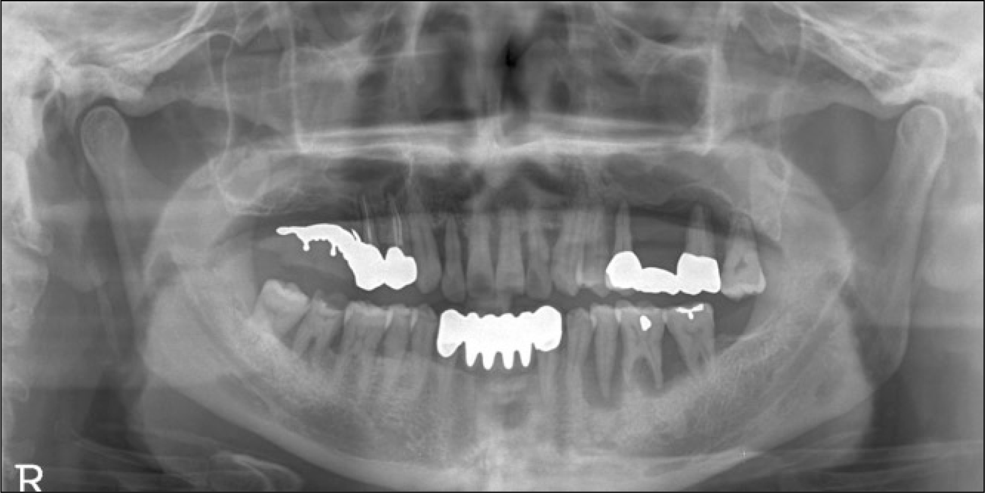
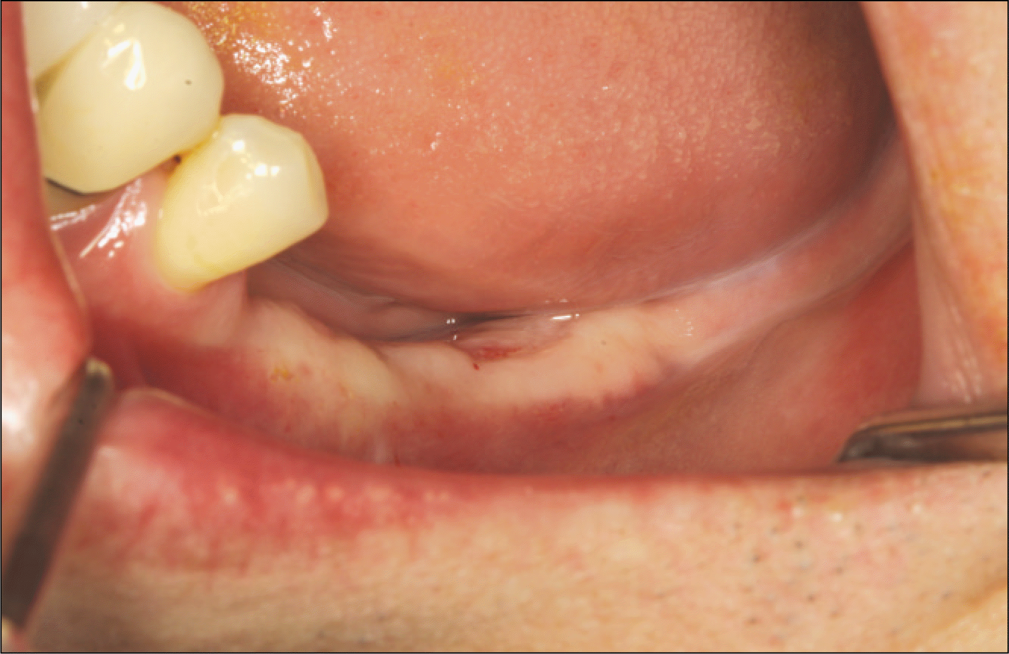
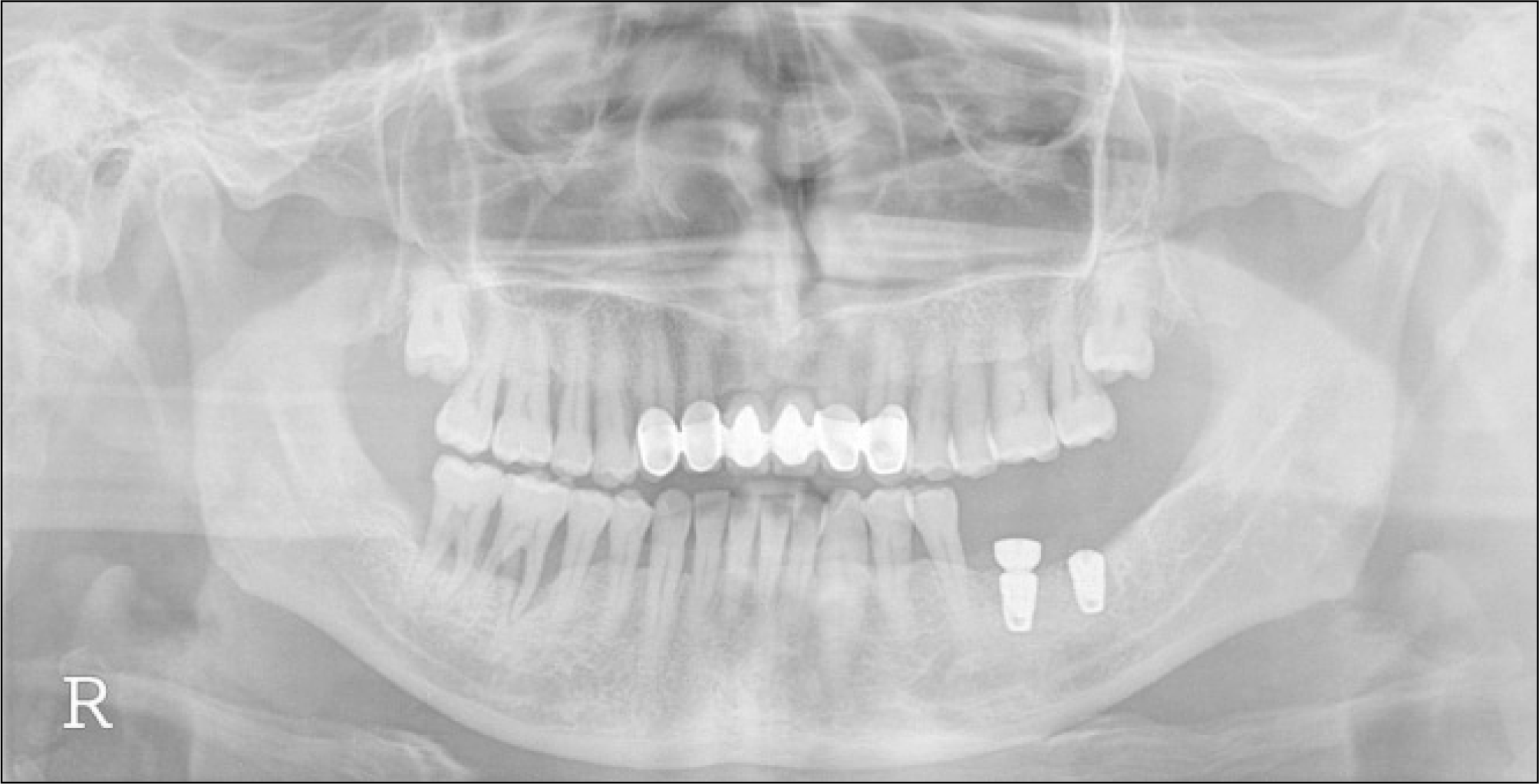
Fig. 3.
Implants were placed and dehiscence defects were covered autogenous tooth bone graft material.
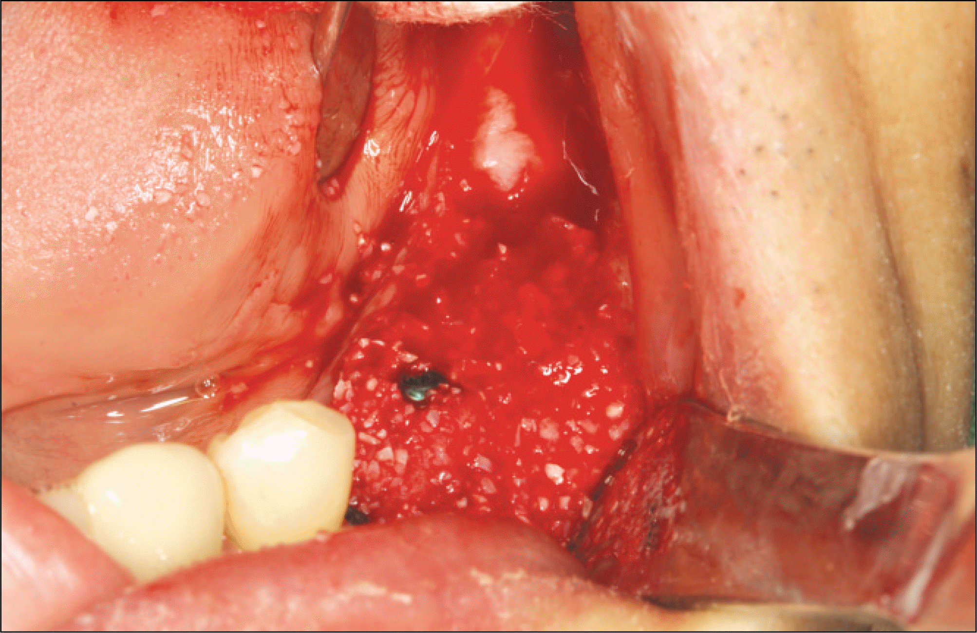
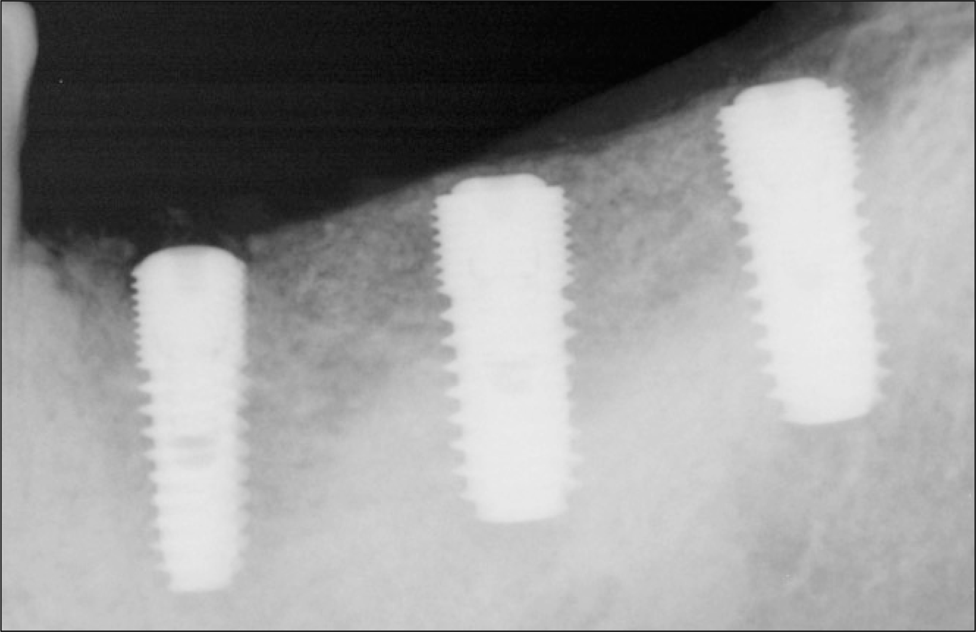
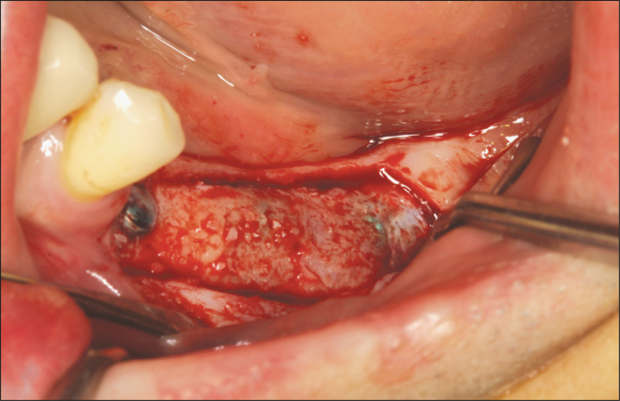
Fig. 6.
The remodeling of new bones formed in the vicinity of graft materials is observed. A was graft materials, B was newly-formed bones, C was bone marrow.(H&E staining, original magnification x100)

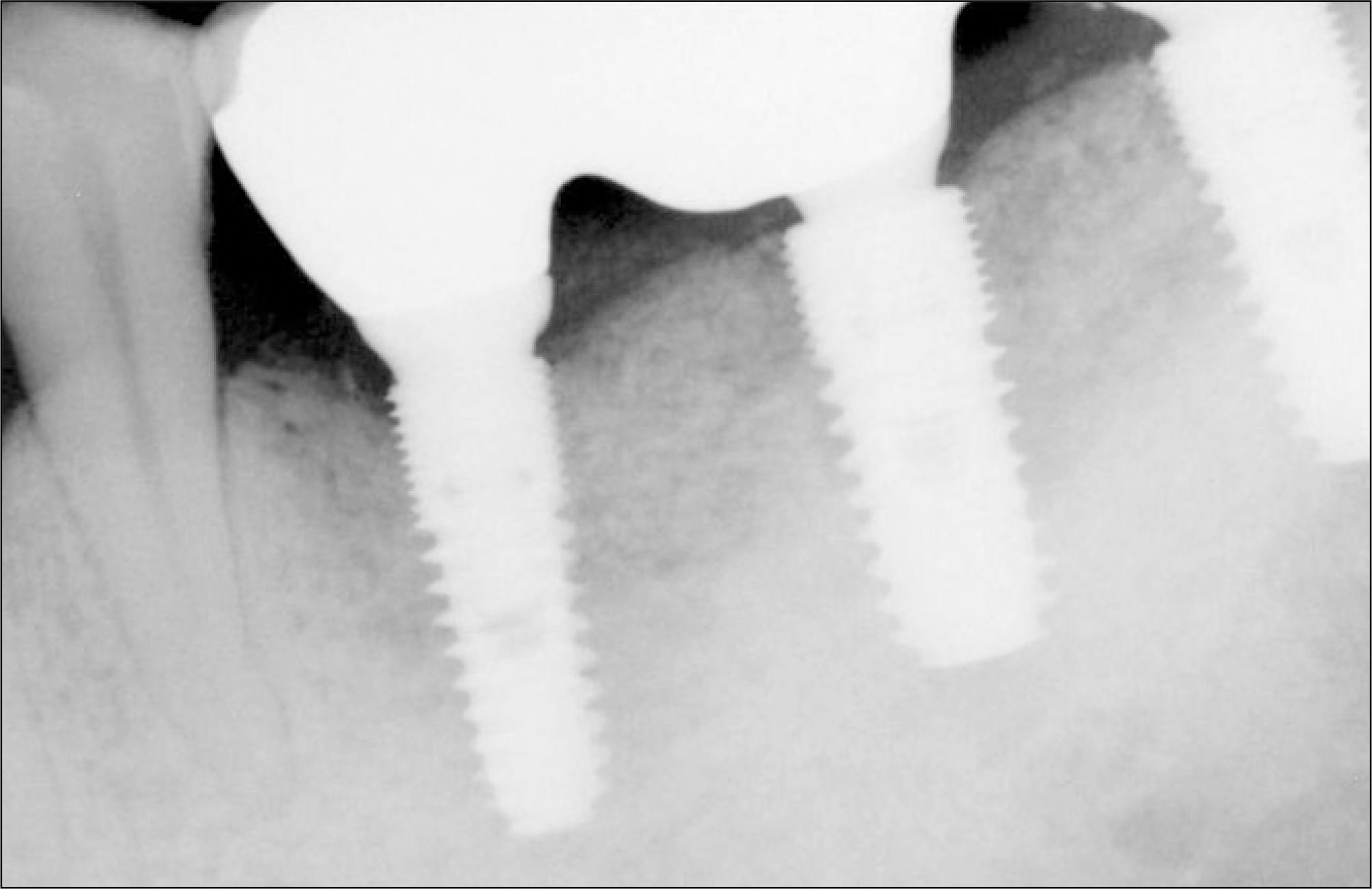
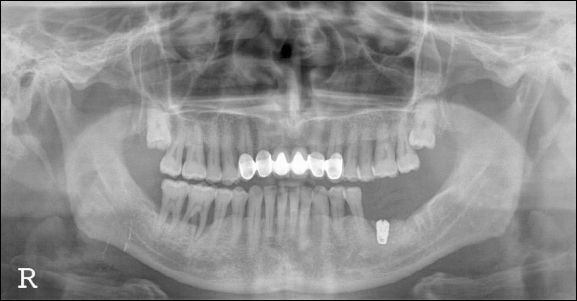
Fig. 8.
Guided bony regeneration (GBR) was performed at right 1st molar area of 49-year old female patient. Autogenous tooth bone graft material and collagen membrane were used.
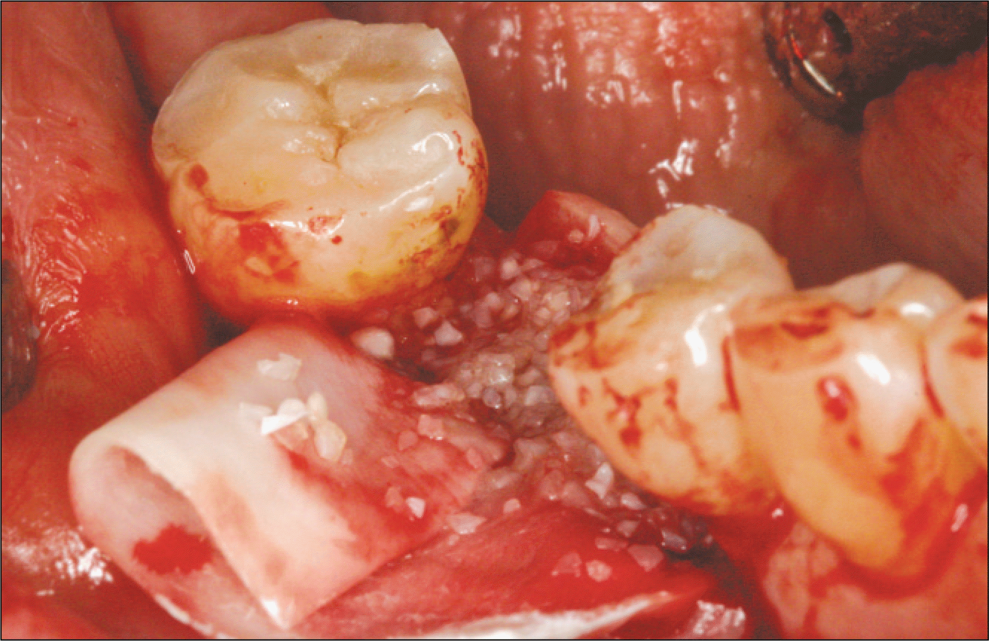
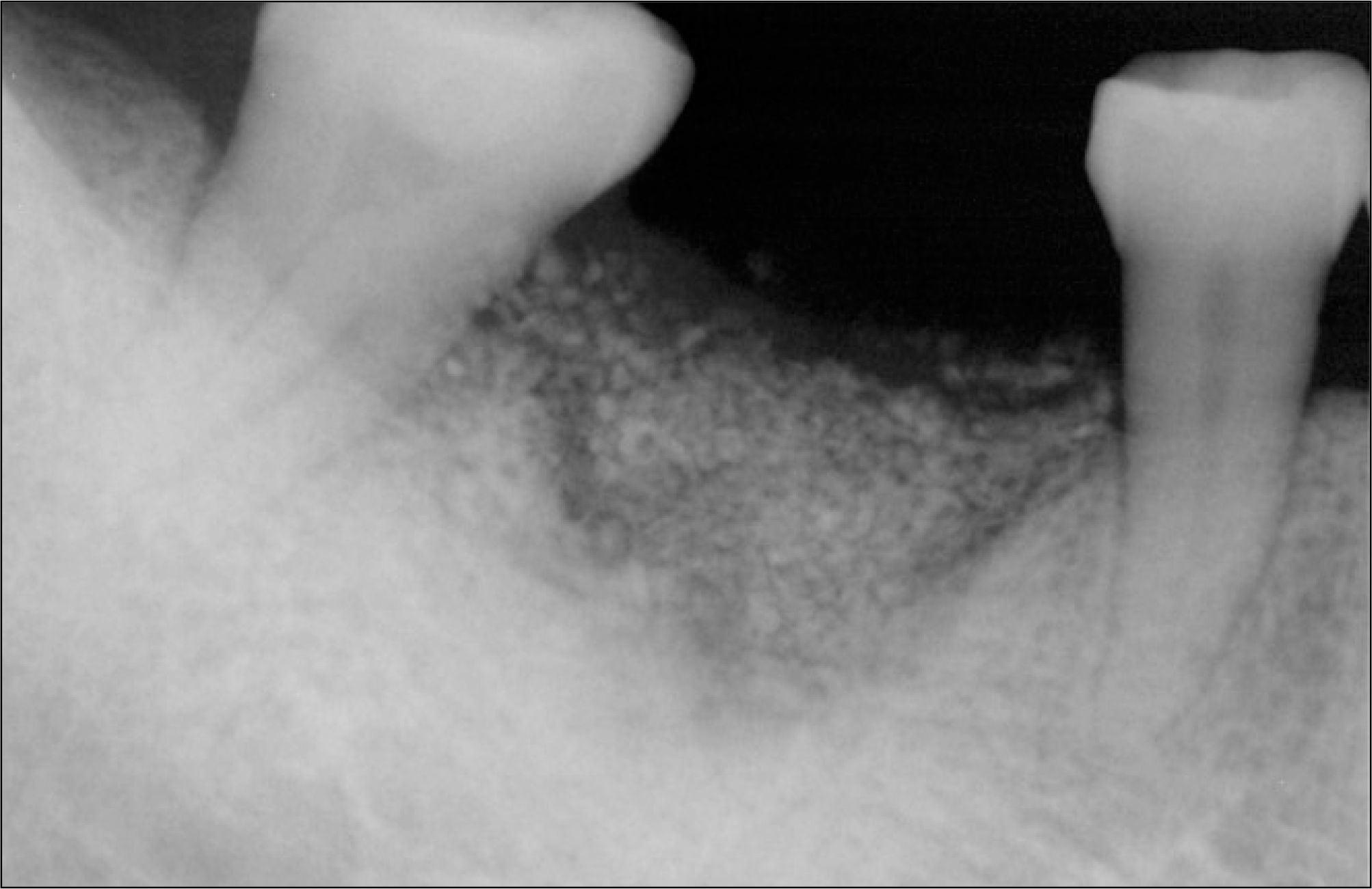
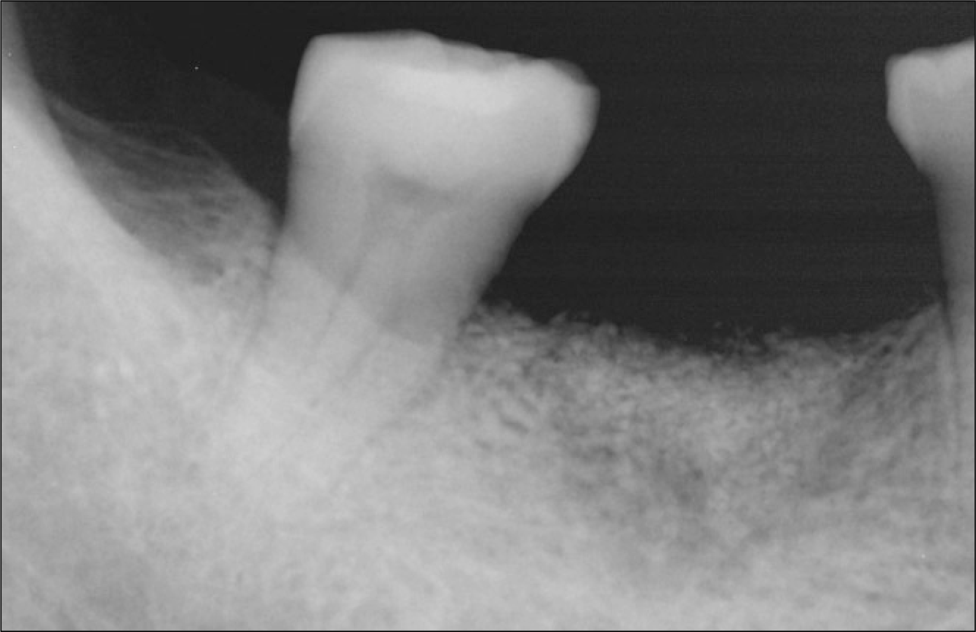
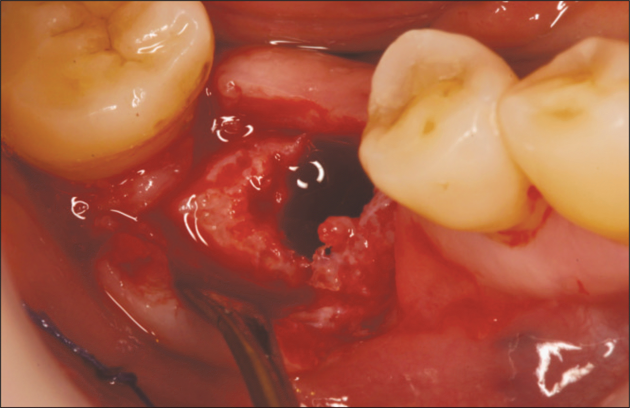
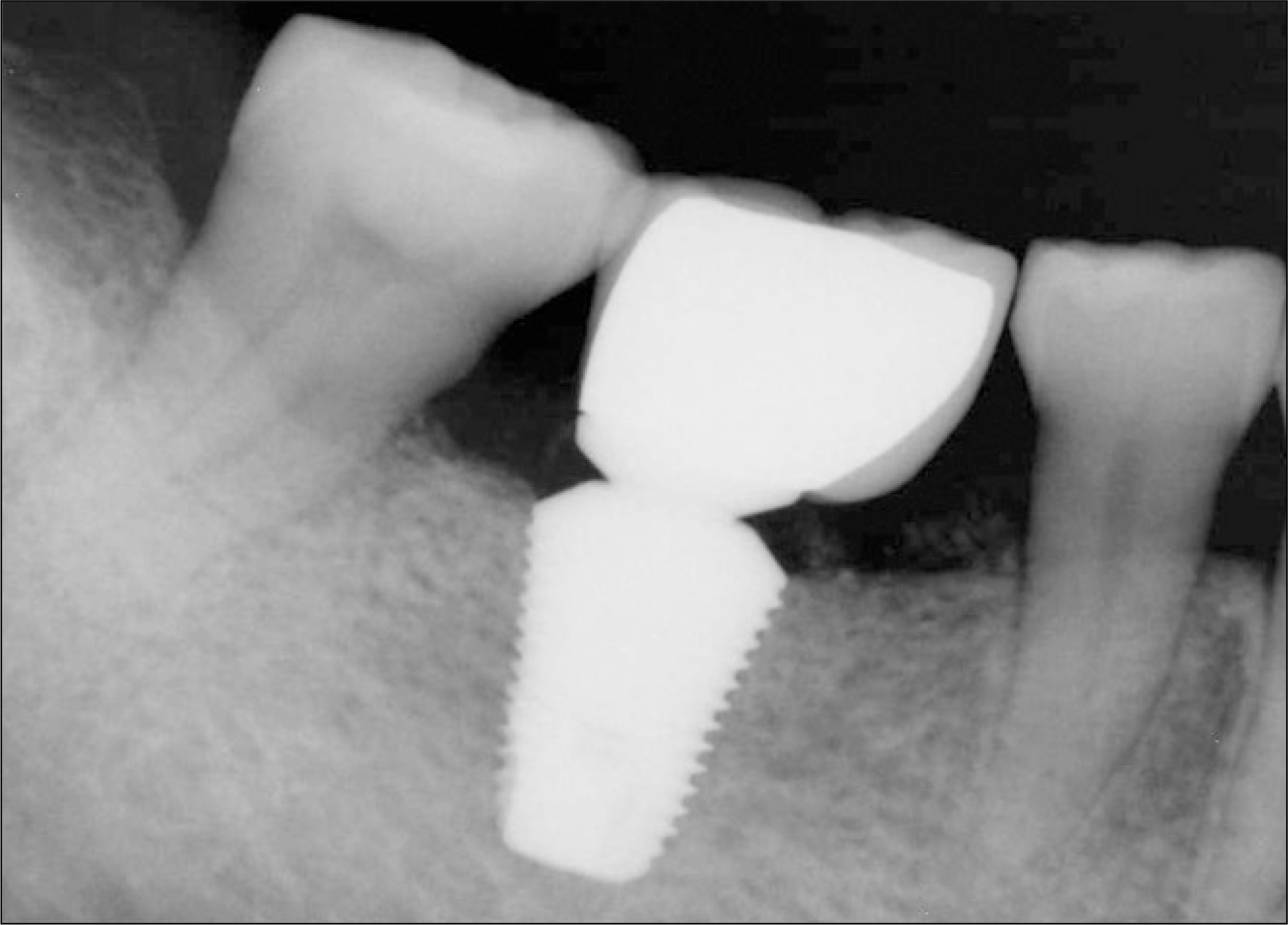
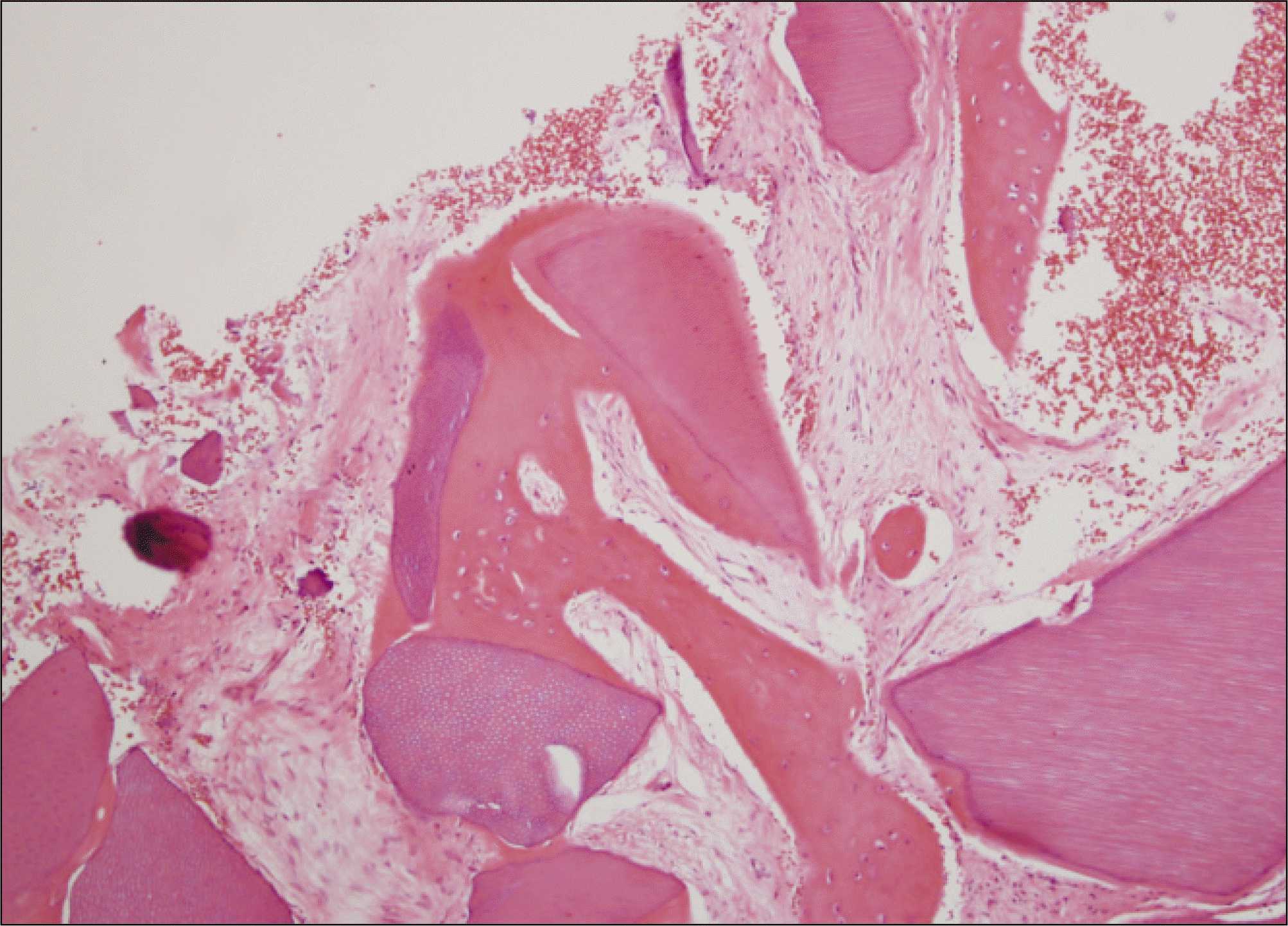
Fig. 13.
Microphotograph 6 months after AutoBT transplantation. Higher magnification demonstrated new bone formation (arrows) around the implant chips (asterisks).(H&E staining, original magnification x100)
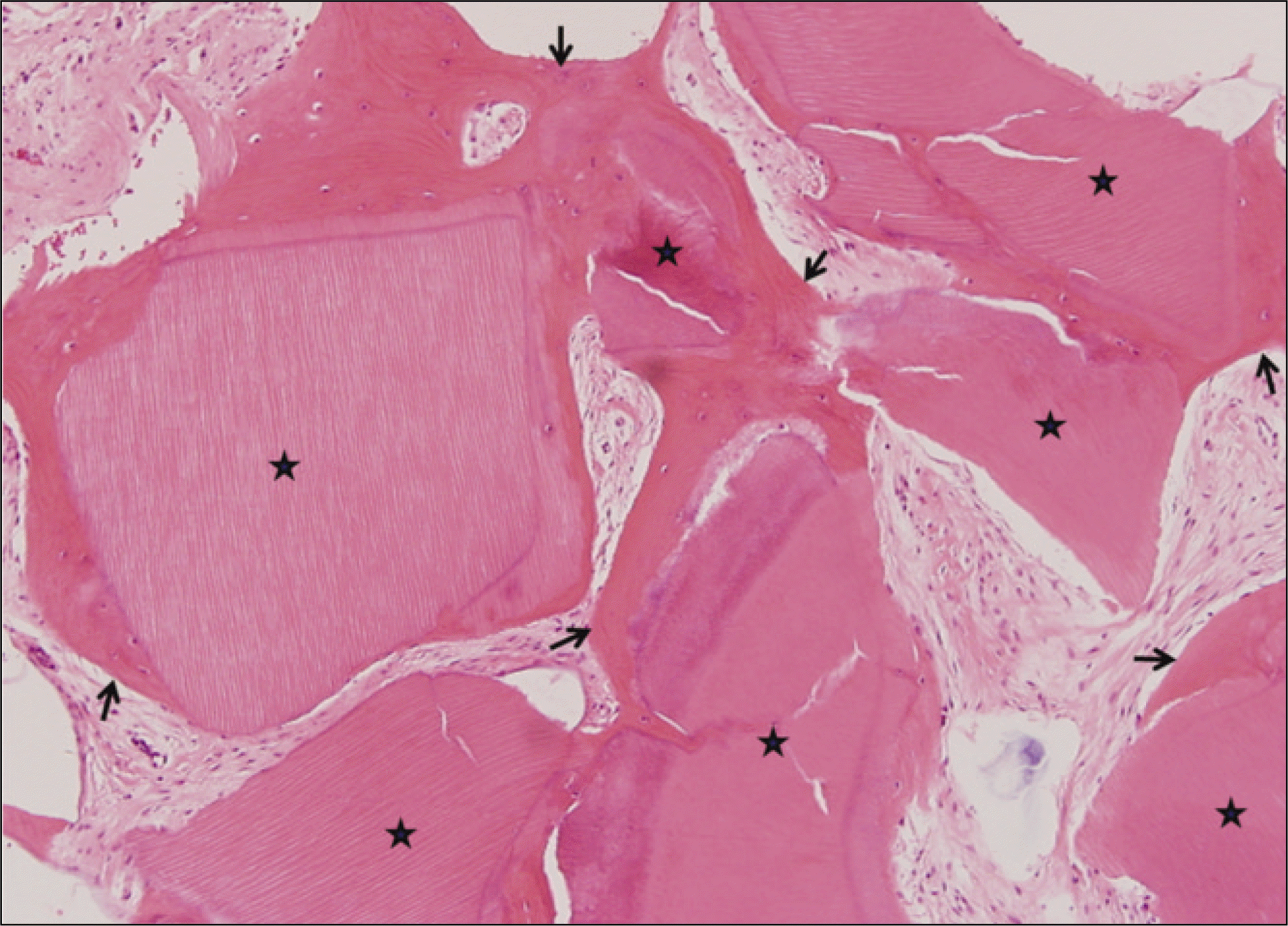
Fig. 14.
Periapical radiography of 50-year old male patient 2 months after extraction of mandibular left 1st molar.
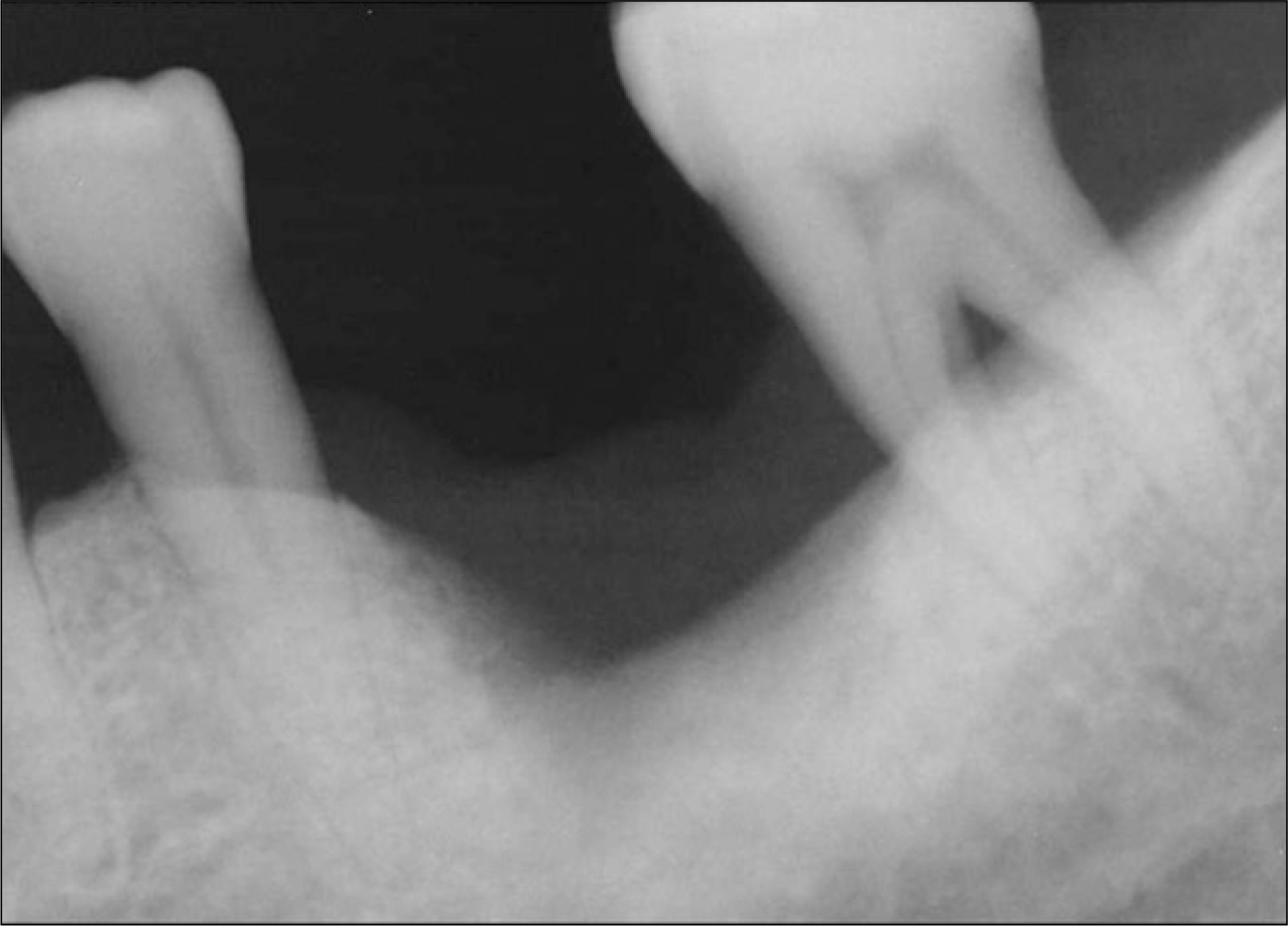

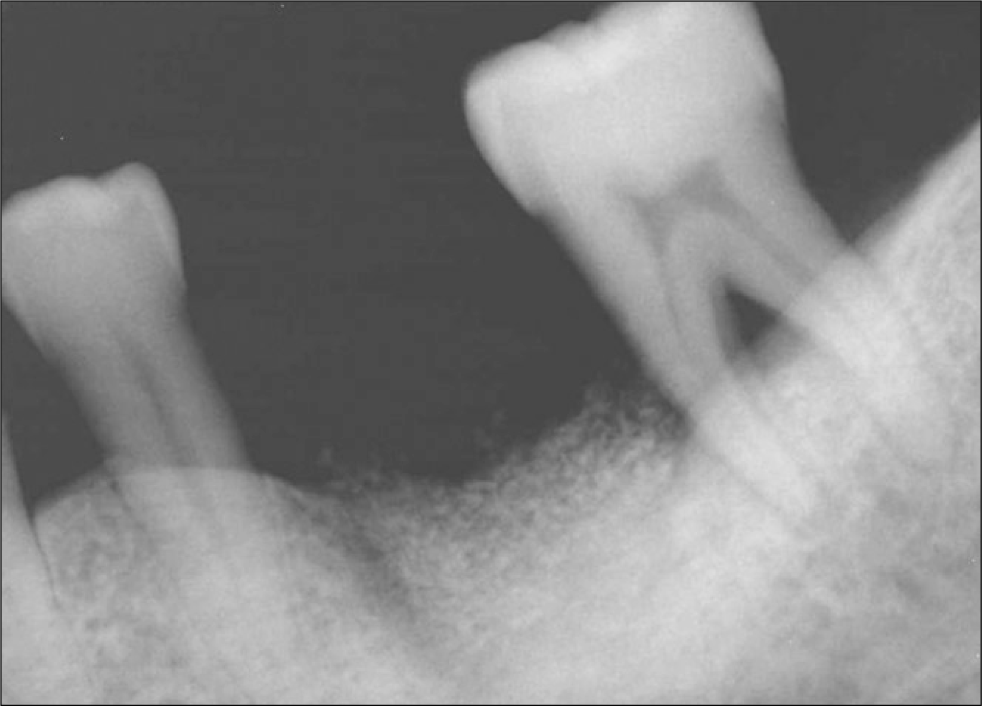
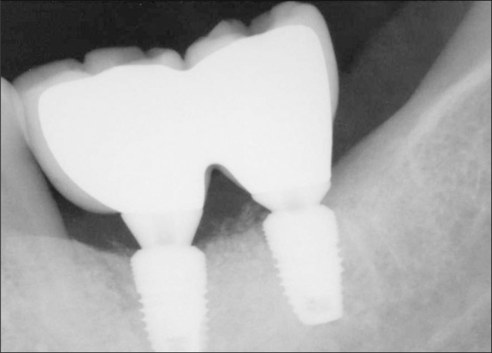
Fig. 16.
Periapical radiography 5 months after autogenous tooth bone graft. Alveolar crestal bone level was stable.
 XML Download