

 PDF
PDF ePub
ePub Citation
 Print
Print


INTRODUCTION
Hidrocystomas are rare, benign, cystic lesions of sweat gland resulting from proliferation of the apocrine secretory coil or eccrine duct1. Apocrine hidrocystoma, also called apocrine cystadenoma, is a benign cystic tumor-like lesion usually presenting as a solitary translucent nodule of cystic consistency2. The apocrine glands are most frequent in the axilla, external auditory canal, eyelids, and on the nipple, and it is not surprising that most apocrine hidrocystomas occur within the head and neck region3. However, involvement of genitalia is extremely rare and a few cases have been reported345678910111213.
CASE REPORT
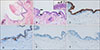
A 39-year-old male patient presented with a mass on the penis that had been growing slowly for 1 year (Fig. 1). On clinical examination, about 4.0×0.3 cm, serpiginous cyst was found. It was not tender, the skin over the cyst was normal and it was not attached to the penile shaft. There were no signs of pain, tenderness, pruritus, or other symptoms. His past medical history was irrelevant. He denied traumatism and risky or vigorous sexual activities. Patient was taken up for surgery and excision of the mass was performed under local anaesthesia. Histological evaluation revealed the presence of a cyst in the dermis (Fig. 2). The inner surface of the cyst and the papillary projections are lined by a row of columnar secretory cells of variable height showing decapitation secretion indicative of apocrine secretion. Peripheral to the layer of secretory cells are elongated myoepithelial cells. Immunohistochemically, the inner cells show cytoplasmic stains for cytokeratin (CK) 7 and outer cells show nuclear stains for p63, respectively. CK20 and carcinoembryonic antigen (CEA) are negative in both cells. A diagnosis of apocrine hidrocystoma of the penis was established.
DISCUSSION
Apocrine hidrocystoma is a benign cyst tumor that arises from the proliferation of apocrine glands in adults between 30 and 70 years of age11. The pathophysiology of hidrocystomas is unclear, though many theories exist. The occlusion or blockage of the sweat duct apparatus, which results in the retention of sweat, and a dilated cystic structure, are considered to be plausible causes14. The apocrine glands are most frequent in the axilla, external auditory canal, eyelids, and on the nipple, and it is not surprising that most apocrine hidrocystomas occur within the head and neck region3. However, involvement of genitalia is extremely rare, and to date twelve cases have been reported (Table 1)345678910111213.
Clinically, Apocrine hidrocystoma usually occurs singly as a unilocular or multilocular, dome-shaped translucent cyst, that is either flesh colored or bluish black and ranges from a few millimeters to more than 1 cm in diameter3. Histopathologically, apocrine hidrocystoma appears as unilocular or multilocular cysts composed of an inner layer of single or double layer of secretory columnar epithelium with decapitation secretion lying above an outer myoepithelial cell layer15. Immunohistochemically, apocrine hidrocystoma is positive for CK7, CK18 and gross cystic disease fluid protein 15 in the inner layer of epithelium, and alpha- smooth muscle actin and p63 in the outer myoepithelial cells116.
For cystic lesions in genitalia, the differential diagnosis also includes congenital cysts of the median raphae, eccrine hidrocystoma, sclerosing lymphangitis, and acquired lymphangioma9. Especially, differentiating apocrine hidrocystoma from median raphe cyst (MRC) could be difficult. In the past, some reports of apocrine hidrocystoma on the penis represented MRC17. However, according to histopathological finding, the lining entirely composed of pseudostratified columnar epithelium with no signs of decapitation secretion318. Immunohistochemically, CEA is expressed in columnar cells presenting in the superficial layer of pseudostratified epithelium19. The presence of myoepithelial cells and decapitation secretion in apocrine hidrocystoma are important in differentiating those two diseases17. In our case, we conclude that apocrine hidrocystoma is more appropriate than MRC because CEA was negative and p63, myoepithelial marker, was positive. On the other hand, discrimination from eccrine hidrocystoma is also important. Eccrine hidrocystomas are true retention cysts that histologically are covered by two layers of cuboidal or flattened cells without myoepithelial cells and decapitation secretion. The immunohistochemical markers that are positive in apocrine hidrocystoma are all negative. Clinically, eccrine hidrocystoma can be seen as multiple lesions of the face area and have seasonal variation13. Sclerosing lymphangitis and acquired lymphangioma are also distinct histologically. Sclerosing lymphangitis shows hypertrophy of lymphatic channels with or without perilymphatic cell infiltration or thrombi. In case of acquired lymphangioma, numerous dilated lymphatic vessels are observed in upper dermis9.
The treatment for apocrine hidrocystoma is excision with narrow margins because of the benign nature of the lesion3. Other methods that have shown success include carbon dioxide laser vaporization and laser treatment15.
In conclusion, although genital apocrine hidrocystoma is rare, we should be considered in the differential diagnosis of a genital cystic lesion. This paper emphasizes the importance of considering this diagnosis when evaluating a cystic lesion in genitalia.


 XML Download