

 PDF
PDF ePub
ePub Citation
 Print
Print


INTRODUCTION
Lipedematous alopecia (LA) is a rare condition of unknown etiology. It most frequently appears in adult African-American women and mainly involves the vertex and occiput1. It is typically a disease of late onset, and the youngest patient reported to date in the pediatric literature was 9 years old2. To our knowledge, congenitally appearing LA has not been described in the literature.
CASE REPORT
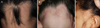
Two girls, aged 6 and 10 years, presented with patchy hair loss on the occipital and left temporal areas of the scalp since birth (Fig. 1). Physical examinations revealed the affected scalp to be non-tender, spongy, and softly swollen. The scalp could easily be pressed down to the bone, but it immediately recovered its initial shape when the pressure was relieved. The overlying skin was slightly erythematous, with no evidence of inflammation or scarring. The 6-year-old girl had a nevus flammeus on the left flank area, and the 10-year-old girl had a café-au-lait spot on her back. No other abnormalities or systemic symptoms were found. Since birth, neither girl had taken any treatment for the patchy alopecia.
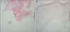
A histological examination of scalp from both patients showed some inflammatory cells in the dermis and prominent increases in mature subcutaneous fat extending into the mid dermis, with decreased numbers of hair follicles (Fig. 2). The findings were consistent with LA.
The condition's course was quite stable in both girls, with only a slight expansion in the 10-year-old patient.
DISCUSSION
LA is characterized by a thick and boggy scalp showing varying degrees of hair loss, mostly because of increased subcutaneous fatty tissue thickness. If no hair abnormalities are present, the condition is called lipedematous scalp (LS)3. The swelling can slowly progress to adjacent areas; hair loss, if present, results in diffuse or patchy alopecia2.
When patchy alopecia appears at birth without scarring, the clinical differential diagnosis includes occipital alopecia of the newborn, triangular alopecia, loose anagen syndrome, alopecia areata, and traumatic alopecia. However, the stable courses of our patients as well as the hair loss patterns did not fit the conditions listed above. In particular, our 10-year-old patient with patchy alopecia on the temporal area could be confused as having triangular alopecia. In triangular alopecia, no edema or inflammation is found and the total number of hair follicles in the biopsy specimen is almost normal4,5. In our patient, although congenital, the scalp was much too boggy for triangular alopecia and the spongy thickening of the underlying scalp along with the expansion of the subcutaneous fat layer and marked decrease in the number of hair follicles were more consistent with LA. Additional differential diagnoses of LA include cutis verticis gyrata, a rare disorder that can be present at birth. However, unlike LA, the lesions of cutis verticis gyrata are furrowed and alopecia is not observed in the involved scalp1.
Though the nature of alopecia in LA is not clearly defined, histological findings in LA have mainly demonstrated an increase in subcutaneous fat tissue and a decrease in the number of follicles. It has been suggested that hair follicles surrounded by adipocytes might provide an inadequate vascular supply and an altered microenvironment, leading to follicular atrophy or destruction6. Computed tomography or magnetic resonance imaging may also reveal increased scalp thickness (10~16 mm) compared to that in normal individuals (5~6 mm)7.
The pathogenesis of LA is still unknown. Several underlying diseases have been reportedly associated with LA, including diabetes mellitus, skin hyperelasticity and joint hyperextensibility, and renal failure2,7. However, such association with underlying medical conditions is controversial because these comorbidities are only observed in limited cases. Based on the main pathology of LA, i.e., an expansion of the subcutaneous fat layer without adipose tissue hypertrophy and dermal edema with occasional lymphatic dilatation, several hypotheses have been described. The frequent development of LA in obese patients can be explained by inadequate lipid distribution and impairment in lymphatic flow7. In addition, Yip et al.8 suggested a potential role for leptin, a hormone that regulates the structure and distribution of adipocytes, in the pathogenesis of LA. An increase in the number of Caucasian and Asian women with the condition has lessened the emphasis on the role of racial factors in the pathogenesis of the disease. However, because most cases are reported in women, hormonal factors seem to contribute to LA and LS. Genetic influences have yet to be elucidated, despite a reported case indicative of genetic transmission6. The congenital nature of both our cases suggests the possibility of a genetic background.
One case of LA on the temporal area and one patient who developed LA in early childhood have been reported previously1,2. In addition to the rarity of LA on the temporal area, congenital cases have not been previously described in the literature.
In conclusion, we report two cases of congenital LA. These distinct clinical features should not be excluded in congenital alopecia. Recognizing congenital cases of LA can be helpful in defining the characteristics of both congenital and acquired types of LA and in providing greater support to the possibility of genetic transmission.
 XML Download