

 PDF
PDF ePub
ePub Citation
 Print
Print


INTRODUCTION
Xanthoma disseminatum (XD) is a rare, benign proliferative dermatologic disorder of unknown etiology1. It is classified as a subset of cutaneous non-Langerhans cell histiocytosis (NLCH). XD typically manifests as hundreds of discrete papules and nodules, which are red-brown to yellow in color1. They chiefly involve the face and trunk, and occur in flexures and folds such as the axillae and groin. XD may also involve the mucous membranes of the mouth, pharynx, larynx, conjunctiva, and cornea. Because of the asymptomatic and self-healing characteristics of NLCH, most forms do not require early and intensive treatments1. Although many conservative treatments are unsatisfying, active intervention may still be warranted for the noninvoluting or disfiguring type for cosmetic reasons or to prevent permanent functional impairments. We report on a persistent and recalcitrant form of XD in a 47-year-old woman, which lead to circumscribed blindness by obscuring her field of vision.
CASE REPORT
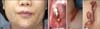
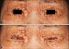
A 47-year-old woman presented with disfiguring yellow to red-brown colored nodules and plaques on her forehead, periocular areas, nasolabial folds, oral mucosa, chin, neck, shoulder, axillary folds, and perianal area. The papules began from both eyelids and perioral area 5 years before the presentation. They had been yellow to flesh red at first and changed to yellow to brown color with time. The papules coalesced into plaques around the mouth, and new lesions had recently developed on the scalp, forehead, oral mucosa, neck, shoulders, axillae, and eventually around the anal region (Fig. 1). They grew larger and larger and were severely pedunculated, especially on the periocular area, neck, and axillae. She usually felt only itching from the newly developing papules, but they were not long lasting. She complained that multiple papules accentuating over the eyelid and eyelashes hindered the blinking of her eyes and obscured some fields of vision (Fig. 2). An ophthalmologic examination showed xerophthalmia but was otherwise normal.
She had been taking antihypertensive medications for 4 years with no other history of skin disorders. There was no drug or family history to explain her cutaneous disorder. She did not have any symptoms indicating systemic dysfunction such as polyuria, polydipsia, fever, or chills. A brain magnetic resonance image (MRI) and chest X-ray were also normal. She did not complain of any difficulty breathing despite the fact that she had similar lesions on her oral and nasal mucosa. Laryngoscopic studies of the pharynx and larynx as well as endoscopy of the gastrointestinal tract showed no signs of obstruction. She had normal values on urinalysis, fasting blood glucose, serum urea, and electrolyte tests. Thyroid- and liver-function tests, lipid profiles such as serum triglyceride, total cholesterol, and high density lipoprotein (HDL) cholesterol were also within normal ranges.
Skin biopsies taken from the axilla and inner canthus revealed intradermal infiltrates of Touton-type giant cells, foam cells, histiocytes and some other inflammatory cells (Fig. 3A, B). Immunohistochemical staining was negative for the CD1a and S-100 proteins but positive for CD68 (Fig. 3C~E). Several large pedunculated nodules on both axillae and the neck were surgically resected, and smaller lesions were removed by CO2 laser. After surgery, she was treated with 20~40 mg of oral prednisolone once per day for 11 weeks with reduced medication for 10 more weeks for prophylactic reasons. However the lesions were recalcitrant and recurring. New lesions on the periocular areas are still emerging, and her visual field continues to narrow.
DISCUSSION
XD is a rare, benign NLCH distinguished by multiple grouped red-brown to yellow papules and nodules involving the skin and mucous membranes. The pathogenesis of NLCH is not clear1; however, there is some information about the epidemiology and clinical findings. First, XD usually starts before the age of 25 years in about 60% of patients1. Second, it is more common in males1. Third, XD may occur anywhere on the body including the scalp, face, trunk, and extremities1. It often involves the eyelids and eyelashes without functional problems. About 50% of cases involve the mucous membranes of the larynx, pharynx, mouth, conjunctiva, and cornea1. Various symptoms are possible according to the location of xanthoma; for example, respiratory obstruction, defecation difficulty, and so on2,3. XD is often related with diabetes insipidus by involving the central nervous system (CNS) in about 40% of patients4-6. Rarely, it may be accompanied by multisystemic involvement7.
Xanthomatous lesions in critical anatomical locations may result in morbidity and mortality. Zak et al.8 reported intracranial involvement of XD, which resulted in death of the patient. Büyükavci et al.9 described a case of XD with hepatic involvement, which resulted in sclerosing cholangitis. Mass effects from xanthomas can create serious problems. Ozcelik et al.2 reported upper airway obstruction with invasion of the respiratory mucosa; Kang and Kim3 reported gastrointestinal complications accompanied by defecation difficulties with invasion of the perianal area. In our case, there was no evidence of vital organ involvement during various evaluations. However, multiple papules accentuating in a coalesced presentation, particularly on the eyelids and eyelashes, screened her field of vision, and made it difficult for her to blink her eyes, because the papules on the eyelashes made her eyelashes stiffer.
Unlike other types of xanthomatous disorders, patients with XD usually show normal lipid profiles10, but slightly elevated levels of serum cholesterol or triglyceride can be seen in a few patients1. Radiologic findings are usually nonspecific, except on a brain MRI when the XD has been associated with intracranial lesions1. In our patient, lipid profiles and thyroid-function test results were normal, and radiologic examinations showed nonspecific findings. As xanthomas were detected on the oral mucosa, an upper and lower gastrointestinal endoscopy and a laryngoscopy were performed, which also revealed nonspecific findings. Histopathological findings of XD can be explained by diffuse dermal infiltration of Touton giant cells, foreign body giant cells, and histiocytes, associated with scattered lymphocytes, neutrophils, and plasma cells11. In early lesions, scalloped macrophages dominate the histology, but most well-developed lesions have a mixture of the above.
Immunohistochemical studies show negative staining for S-100, CD1a, and Birbeck granules, and positive staining for the surface markers CD68 and factor XIIIa12. Rough endoplasmic reticulum, fat droplets in histiocytes, and Touton giant cells can be seen on electron microscopy13.
XD can be classified into three groups by its evolution and prognosis: a self-healing form with spontaneous resolution of lesions; a common persistent form in which lesions may never resolve; and a very rare progressive form with organ dysfunction and CNS involvement14. Though many subtypes among these are possible, the persistent form is the most common type. The prognosis of XD is usually good1, although it can be worse if vital organs are involved. We considered our case as a persistent form from her poor response to systemic steroid treatment and the continuously spreading clinical course without any signs of organ dysfunction.
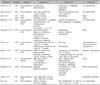
There are various treatments for XD, such as vasopressin, radiotherapy, cryotherapy, corticosteroids, and antiblastic chemotherapy1, but no single treatment is universally successful (Table 1)2,3,8,9,14-19. For example, Büyükavci et al.9 used mainly oral prednisolone (2 mg/kg/day) and oral azathioprine (2 mg/kg/day), but the patient failed to respond. Further, some have had successful results using a combination of lipid lowering agents or azathioprine and cyclophosphamide16,18. In some cases, treatments with oral steroids, clofibrate, and chemotherapy are effective, but it is difficult to distinguish the effect of such drugs from that of spontaneous resolution3. Our patient was treated with oral corticosteroid after two surgical excisions and several CO2 laser therapies. Until recently, the disease was not well controlled. Because new lesions are still emerging, and her visual field is continuously narrowing, we are planning to use lipid lowering agents such as a series of statins and perform several surgical excisions to retain her vision.

 XML Download