Journal List > J Cardiovasc Imaging > v.27(1) > 1115530

Kalra, Park, Hemu, and Goldberg: Loeffler Endocarditis: A Diagnosis Made with Cardiovascular Magnetic Resonance
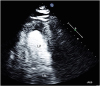
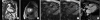
A 61-year-old Caucasian man with recently diagnosed eosinophilic chronic myelomonocytic leukemia presented with fevers and right-sided heart failure that failed to respond to diuretics. He denied any history of travel to the tropics. Exam was remarkable for pallor, edema and S4 gallop. He had leukocytosis (54 × 109/L; 55% eosinophilic predominance), anemia and thrombocytopenia (3 × 10⁹/L). Echocardiogram showed grade 2 diastolic dysfunction, moderate mitral regurgitation and elevated right heart pressure. There was apical left ventricular thickening concerning for tumor, laminated thrombus or focal hypertrophy (Figure 1, Movie 1). Given severe thrombocytopenia, endomyocardial biopsy and anticoagulation were contraindicated. Cardiovascular magnetic resonance (CMR, Figure 2, Movie 2, 3) showed increased focal apical wall thickness of 22 mm with hypokinesis of this segment (Figure 2A). First pass perfusion revealed hypoperfused apical and lateral endocardium with a layer of superimposed clot (Figure 2B). Late gadolinium enhancement images showed hyperintense apical fibrosis extending from the endocardium to mid myocardium, covered by a cap where laminated clot was present producing a “double V” sign (Figure 2C). This was also confirmed on the 3-chamber early gadolinium enhancement image with long TI of 600 ms that showed apical thrombus distinct from the underlying myocardium (Figure 2D).
Loeffler endocarditis is a rare type of restrictive cardiomyopathy that follows a two-stage process.1)2) Early manifestations include symptoms of fevers, dyspnea and swelling. During this phase, infiltration of myocardium and release of eosinophilic major basic protein and reactive oxygen species promote endothelial and myocyte injury.3) The later stages are marked by myocardial fibrosis and thrombus formation causing a restrictive cardiomyopathy.4)5) Our patient was started on imatinib and steroids and his symptoms improved over 2 weeks. Unfortunately, he died from bacterial sepsis after that.

Figures and Tables

Figure 1

Echo (apical 4 chamber) with perflutren contrast showing a 2.4 × 3.8 cm non-perfused mass at the LV apex suggestive of thrombus or infiltrative tumor. LA: left atrium, LV: left ventricle, RV: right ventricle.

jcvi-27-70-g001
Figure 2

(A) Cardiovascular magnetic resonance – Steady State Free Precession (SSFP) still image (apical 2 chamber) showing normal chamber sizes with increased focal apical wall thickness of 22 mm and hypokinesis of this segment. A small pericardial effusion is also seen. (B) First pass perfusion shows hypoperfused apical and lateral endocardium along with a layer of clot lining these segments. (C) Three-chamber late gadolinium enhancement shows fibrosis in the apex in the endocardium extending to the mid myocardium in the shape of a cap. The laminated hypointense clot (arrowhead) produces a “double V” sign when contrasted with the underlying hyperintense fibrosis (asterisk). (D) Three-chamber early gadolinium enhancement with long TI of 600 ms shows dark hypointense thrombus at the apex distinct from the underlying scarred myocardium. LA: left atrium, LV: left ventricle, RV: right ventricle.

jcvi-27-70-g002

Notes

Conflict of Interest The authors have no financial conflicts of interest.

References

1. Mankad R, Bonnichsen C, Mankad S. Hypereosinophilic syndrome: cardiac diagnosis and management. Heart. 2016; 102:100–106.
crossref pmid
2. Ogbogu PU, Rosing DR, Horne MK 3rd. Cardiovascular manifestations of hypereosinophilic syndromes. Immunol Allergy Clin North Am. 2007; 27:457–475.
crossref pmid pmc
3. Spry CJ, Tai PC, Davies J. The cardiotoxicity of eosinophils. Postgrad Med J. 1983; 59:147–153.
crossref pmid pmc
4. Beedupalli J, Modi K. Early-stage Loeffler's endocarditis with isolated right ventricular involvement: management, long-term follow-up, and review of literature. Echocardiography. 2016; 33:1422–1427.
crossref pmid
5. Kleinfeldt T, Ince H, Nienaber CA. Hypereosinophilic syndrome: a rare case of Loeffler's endocarditis documented in cardiac MRI. Int J Cardiol. 2011; 149:e30–e32.
crossref

SUPPLEMENTARY MATERIALS

Movie 1

Apical 4-chamber echo showing laminated mass at left ventricular apex.

Movie 2

Cardiovascular magnetic resonance – 2-chamber Steady State Free Precession cine imaging showing Focal thickening at the left ventricular apex (22 mm) along with hypokinesis.

Movie 3

Cardiovascular magnetic resonance – Apical short-axis first pass perfusion imaging demonstrating hypoperfusion of the subendocardium and a layer of overlying thrombus.
TOOLS
ORCID iDs

Dinesh K Kalra
https://orcid.org/0000-0001-5254-4067

Jeffrey Park
https://orcid.org/0000-0002-2117-7118

Mohamad Hemu
https://orcid.org/0000-0003-3059-7437

Alan Goldberg
https://orcid.org/0000-0003-2257-119X

Similar articles