Abstract
May-Thurner syndrome, compression of the left common iliac vein by the right common iliac artery, or intimal hypertrophy of the vein resulting from chronic pulsatile force of the right common iliac artery, may results in deep vein thrombosis on the left lower extremity. A patient presented to our facility with deep vein thrombosis caused by May-Thurner syndrome, and showed postoperative fever, pain, and tenderness over the left leg, showing severe lumbar disc herniation with acute cauda equina syndrome. This syndrome should be considered as one of the causes of deep vein thrombosis in the left lower extremity.
REFERENCES
1). May R, Thurner J. The cause of the predominantly sinis-tral occurrence of thrombosis of the pelvic veins. Angiolo-gy. 1957; 8:419–427.

2). Patel NH, Stookey KR, Kertcham DB, Cragg AH. Endovascular management of acute extensive iliofemoral deep venous thrombosis caused by May-Thurner syndrome. J Vasc Interv Radiol. 2000; 11:1297–1302.

3). Taheri SA, Williams J, Powell S, et al. .:. Iliocaval compression syndrome. Am J Surg. 1987; 154:169–172.

4). Turner JA, Ersek M, Herron L, et al. .:. Patient outcomes after lumbar spinal fusion. JAMA. 1992; 268:907–911.
6). Markel A, Manzo RA, Bergelin RO, Strandness DE Jr. Pattern and distribution of thrombi in acute venous thrombosis. Arch Surg. 1992; 127:305–309.

7). Mickley V, Schwagierek R, Rilinger N, Gorich J, Sun-der-Plassmann L. Left iliac venous thrombosis caused by venous spur: treatment with thrombectomy and stent implantation. J Vasc Surg. 1998; 28:492–497.

8). Lee HM, Suk KS, Moon SH, Kim DJ, Wang JM, Kim NH. Deep vein thrombosis after major spinal surgery: Incidence in an East Asian population. Spine. 2000; 25:1827–1830.
9). Geerts WH, Heit JA, Clagett GP, at al. .:. Prevention of venous thromboembolism. Chest. 2001; 119:132–175.
10). Salzman EW, Harris WH. Prevention of venous thromboembolism in orthopedic patients. J Bone and Joint Surg. 1976. 903–913.
11). Lehr S, Vormittag R, Vukovich T, at al. .:. Basal high-sensitivity-C-reactive protein levels in patients with spontaneous venous thromboembolism, Thromb haemost. 2005. 488–493.
12). Goldhaber SZ. Pulmonary Embolism and Deep Vein Thrombosis. Philadelphia: WB Saunders Co;79-97. 1985.
13). Kearon C, Julian JA, Newman TE, Ginsberg JS. Non-invasive diagnosis of deep vein thrombosis. McMaster Diagnostic Imaging Practice Guidelines Initiative. Ann Inter Med. 1998; 128:663–677.
14). White RH, Goulet JA, Bray TJ, Daschbach MM, McGahan JP, Hartling RP. Deep-vein thrombosis after fracture of the pelvis; assessment with serial duplex-ultra- sound screening. J Bone and Joint Surg. 1990. 495–500.
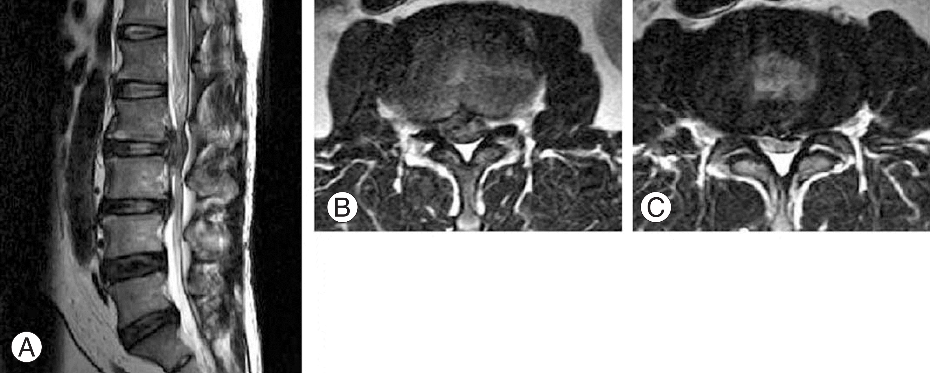
Fig. 1.
MRI finding of Lumbar spine. (A) Sagittal T2-weighted MRI of lumbar spine shows severe herniated lumbar disc in lumbar area. (B) Axial T2-weighted MRI of lumbar spine shows severe lumbar disc herniation between L2 and L3. (C) Axial T2-weighted MRI of lumbar spine shows severe lumbar disc herniation between L3 and L4.


 PDF
PDF ePub
ePub Citation
 Print
Print


 XML Download