

 PDF
PDF ePub
ePub Citation
 Print
Print


References
1. Cronkhite LW Jr, Canada WJ. Generalized gastrointestinal polyposis; an unusual syndrome of polyposis, pigmentation, alopecia and onychotrophia. New Engl J Med. 1955; 252:1011–1015.
2. Watanabe C, Komoto S, Tomita K, et al. Endoscopic and clinical evaluation of treatment and prognosis of Cronkhite-Canada syndrome: a Japanese nationwide survey. J Gastroenterol. 2016; 51:327–336.

3. Wen XH, Wang L, Wang YX, Qian JM. Cronkhite-Canada syndrome: report of six cases and review of literature. World J Gastroenterol. 2014; 20:7518–7522.
4. Kang DU, Yang DH, Choi Y, et al. A case of Cronkhite-Canada syndrome showing spontaneous remission. Intest Res. 2013; 11:317–322.
5. Yun SH, Cho JW, Kim JW, et al. Cronkhite-Canada syndrome associated with serrated adenoma and malignant polyp: a case report and a literature review of 13 Cronkhite-Canada syndrome cases in Korea. Clin Endosc. 2013; 46:301–305.
6. Daniel ES, Ludwig SL, Lewin KJ, Ruprecht RM, Rajacich GM, Schwabe AD. The Cronkhite-Canada syndrome. An analysis of clinical and pathologic features and therapy in 55 patients. Medicine (Baltimore). 1982; 61:293–309.
7. Sweetser S, Ahlquist DA, Osborn NK, et al. Clinicopathologic features and treatment outcomes in Cronkhite-Canada syndrome: support for autoimmunity. Dig Dis Sci. 2012; 57:496–502.
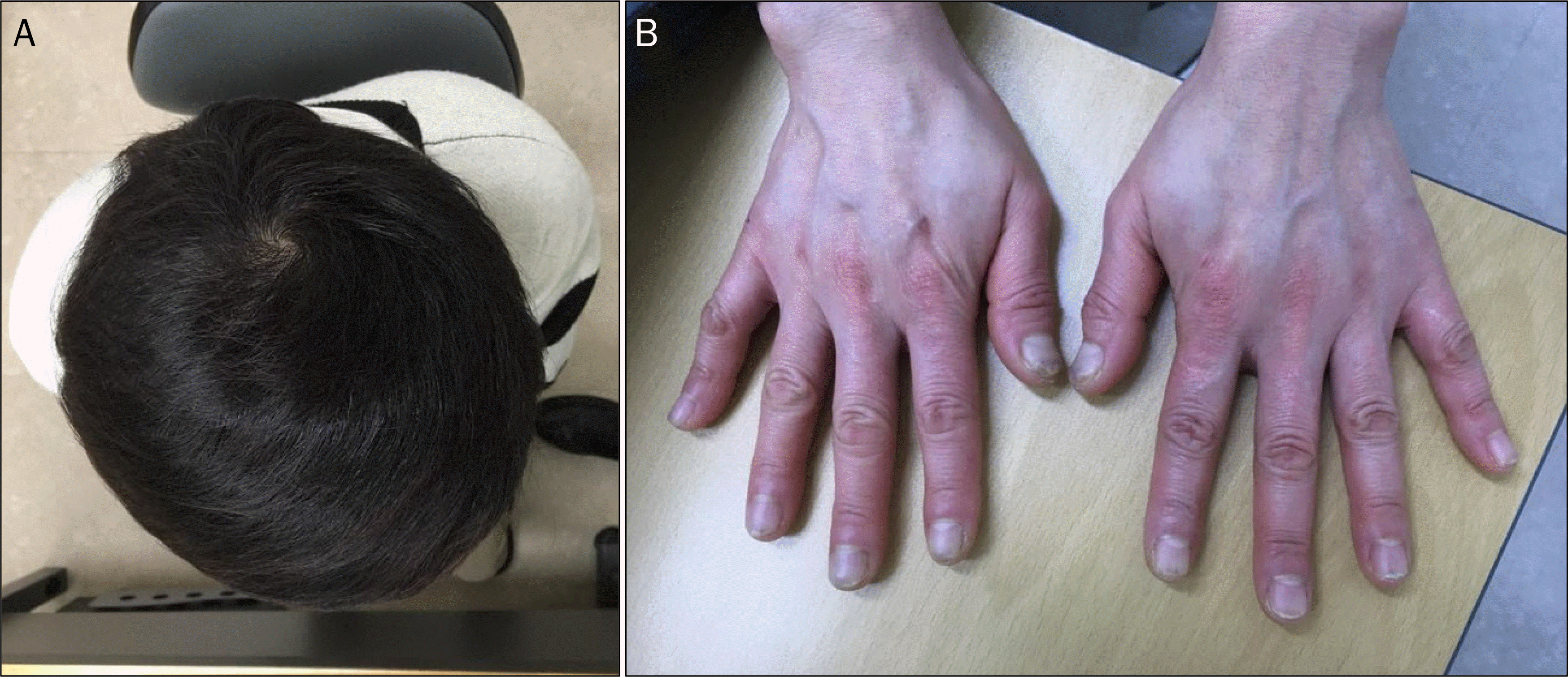
Fig. 1.
Clinical manifestation of the patient at the initial presentation. (A) Hair loss. (B) Skin pigmentation and nail dystrophy. (C) Tongue swelling and mucosal atrophy.

Fig. 2.
Initial upper and lower endoscopic findings. Numerous variable-sized sessile polyps with mucosal edema and hyperemia were observed in the (A, B) stomach, and (C, D) colon.

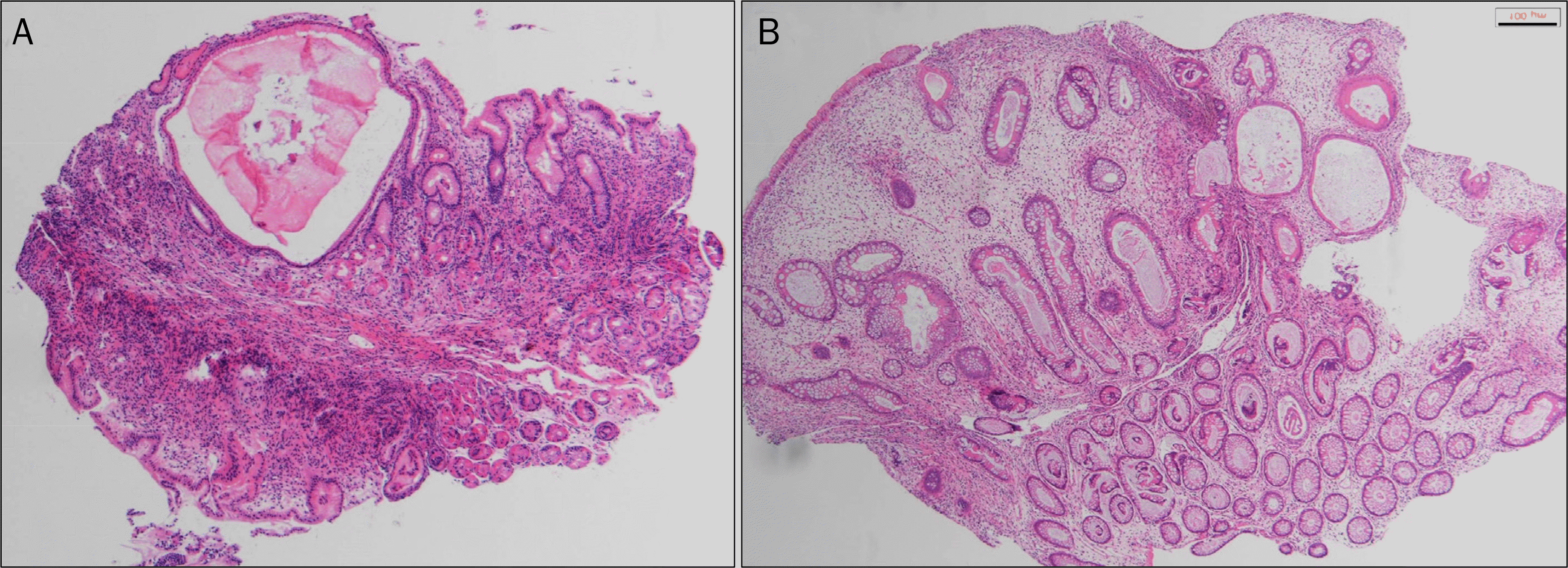
Fig. 3.
Histologic findings. (A) Dilatation of gland, and inflammatory cell infiltration in gastric mucosa (H&E, ×40). (B) Dilatation of gland, cystic dilatation of crypt, and edema of lamina propria in colonic mucosa (H&E, ×40).

 XML Download