Journal List > Nutr Res Pract > v.8(3) > 1051364

Hardman: Diet components can suppress inflammation and reduce cancer risk

Abstract

Epidemiology studies indicate that diet or specific dietary components can reduce the risk for cancer, cardiovascular disease and diabetes. An underlying cause of these diseases is chronic inflammation. Dietary components that are beneficial against disease seem to have multiple mechanisms of action and many also have a common mechanism of reducing inflammation, often via the NFκB pathway. Thus, a plant based diet can contain many components that reduce inflammation and can reduce the risk for developing all three of these chronic diseases. We summarize dietary components that have been shown to reduce cancer risk and two studies that show that dietary walnut can reduce cancer growth and development. Part of the mechanism for the anticancer benefit of walnut was by suppressing the activation of NFκB. In this brief review, we focus on reduction of cancer risk by dietary components and the relationship to suppression of inflammation. However, it should be remembered that most dietary components have multiple beneficial mechanisms of action that can be additive and that suppression of chronic inflammation should reduce the risk for all three chronic diseases.

INTRODUCTION

In 1981, Doll and Peto published the results of an epidemiology study to estimate the causes of cancer in the United States [1]. The conclusion of that study was that between 1/3 and 2/3 of all human cancers are preventable. Certainly smoking is the cause of about 30% of cancers but they also estimated that in addition to stopping smoking, dietary change was a major strategy for preventing cancer [1].
The important influence of diet on carcinogenesis is further supported by the more recent literature surveys sponsored by the American Institute for Cancer Research (AICR)/World Cancer Research Fund [2,3]. These reports were produced by a team of distinguished scientists who surveyed the existing literature, including both basic science and epidemiology reports, on diet and cancer. A conclusion of the first report [2], released in 1997, was that 'inappropriate diets cause about one-third of all cancer deaths' (p.7, [2]) and it was estimated that 30% to 40% of cancer could be prevented by a recommended diet change, physical activity and appropriate body mass (p.7, [2]).
During the next 10 years, the scientific literature reporting the results of research on diet and cancer dramatically increased, thus, a committee was commissioned to update the 1997 AICR report. These findings, published in 2007 [3], again support the notion that cancer is largely a preventable disease. In addition, the scientists compiling this report [3] chose to assess the effects of diet and lifestyle choices on other chronic diseases, including Type II diabetes and cardiovascular diseases, thus, making a recommendation for overall 'health promotion'. It was reported that a plant based diet coupled with exercise and maintenance of a healthy weight could reduce risk for all of these chronic diseases [3]. What common mechanism contributes to these common, chronic diseases that can be addressed by diet and lifestyle changes?

Inflammation increases risk for cancer and other chronic diseases

The link between inflammation and cancer was first noted as an association between inflammatory diseases and increased risk for cancer of that organ. For example, inflammatory bowel disease increases the risk for colon cancer [4], human papilloma virus increases risk for cervical cancer [5], schistosoma infection increases risk for bladder cancer [6] and Helicobacter pylori (and stomach ulcers) increase risk for stomach cancer [7]. The presence of chronic inflammation also contributes to the development of type 2 diabetes and cardiovascular disease [8, 9,10,11,12,13]. An underlying source of chronic inflammation can be obesity [13] effectively tying obesity into these chronic diseases. We now know that excess adipose tissue produces inflammation inducing cytokines [13], increases oxidative damage [9] and directly alters gene transcription [14]. All of these mechanisms can contribute to cancer development.
Chronic inflammation can have both positive [15] and negative [16] influences on tumor development. The cytokines produced by immune cells [such as macrophages, cytotoxic T cells (CTLs) and natural killer (NK) cells) can promote tumor immunity or promote the development of chronic inflammation [17]. However, the microenvironment of the tumor lacks normal immune-protective mechanisms and promotion of chronic inflammation dominates [17]. Cytokines found in the milieu of the inflammatory state can promote tumor cell growth [tumor necrosis factor α (TNFα), interleukin 6 (IL-6)], enhance tumor cell invasion [transforming growth factor β (TGFβ)], enhance metastasis (TNF α, IL-6, TGFβ), enhance angiogenesis (TNFα, IL-17, TGFβ) and impair the function of NK cells, CTLs and macrophages [17].

Inflammation and NFκB

Activation of the NFκB pathway provides a key molecular link between inflammation and carcinogenesis [18]. NFκB is a transcription factor that activates expression of multiple genes related to inflammation. It is also known as an 'oxidative stress responsive transcription factor' and is upregulated in response to oxidative stress. The mammalian NFκB family has five members, RelA (p65), c-Rel, RelB, p50 (NFKB1), and p52(NFKB2) [19]. The active form is a dimer of combinations of these five members. In the cytoplasm the dimer is found bound to IκB, (inhibitor of NFκB). On activation, the inhibitor is released and degraded in the proteosome, the dimer is phosphorylated and moves into the nucleus to bind DNA and to activate gene transcription [16,20]. Activation of NFκB provokes an autocrine, self-perpetuating response, that is, the products of many of the genes that are activated by NFκB (such as COX2 or TNFα) feedback and increase activation of NFκB [18,21].
Activation of NFκB is critical for normal immune response and for organogenesis [22], however NFκB has been found to be hyperactivated and to contribute to progression of multiple tumor types [16]. In organogenesis or in inflammatory responses the genes for cell survival, proliferation, angiogenesis and invasion that are activated by NFκB are critical for organ growth, development and wound healing [23]. However, when activated in a cancer, these same genes contribute to cancer growth and progression. Multiple carcinogens, inflammatory agents and factors in the tumor microenvironment have been found to activate NFκB [16]. Breaking the cycle of NFκB activation has been shown to slow tumor growth [24, 25,26]. Thus, one critical mechanism by which dietary components can suppress cancer growth and progression is by suppressing the activation of NFκB.

Dietary components that suppress NFκB and/or carcinogenesis

Dietary change is a key mechanism for reducing obesity and for providing components to suppress inflammation, decrease oxidative damage and change gene expression. It has been shown that at least part of the mechanism for suppression of carcinogenesis of many dietary components is by the suppressing the activation of NFκB. Some of these beneficial dietary components are reviewed below.
Fruits, vegetables, whole grains and nuts: Plants produce multiple nutraceuticals that have been shown to modify carcinogenesis via multiple pathways [27] thus increased consumption of plant products has been recommended to reduce risk for cancer [28,29].
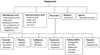
Polyphenols: Plants produce varying forms of polyphenols, a large, diverse class of compounds, many with antioxidant properties [30]. Structurally, polyphenols all have one or more aromatic (phenolic) rings with different structural elements that allows classification into subgroups [30]. In addition to (or perhaps because of) their antioxidant properties, most polyphenols have a common mechanism of decreasing the activation of NFκB. As such, a diet with generous amounts of fruit, vegetables, whole grains and nuts will provide multiple polyphenols for suppression of NFκB. See Fig. 1 for a diagram of polyphenol classes. Some especially well researched polyphenols are highlighted below.

Isoflavones

Isoflavones comprise a subclass of naturally occurring phenolic flavonoids and have a structure similar to estrogen. Isoflavones are produced almost exclusively by the members of the Fabaceae (i.e., Leguminosae, or bean) family. Soybeans (which contain high amounts of the isoflavone, genistein) are a common source of isoflavones in human food. Genistein can act as a phytoestrogen in mammals and, though they are not steroids, can alter activity of the estrogen receptor [28,30,31]. Some isoflavones are termed antioxidants because of their ability to trap singlet oxygen. Genistein has been shown to suppress the activity of NFκB when arachidonic acid (an omega 6 fatty acid which usually promotes inflammation) is included in the diet and to complement the activity of docosahexaenoic acid (DHA, which is usually anti-inflammatory) against breast cancer [32]. Diadzein, another isoflavone, has been shown to decrease proinflammatory cytokines and suppress chronic inflammation in obese mice [33].

Flavonols

The subclass flavonols are the most ubiquitous of flavonoids in food [30]. Flavonol synthesis is stimulated by light so there can be large differences in flavonol concentration between different pieces of the same fruit depending on light exposure [30]. Quercetin has been shown to have anticancer activity by multiple mechanisms including: induction of apoptosis [34], inhibition of COX2 activity [35], enhancing natural killer cell activity [36] and inhibiting NFκB [27]. Quercetin has also been shown to enhance the activity and to reduce the detrimental side effects of doxorubicin [37]. Kaempferol has been shown to promote antioxidant defense (which would help suppress activation of NFκB), to prevent cancer and to slow growth of existing cancer [38].

Flavanols

The flavanols include catechins and gallocatechins that are found in many types of fruit, in green tea, red wine and chocolate [30]. This class of compounds has been found to suppress proliferation of many cell types and to suppress activation of NFκB and the associated inflammation [39,40]. Epigallocatechin, found in green tea, is one of the best known of these compounds and has been found to suppress carcinogenesis by multiple mechanisms including suppression of NFκB activation [41,42,43].

Stilbenes

Resveratrol is found in red wine. Results of studies on cell culture and animal models have mostly shown that resveratrol slows cancer growth, reduces inflammation [44] and decreases the activation of NFκB [45]. However, due to the poor bioavailability of resveratrol, the applicability in human cancer prevention is uncertain. Studies are ongoing to find methods to increase the bioavailability of resveratrol. There is indication that the multiple polyphenols found in red wine (anthocyanins, flavanols, flavonols, catechins, resveratrol) can act synergistically to suppress carcinogenesis more than would be expected from the amounts of the individual components [46].

Hydrobenzoic Acids

Ellagitannins and gallotannins (hydrolysable tannins) are converted to ellagic acid in the upper gastrointestinal tract and then to urolithins by gut microflora in the lower tract [47]. The urolithins are then bioavailable and have potent antioxidant activity. Ellagitannins and gallotannins are primarily consumed in berries [48] and other fruits including pomegranate [49]. These tannins have been found to have multiple health benefits including cancer suppression [48,50,51].

Hydroxycinnamic acids

Curcumin, a polyphenolic derivative of hydroxycinnamic acid [52], has been shown to have benefit against multiple diseases, at least partly by protecting against oxidative stress and inflammatory damage [53,54]. The clinical use of curcumin is limited by its poor bioavailability thus there is an active search for curcumin analogues with better bioavailability [54]. One mechanism for the activity of curcumin is to suppress NFκB [53].

Terpenes

Terpenes are isoprenoids which form the building blocks for the synthesis of molecules such as retinoids, tocopherols and lycopene. Both retinoids and tocopherols are well known antioxidants but they also have specific anticancer activity.

Retinoids

Retinoids are a class of compounds related to carotenoids, the dark green and deep yellow pigments of fruits and vegetables. They mediate anticancer effects via the RAR (retinoic acid receptor) and RXR (retinoid X receptor) pathways [55]. These pathways regulate cell proliferation, differentiation, and apoptosis and are essential for normal cell differentiation [55]. Disruption of retinoid signaling has been linked to multiple human cancers [55]. In humans, the retinoid, fenretinide, has been used as 'differentiation therapy' in combination with tamoxifen to help prevent recurrence of breast cancer [56]. Plasma carotenoid levels have been inversely correlated with human breast cancer risk [57]. The potential toxicity of high levels of synthetic retinoids is not a problem when carotenoids are consumed naturally in fruits and vegetables.

Lycopenes

Lycopene is a carotenoid that has antioxidant activity but does not have vitamin A activity; it is responsible for the red color of fruits and vegetables including tomatoes, watermelon and pink grapefruit [58]. Some studies indicate that lycopene is better absorbed after cooking and in the presence of fat, i.e., lycopene in spaghetti sauce may be more bioavailable than lycopene in raw tomatoes [58]. Lycopene has been shown to have anticancer activity against breast cancers by a variety of mechanisms and with varied potency depending on the genetic mutations of the cancer [59,60]. Dietary lycopene has been associated with decreased risk of death from prostate cancer [61].

Tocopherols

Vitamin E is also a well-known, lipid soluble antioxidant and is represented by 8 different molecules - four different tocopherols: alpha, beta, gamma and delta-tocopherol and 4 different tocotrienols: alpha, beta, gamma and delta tocotrienols [62]. In addition to its antioxidant activity, tocopherols can directly modulate gene expression [63,64]. The major dietary form of Vitamin E is gamma-tocopherol whereas synthetic tocopherol is usually a mix of isoforms. Gamma-tocopherol has been found to be effective at slowing growth of multiple cancer types, whereas alpha-tocopherol (high in vitamin E supplements) is largely ineffective [65,66].

Sulphur containing compounds

Sulphur containing compounds include isothiocyanates [found in cruciferous vegetables (broccoli, cauliflower, cabbage)] and the organosulfur compounds found vegetables of the allium family (onion, garlic).

Isothiocyanates

The isothiocyanates impart the characteristic sharp taste of wasabi, mustard, watercress, broccoli and other cruciferous vegetables. The isothiocyanates include allyl-, benzyl- and-phenethyl-isothiocyanate and sulforaphane. These molecules have been found to induce cancer cell death and to increase sensitivity to chemotherapy in multiple models [67,68,69,70,71,72]. The identified mechanisms were varied and range from induction of free radical mediated oxidative stress in some systems [67,72] to being antioxidative in other systems [68,73]. However, whatever the identified mechanism, the isothiocyanates protected against cancer development.

Organosulfur compounds

The consumption of allium vegetables, especially garlic have been linked to reduced risk of multiple cancers [74] especially prostate cancer [75,76]. As for many of the other natural compounds, multiple mechanisms of action, including suppressing inflammation, suppressing activity of NfκB and altering cell signaling pathways have been identified for the organosulfur compounds [74,76].

Melatonin

Melatonin is a potent antioxidant produced naturally by humans in the pineal gland and it is also produced by plants. In humans, melatonin has a wide variety of endocrine, neural and immune functions [77]. Melatonin has been shown to be chemoprotective against prostate [78], pancreatic [79], breast [80] and colon [81] cancer by multiple mechanisms [82] including reducing oxidative stress and inflammation [81,83], inducing apoptosis [82] and inhibiting angiogenesis [79].

Omega 3 fatty acids

Omega 3 fatty acids are commonly found in fish and fish oils. However, the 18 carbon omega 3 fat, α-linolenic acid (ALA) is made by plants and can be found in many plant foods. Canola and flax oil are especially high in ALA with low amounts of the corresponding omega 6 oil, linoleic acid. Omega 3 fats, not only ALA but also the longer chain eicosapentaenoic acid (EPA, 20 carbons) and docosahexaenoic acid (DHA, 22 carbons) have been studied as interventions in the growth of many cancer types. Suppression of the activation of NFκB has been identified as one mechanism of action of omega 3 fatty acids against multiple cancers (examples: [84,85,86,87,88,89,90,91,92,93,94,95,96,97]).

Use of walnuts as an intervention for cancer

Walnuts (English walnuts or Juglans regia) have multiple ingredients that individually (as described above) have been shown to have benefit for cancer prevention or suppression of growth of existing cancer. These ingredients in a single serving (1 ounce, 28.5g of walnuts) include ([98] except as noted): the highest amount of omega 3 fatty acid of any nut (2.5 g), polyphenols mostly anthocyanidins (0.8 mg) and proanthocyanidin (about 19 mg) and other antioxidants including vitamin A (6 IU), melatonin (3.5 ng/g [99]), gamma-tocopherol (5.9 mg), β-carotene (3 µg) and ellagic acid monomers [100], phytosterols (18.3 mg/100 g) and dietary fiber (1.9 g). There had been few to no studies using walnuts as an intervention for cancer prior to our work [101], but, with this mix of ingredients, we thought walnuts might be a whole food that would show benefit against cancer.
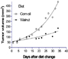
Our first study was to assess the effects of including walnut in the diet on the growth of existing cancer. We used a nude mouse model in which MDA-MB 231 human breast cancer cells were implanted between the scapulae. After the tumors had grown to measurable size, the mice were divided into two groups. One group was placed on a diet containing the AIN-76 diet modified to contain 10% total fat and to include human equivalent of 2 ounces per day of walnuts, the other group continued on the AIN-76 diet containing 10% corn oil as the fat. Diets were formulated to be isocaloric and isonutrient (see [101] for details). All animal work received approval from the Marshall University Institutional Animal Care Use and Committee prior to initiation.
As shown in Fig. 2 the tumors of the mice that consumed walnut in the diet grew significantly slower than the tumors of the mice that did not consume walnut. We found that the slower tumor growth could be related to a significant decrease in cell proliferation (proliferating cell nuclear antigen positive cells) and a slight but not significant increase in apoptotic cells. The omega 3 content of the walnut diet was reflected in the significant decrease in arachidonic acid (omega 6 fatty acid) and significant increases in the omega 3 fatty acids, eicosapentaenoic and docosahexaenoic acids in the liver [101].
In our second study (Fig. 3), we used a transgenic mouse model that develops mammary gland cancers to determine if regular consumption of walnuts could help prevent mammary gland cancers (all details in [102]). This was a transgenerational study; we started half of the mother mice on the walnut diet then assessed mammary gland cancer development in the female offspring that were randomized to control or walnut containing diets. The diets were the same as described in the first study (above). The results indicated that consumption of walnut after weaning reduced the risk for mammary gland cancer, however if the mother also consumed walnut (as would happen in a population that routinely includes walnut in the diet) then there was significant suppression of tumor development in the pups. Analyses of gene expression and protein in mammary glands without tumor indicated that reduced tumor risk was associated with suppression of NFκB activity by the walnut containing diet. We did not identify a single component of walnut that was responsible for decreasing risk for cancer development but rather proposed that it was the combination of ingredients in the walnut that reduced mammary gland cancer growth and development in mice.

CONCLUSIONS

Epidemiology studies have indicated that there is an environmental influence to cancer development and that overall diet or specific dietary components can increase or decrease cancer risk. Cell culture and animal studies have shown suppression of carcinogenesis by multiple components found in the fruits, vegetables, whole grains and nuts that would be consumed as part of a plant based diet. It seems difficult for humans to consume any single component that has been shown to be beneficial at a level and continuously enough to suppress carcinogenesis. However, the effects of the individual dietary components can be summed and increased when people eat a varied, plant based diet. The work of many scientists has shown that the individual components tested can reduce the production of inflammatory cytokines in animal and cell culture models. Since chronic inflammation contributes to the development of cancer, cardiovascular disease and diabetes, consumption of a varied plant-based diet, as recommended by multiple public health agencies, could effectively reduce the incidence of cancer and other chronic diseases.

Figures and Tables

Fig. 1
Polyphenol classifications. Classes of polyphenols and their relationships to each other. The top line of each class gives the general class name, examples of the class follow.
nrp-8-233-g001
Fig. 2
Growth of MDA-MB231 human breast cancers in nude mice consuming diets with or without walnut. Graph shows the average volume of MDA-MB 231 human breast cancers implanted in nude mice consuming a diet either with or without walnuts. Tumor were grown to about 50mm3 volume before diet change. Day 0 is the day of diet change. The 'corn oil' group consumed the AIN-76 based diet, the 'walnut' group had the AIN-76 formula adjusted to include a human equivalent of 2 ounces of walnut per day. N = 11 per group. Non-linear regression analyses (Prism©) showed that tumors of the 'corn oil' group grew significantly faster than those of the group that consumed walnut, P = 0.001. The doubling time of the corn oil group was 16 days whereas the doubling time of the walnut group was 23.3 days.
nrp-8-233-g002
Fig. 3
Tumor incidence and multiplicity at 145 days of age in mice that consumed diets that did or did not contain walnut. Tumor incidence (fraction of mice with any tumor) and tumor multiplicity (number of mammary glands with any tumor) in C3(1) TAg mice. The group designations are mother diet/pup diet thus: CO/CO - mother and pup consumed corn oil diet; CO/walnut - mother consumed corn oil diet/pup consumed walnut containing diet; Walnut/Walnut - mother and pup consumed walnut containing diet; Walnut/CO - mother consumed walnut diet, pup consumed corn oil diet. N = 10 to 13 mice per group. Pups that were exposed to walnut in the diet throughout life had significantly fewer tumors than those that did not receive walnut. Groups with different letters are significantly different, P < 0.05, (Prism© was used for statistical analyses see [102] for details.).
nrp-8-233-g003

Notes

The author gratefully acknowledges financial support from: Two matching grants from the American Institute for Cancer Research and the California Walnut Commission, an unnumbered grant and MG 07A001. Laboratory and personnel support from NIH R01CA114018 and W81XWH-04-1-0450 from the Department of Defense Breast Cancer Research Program to WEH. Genomics core work was supported by grants NIH grants: P20RR020180 and P20RR016477. No funding agency had input on experimental design, collection or interpretation of the data. Any opinions are solely those of the author and are not of any funding agency or of Marshall University. Pentresation at the Korean Nutrition Society meeting was sponsored by the California Walnut Commission (CWC). The CWC had no input or influence on the content of this paper.

References

1. Doll R, Peto R. The causes of cancer: quantitative estimates of avoidable risks of cancer in the United States today. J Natl Cancer Inst. 1981; 66:1191–1308.
crossref
2. World Cancer Research Fund. American Institute for Cancer Research (US). Food, Nutrition and the Prevention of Cancer: a Global Perspective. Washington, D.C.: American Institute for Cancer Research;1997.
3. World Cancer Research Fund. American Institute for Cancer Research (US). Food, Nutrition, Physical Activity and the Prevention of Cancer: a Global Perspective. Washington, D.C.: American Institute for Cancer Research;2007.
4. Rhodes JM. Unifying hypothesis for inflammatory bowel disease and associated colon cancer: sticking the pieces together with sugar. Lancet. 1996; 347:40–44.
crossref
5. Grce M, Mravak-Stipetić M. Human papillomavirus-associated diseases. Clin Dermatol. 2014; 32:253–258.
crossref
6. Rambau PF, Chalya PL, Jackson K. Schistosomiasis and urinary bladder cancer in North Western Tanzania: a retrospective review of 185 patients. Infect Agent Cancer. 2013; 8:19.
crossref
7. Qadri Q, Rasool R, Gulzar GM, Naqash S, Shah ZA. H. pylori infection, inflammation and gastric cancer. J Gastrointest Cancer. Forthcoming 2014.
crossref
8. Donath MY, Dalmas É, Sauter NS, Böni-Schnetzler M. Inflammation in obesity and diabetes: islet dysfunction and therapeutic opportunity. Cell Metab. 2013; 17:860–872.
crossref
9. Crujeiras AB, Díaz-Lagares A, Carreira MC, Amil M, Casanueva FF. Oxidative stress associated to dysfunctional adipose tissue: a potential link between obesity, type 2 diabetes mellitus and breast cancer. Free Radic Res. 2013; 47:243–256.
crossref
10. Balkwill F, Mantovani A. Inflammation and cancer: back to Virchow? Lancet. 2001; 357:539–545.
crossref
11. Coussens LM, Werb Z. Inflammation and cancer. Nature. 2002; 420:860–867.
crossref
12. Aroor AR, McKarns S, Demarco VG, Jia G, Sowers JR. Maladaptive immune and inflammatory pathways lead to cardiovascular insulin resistance. Metabolism. 2013; 62:1543–1552.
crossref
13. Lee H, Lee IS, Choue R. Obesity, inflammation and diet. Pediatr Gastroenterol Hepatol Nutr. 2013; 16:143–152.
crossref
14. Tarantino G, Capone D. Inhibition of the mTOR pathway: a possible protective role in coronary artery disease. Ann Med. 2013; 45:348–356.
crossref
15. Chen F, Castranova V. Nuclear factor-kappaB, an unappreciated tumor suppressor. Cancer Res. 2007; 67:11093–11098.
16. Aggarwal BB. Nuclear factor-kappaB: the enemy within. Cancer Cell. 2004; 6:203–208.
17. Lin WW, Karin M. A cytokine-mediated link between innate immunity, inflammation, and cancer. J Clin Invest. 2007; 117:1175–1183.
crossref
18. Karin M, Greten FR. NF-kappaB: linking inflammation and immunity to cancer development and progression. Nat Rev Immunol. 2005; 5:749–759.
crossref
19. Miyamoto S. Nuclear initiated NF-kappaB signaling: NEMO and ATM take center stage. Cell Res. 2011; 21:116–130.
crossref
20. Bours V, Bonizzi G, Bentires-Alj M, Bureau F, Piette J, Lekeux P, Merville M. NF-kappaB activation in response to toxical and therapeutical agents: role in inflammation and cancer treatment. Toxicology. 2000; 153:27–38.
crossref
21. Clevers H. At the crossroads of inflammation and cancer. Cell. 2004; 118:671–674.
crossref
22. Cao Y, Karin M. NF-kappaB in mammary gland development and breast cancer. J Mammary Gland Biol Neoplasia. 2003; 8:215–223.
23. Karin M, Cao Y, Greten FR, Li ZW. NF-kappaB in cancer: from innocent bystander to major culprit. Nat Rev Cancer. 2002; 2:301–310.
crossref
24. Jin RJ, Lho Y, Connelly L, Wang Y, Yu X, Saint Jean L, Case TC, Ellwood-Yen K, Sawyers CL, Bhowmick NA, Blackwell TS, Yull FE, Matusik RJ. The nuclear factor-kappaB pathway controls the progression of prostate cancer to androgen-independent growth. Cancer Res. 2008; 68:6762–6769.
crossref
25. Kim HJ, Hawke N, Baldwin AS. NF-kappaB and IKK as therapeutic targets in cancer. Cell Death Differ. 2006; 13:738–747.
26. Bharti AC, Shishodia S, Reuben JM, Weber D, Alexanian R, Raj-Vadhan S, Estrov Z, Talpaz M, Aggarwal BB. Nuclear factor-kappaB and STAT3 are constitutively active in CD138 + cells derived from multiple myeloma patients, and suppression of these transcription factors leads to apoptosis. Blood. 2004; 103:3175–3184.
crossref
27. Gupta SC, Kim JH, Prasad S, Aggarwal BB. Regulation of survival, proliferation, invasion, angiogenesis, and metastasis of tumor cells through modulation of inflammatory pathways by nutraceuticals. Cancer Metastasis Rev. 2010; 29:405–434.
crossref
28. Potter JD, Steinmetz K. Vegetables, fruit and phytoestrogens as preventive agents. IARC Sci Publ. 1996; 61–90.
29. Chan JM, Gann PH, Giovannucci EL. Role of diet in prostate cancer development and progression. J Clin Oncol. 2005; 23:8152–8160.
crossref
30. Manach C, Scalbert A, Morand C, Rémésy C, Jiménez L. Polyphenols: food sources and bioavailability. Am J Clin Nutr. 2004; 79:727–747.
crossref
31. Oseni T, Patel R, Pyle J, Jordan VC. Selective estrogen receptor modulators and phytoestrogens. Planta Med. 2008; 74:1656–1665.
crossref
32. Horia E, Watkins BA. Complementary actions of docosahexaenoic acid and genistein on COX-2, PGE2 and invasiveness in MDA-MB-231 breast cancer cells. Carcinogenesis. 2007; 28:809–815.
crossref
33. Sakamoto Y, Naka A, Ohara N, Kondo K, Iida K. Daidzein regulates proinflammatory adipokines thereby improving obesity-related inflammation through PPARγ. Mol Nutr Food Res. 2014; 58:718–726.
crossref
34. Brusselmans K, Vrolix R, Verhoeven G, Swinnen JV. Induction of cancer cell apoptosis by flavonoids is associated with their ability to inhibit fatty acid synthase activity. J Biol Chem. 2005; 280:5636–5645.
crossref
35. O'Leary KA, de Pascual-Teresa S, Needs PW, Bao YP, O'Brien NM, Williamson G. Effect of flavonoids and vitamin E on cyclooxygenase-2 (COX-2) transcription. Mutat Res. 2004; 551:245–254.
36. Bae JH, Kim JY, Kim MJ, Chang SH, Park YS, Son CH, Park SJ, Chung JS, Lee EY, Kim SH, Kang CD. Quercetin enhances susceptibility to NK cell-mediated lysis of tumor cells through induction of NKG2D ligands and suppression of HSP70. J Immunother. 2010; 33:391–401.
crossref
37. Wang G, Zhang J, Liu L, Sharma S, Dong Q. Quercetin potentiates doxorubicin mediated antitumor effects against liver cancer through p53/Bcl-xl. PLoS One. 2012; 7:e51764.
crossref
38. Chen AY, Chen YC. A review of the dietary flavonoid, kaempferol on human health and cancer chemoprevention. Food Chem. 2013; 138:2099–2107.
crossref
39. Delgado L, Fernandes I, González-Manzano S, de Freitas V, Mateus N, Santos-Buelga C. Anti-proliferative effects of quercetin and catechin metabolites. Food Funct. 2014; 5:797–803.
crossref
40. Al-Hanbali M, Ali D, Bustami M, Abdel-Malek S, Al-Hanbali R, Alhussainy T, Qadan F, Matalka KZ. Epicatechin suppresses IL-6, IL-8 and enhances IL-10 production with NF-kappaB nuclear translocation in whole blood stimulated system. Neuro Endocrinol Lett. 2009; 30:131–138.
41. Shan X, Li Y, Meng X, Wang P, Jiang P, Feng Q. Curcumin and (-)-epigallocatechin-3-gallate attenuate acrylamide-induced proliferation in HepG2 cells. Food Chem Toxicol. 2014; 66:194–202.
crossref
42. Saldanha SN, Kala R, Tollefsbol TO. Molecular mechanisms for inhibition of colon cancer cells by combined epigenetic-modulating epigallocatechin gallate and sodium butyrate. Exp Cell Res. Forthcoming 2014.
crossref
43. Thangapazham RL, Singh AK, Sharma A, Warren J, Gaddipati JP, Maheshwari RK. Green tea polyphenols and its constituent epigallocatechin gallate inhibits proliferation of human breast cancer cells in vitro and in vivo. Cancer Lett. 2007; 245:232–241.
crossref
44. Carter LG, D'Orazio JA, Pearson KJ. Resveratrol and cancer: a focus on in vivo evidence. Endocr Relat Cancer. Forthcoming 2014.
45. Mezzanotte L, An N, Mol IM, Löwik CW, Kaijzel EL. A new multicolor bioluminescence imaging platform to investigate NF-κB activity and apoptosis in human breast cancer cells. PLoS One. 2014; 9:e85550.
crossref
46. Mazué F, Delmas D, Murillo G, Saleiro D, Limagne E, Latruffe N. Differential protective effects of red wine polyphenol extracts (RWEs) on colon carcinogenesis. Food Funct. 2014; 5:663–670.
crossref
47. Garcia-Muñoz C, Vaillant F. Metabolic fate of ellagitannins: implications for health, and research perspectives for innovative functional foods. Crit Rev Food Sci Nutr. 2014; 54:1584–1598.
crossref
48. Rodriguez-Mateos A, Heiss C, Borges G, Crozier A. Berry (Poly) phenols and cardiovascular health. J Agric Food Chem. Forthcoming 2013.
49. Nuñez-Sánchez MA, García-Villalba R, Monedero-Saiz T, García-Talavera NV, Gómez-Sánchez MB, Sánchez-Álvarez C, García-Albert AM, Rodríguez-Gil FJ, Ruiz-Marín M, Pastor-Quirante FA, Martínez-Díaz F, Yáñez-Gascón MJ, González-Sarrías A, Tomás-Barberán FA, Espín JC. Targeted metabolic profiling of pomegranate polyphenols and urolithins in plasma, urine and colon tissues from colorectal cancer patients. Mol Nutr Food Res. Forthcoming 2014.
crossref
50. Banerjee N, Kim H, Talcott S, Mertens-Talcott S. Pomegranate polyphenolics suppressed azoxymethane-induced colorectal aberrant crypt foci and inflammation: possible role of miR-126/VCAM-1 and miR-126/PI3K/AKT/mTOR. Carcinogenesis. 2013; 34:2814–2822.
crossref
51. González-Sarrías A, Miguel V, Merino G, Lucas R, Morales JC, Tomás-Barberán F, Alvarez AI, Espín JC. The gut microbiota ellagic acid-derived metabolite urolithin A and its sulfate conjugate are substrates for the drug efflux transporter breast cancer resistance protein (ABCG2/BCRP). J Agric Food Chem. 2013; 61:4352–4359.
crossref
52. Kita T, Imai S, Sawada H, Kumagai H, Seto H. The biosynthetic pathway of curcuminoid in turmeric (Curcuma longa) as revealed by 13C-labeled precursors. Biosci Biotechnol Biochem. 2008; 72:1789–1798.
crossref
53. Aggarwal BB, Sung B. Pharmacological basis for the role of curcumin in chronic diseases: an age-old spice with modern targets. Trends Pharmacol Sci. 2009; 30:85–94.
crossref
54. Anand P, Thomas SG, Kunnumakkara AB, Sundaram C, Harikumar KB, Sung B, Tharakan ST, Misra K, Priyadarsini IK, Rajasekharan KN, Aggarwal BB. Biological activities of curcumin and its analogues (Congeners) made by man and Mother Nature. Biochem Pharmacol. 2008; 76:1590–1611.
crossref
55. Liu T, Bohlken A, Kuljaca S, Lee M, Nguyen T, Smith S, Cheung B, Norris MD, Haber M, Holloway AJ, Bowtell DD, Marshall GM. The retinoid anticancer signal: mechanisms of target gene regulation. Br J Cancer. 2005; 93:310–318.
crossref
56. Moon RC, Constantinou AI. Dietary retinoids and carotenoids in rodent models of mammary tumorigenesis. Breast Cancer Res Treat. 1997; 46:181–189.
crossref
57. Tamimi RM, Hankinson SE, Campos H, Spiegelman D, Zhang S, Colditz GA, Willett WC, Hunter DJ. Plasma carotenoids, retinol, and tocopherols and risk of breast cancer. Am J Epidemiol. 2005; 161:153–160.
crossref
58. Rao AV, Rao LG. Carotenoids and human health. Pharmacol Res. 2007; 55:207–216.
crossref
59. Wang L, Li B, Pan MX, Mo XF, Chen YM, Zhang CX. Specific carotenoid intake is inversely associated with the risk of breast cancer among Chinese women. Br J Nutr. 2014; 111:1686–1695.
crossref
60. Takeshima M, Ono M, Higuchi T, Chen C, Hara T, Nakano S. Anti-proliferative and apoptosis-inducing activity of lycopene against three subtypes of human breast cancer cell lines. Cancer Sci. 2014; 105:252–257.
crossref
61. Zu K, Mucci L, Rosner BA, Clinton SK, Loda M, Stampfer MJ, Giovannucci E. Dietary lycopene, angiogenesis, and prostate cancer: a prospective study in the prostate-specific antigen era. J Natl Cancer Inst. 2014; 106:djt430.
crossref
62. Brigelius-Flohé R, Traber MG. Vitamin E: function and metabolism. FASEB J. 1999; 13:1145–1155.
crossref
63. Campbell SE, Stone WL, Whaley SG, Qui M, Krishnan K. Gamma (γ) tocopherol upregulates peroxisome proliferator activated receptor (PPAR) gamma (γ) expression in SW 480 human colon cancer cell lines. BMC Cancer. 2003; 3:25.
crossref
64. Lee HJ, Ju J, Paul S, So JY, DeCastro A, Smolarek A, Lee MJ, Yang CS, Newmark HL, Suh N. Mixed tocopherols prevent mammary tumorigenesis by inhibiting estrogen action and activating PPAR-gamma. Clin Cancer Res. 2009; 15:4242–4249.
crossref
65. Dolfi SC, Yang Z, Lee MJ, Guan F, Hong J, Yang CS. Inhibitory effects of different forms of tocopherols, tocopherol phosphates, and tocopherol quinones on growth of colon cancer cells. J Agric Food Chem. 2013; 61:8533–8540.
crossref
66. Lu G, Xiao H, Li GX, Picinich SC, Chen YK, Liu A, Lee MJ, Loy S, Yang CS. A gamma-tocopherol-rich mixture of tocopherols inhibits chemically induced lung tumorigenesis in A/J mice and xenograft tumor growth. Carcinogenesis. 2010; 31:687–694.
crossref
67. Park HS, Han MH, Kim GY, Moon SK, Kim WJ, Hwang HJ, Park KY, Choi YH. Sulforaphane induces reactive oxygen species-mediated mitotic arrest and subsequent apoptosis in human bladder cancer 5637 cells. Food Chem Toxicol. 2014; 64:157–165.
crossref
68. Hussain A, Mohsin J, Prabhu SA, Begum S, Nusri Qel A, Harish G, Javed E, Khan MA, Sharma C. Sulforaphane inhibits growth of human breast cancer cells and augments the therapeutic index of the chemotherapeutic drug, gemcitabine. Asian Pac J Cancer Prev. 2013; 14:5855–5860.
crossref
69. Lenzi M, Fimognari C, Hrelia P. Sulforaphane as a promising molecule for fighting cancer. Cancer Treat Res. 2014; 159:207–223.
crossref
70. Wolf MA, Claudio PP. Benzyl isothiocyanate inhibits HNSCC cell migration and invasion, and sensitizes HNSCC cells to cisplatin. Nutr Cancer. 2014; 66:285–294.
crossref
71. Tse G, Eslick GD. Cruciferous vegetables and risk of colorectal neoplasms: a systematic review and meta-analysis. Nutr Cancer. 2014; 66:128–139.
crossref
72. Tusskorn O, Senggunprai L, Prawan A, Kukongviriyapan U, Kukongviriyapan V. Phenethyl isothiocyanate induces calcium mobilization and mitochondrial cell death pathway in cholangiocarcinoma KKU-M214 cells. BMC Cancer. 2013; 13:571.
crossref
73. Liu H, Talalay P. Relevance of anti-inflammatory and antioxidant activities of exemestane and synergism with sulforaphane for disease prevention. Proc Natl Acad Sci U S A. 2013; 110:19065–19070.
crossref
74. Schäfer G, Kaschula CH. The immunomodulation and anti-inflammatory effects of garlic organosulfur compounds in cancer chemoprevention. Anticancer Agents Med Chem. 2014; 14:233–240.
crossref
75. Zhou XF, Ding ZS, Liu NB. Allium vegetables and risk of prostate cancer: evidence from 132,192 subjects. Asian Pac J Cancer Prev. 2013; 14:4131–4134.
crossref
76. Borkowska A, Knap N, Antosiewicz J. Diallyl trisulfide is more cytotoxic to prostate cancer cells PC-3 than to noncancerous epithelial cell line PNT1A: a possible role of p66Shc signaling axis. Nutr Cancer. 2013; 65:711–717.
crossref
77. García JJ, López-Pingarrón L, Almeida-Souza P, Tres A, Escudero P, García-Gil FA, Tan DX, Reiter RJ, Ramírez JM, Bernal-Pérez M. Protective effects of melatonin in reducing oxidative stress and in preserving the fluidity of biological membranes: a review. J Pineal Res. 2014; 56:225–237.
crossref
78. Reiter RJ, Tan DX, Manchester LC, Korkmaz A, Fuentes-Broto L, Hardman WE, Rosales-Corral SA, Qi W. A walnut-enriched diet reduces the growth of LNCaP human prostate cancer xenografts in nude mice. Cancer Invest. 2013; 31:365–373.
crossref
79. Lv D, Cui PL, Yao SW, Xu YQ, Yang ZX. Melatonin inhibits the expression of vascular endothelial growth factor in pancreatic cancer cells. Chin J Cancer Res. 2012; 24:310–316.
crossref
80. Srinivasan V, Spence DW, Pandi-Perumal SR, Trakht I, Esquifino AI, Cardinali DP, Maestroni GJ. Melatonin, environmental light, and breast cancer. Breast Cancer Res Treat. 2008; 108:339–350.
crossref
81. García-Navarro A, González-Puga C, Escames G, López LC, López A, López-Cantarero M, Camacho E, Espinosa A, Gallo MA, Acuña-Castroviejo D. Cellular mechanisms involved in the melatonin inhibition of HT-29 human colon cancer cell proliferation in culture. J Pineal Res. 2007; 43:195–205.
crossref
82. Rodriguez C, Martín V, Herrera F, García-Santos G, Rodriguez-Blanco J, Casado-Zapico S, Sánchez-Sánchez AM, Suárez S, Puente-Moncada N, Anítua MJ, Antolín I. Mechanisms involved in the pro-apoptotic effect of melatonin in cancer cells. Int J Mol Sci. 2013; 14:6597–6613.
crossref
83. Wu KK. Control of cyclooxygenase-2 transcriptional activation by pro-inflammatory mediators. Prostaglandins Leukot Essent Fatty Acids. 2005; 72:89–93.
crossref
84. Akinsete JA, Ion G, Witte TR, Hardman WE. Consumption of high omega-3 fatty acid diet suppressed prostate tumorigenesis in C3(1) Tag mice. Carcinogenesis. 2012; 33:140–148.
crossref
85. Badawi AF, El-Sohemy A, Stephen LL, Ghoshal AK, Archer MC. The effect of dietary n-3 and n-6 polyunsaturated fatty acids on the expression of cyclooxygenase 1 and 2 and levels of p21ras in rat mammary glands. Carcinogenesis. 1998; 19:905–910.
crossref
86. Berquin IM, Edwards IJ, Chen YQ. Multi-targeted therapy of cancer by omega-3 fatty acids. Cancer Lett. 2008; 269:363–377.
crossref
87. Biondo PD, Brindley DN, Sawyer MB, Field CJ. The potential for treatment with dietary long-chain polyunsaturated n-3 fatty acids during chemotherapy. J Nutr Biochem. 2008; 19:787–796.
crossref
88. Blok WL, Katan MB, van der Meer JW. Modulation of inflammation and cytokine production by dietary (n-3) fatty acids. J Nutr. 1996; 126:1515–1533.
crossref
89. Bougnoux P, Germain E, Chajès V, Hubert B, Lhuillery C, Le Floch O, Body G, Calais G. Cytotoxic drugs efficacy correlates with adipose tissue docosahexaenoic acid level in locally advanced breast carcinoma. Br J Cancer. 1999; 79:1765–1769.
crossref
90. Calviello G, Serini S, Piccioni E. N-3 polyunsaturated fatty acids and the prevention of colorectal cancer: molecular mechanisms involved. Curr Med Chem. 2007; 14:3059–3069.
crossref
91. Cavazos DA, Price RS, Apte SS, deGraffenried LA. Docosahexaenoic acid selectively induces human prostate cancer cell sensitivity to oxidative stress through modulation of NF-kappaB. Prostate. 2011; 71:1420–1428.
crossref
92. Fahrmann JF, Ballester OF, Ballester G, Witte TR, Salazar AJ, Kordusky B, Cowen KG, Ion G, Primerano DA, Boskovic G, Denvir J, Hardman WE. Inhibition of nuclear factor kappa B activation in early-stage chronic lymphocytic leukemia by omega-3 fatty acids. Cancer Invest. 2013; 31:24–38.
crossref
93. Ion G, Akinsete JA, Hardman WE. Maternal consumption of canola oil suppressed mammary gland tumorigenesis in C3(1) TAg mice offspring. BMC Cancer. 2010; 10:81.
crossref
94. Reddy BS. Studies with the azoxymethane-rat preclinical model for assessing colon tumor development and chemoprevention. Environ Mol Mutagen. 2004; 44:26–35.
crossref
95. Rose DP, Connolly JM, Rayburn J, Coleman M. Influence of diets containing eicosapentaenoic or docosahexaenoic acid on growth and metastasis of breast cancer cells in nude mice. J Natl Cancer Inst. 1995; 87:587–592.
crossref
96. Ross JA, Maingay JP, Fearon KC, Sangster K, Powell JJ. Eicosapentaenoic acid perturbs signalling via the NFkappaB transcriptional pathway in pancreatic tumour cells. Int J Oncol. 2003; 23:1733–1738.
97. Sauer LA, Dauchy RT, Blask DE. Mechanism for the antitumor and anticachectic effects of n-3 fatty acids. Cancer Res. 2000; 60:5289–5295.
98. United States Department of Agriculture (US). USDA Nutrient Database, walnut composition [Internet]. Washington, D.C.: United States Department of Agriculture;2011. cited 2014 March 14. Available from: http://ndb.nal.usda.gov/.
99. Reiter RJ, Manchester LC, Tan DX. Melatonin in walnuts: influence on levels of melatonin and total antioxidant capacity of blood. Nutrition. 2005; 21:920–924.
crossref
100. McKay DL, Chen CY, Yeum KJ, Matthan NR, Lichtenstein AH, Blumberg JB. Chronic and acute effects of walnuts on antioxidant capacity and nutritional status in humans: a randomized, cross-over pilot study. Nutr J. 2010; 9:21.
crossref
101. Hardman WE, Ion G. Suppression of implanted MDA-MB 231 human breast cancer growth in nude mice by dietary walnut. Nutr Cancer. 2008; 60:666–674.
crossref
102. Hardman WE, Ion G, Akinsete JA, Witte TR. Dietary walnut suppressed mammary gland tumorigenesis in the C(3)1 TAg mouse. Nutr Cancer. 2011; 63:960–970.
crossref
TOOLS
Similar articles