

 PDF
PDF ePub
ePub Citation
 Print
Print


Introduction
Several lines of evidence suggest that soy isoflavones exert anti-atherogenic effects in mice, rabbits, monkeys, and humans [1-4]. Antihypertensive effects of isoflavones have also been proposed in spontaneously hypertensive rats and humans with mild to moderate essential hypertension [5-7]. The early stage of atherosclerosis is characterized by increased levels of inflammatory cytokines, oxidative stress, and the expression of adhesion molecules on vascular endothelium. Various cell adhesion molecules (CAMs), such as vascular cell adhesion molecule (VCAM)-1, intercellular adhesion molecule (ICAM)-1, and monocyte chemoattractant protein (MCP)-1, have been identified in endothelial cells [8,9]. Expression of CAMs and MCP-1 in endothelial cells is stimulated by interleukin (IL)-1β, tumor necrosis factor (TNF)-α, and lipopolysaccharide (LPS), which serve as signals to facilitate nuclear factor (NF)κB transactivation [10-12]. CAM expression is closely related to NFκB upregulation [8,13]. The endothelium also produces nitric oxide (NO), synthesized by endothelial nitric oxide synthase (eNOS), which is a vasoprotective and anti-atherosclerotic agent [5,6,14]. An eNOS isoform is constitutively expressed and can be activated and inhibited by phosphorylation through various protein kinases [5,15-17]. Therefore, any chemicals that suppress CAMs or enhance eNOS activity and expression in the vascular endothelium may be usable as therapeutic agents against the development of atherosclerosis.
Soy isoflavones are comprised of genistein (4',5,7-trihydroxyisoflavone), daidzein (4',7-dihydroxyisoflavone), glycitein (6-methoxydaidzein), and their glycosides. The molecular mechanisms of how these isoflavones affect the development and progression of atherosclerosis has not been thoroughly studied. However, genistein, a protein kinase inhibitor, exhibits anti-oxidative and anti-inflammatory activities, such as scavenging free radicals to prevent reactive oxygen species (ROS)-mediated DNA damage [18-21] and suppression of NO production through down-regulation of inducible NOS (iNOS) expression and NFκB transactivation [16,17,22]. However, contradicting data have been reported on the effect of genistein on CAM activities and mRNA expression induced by TNF-α in endothelial cells [11,12,23]. Furthermore, little information is available about the effects of daidzein, another major component of soy isoflavones, on NFκB activity and eNOS expression, which regulate CAM expression and bioactive NO production in endothelial cells. These questions led us to compare the effects of genistein and daidzein on MCP-1, eNOS, and CAM expression in TNF-α-stimulated human umbilical vascular endothelial cells (HUVECs) and to investigate possible mechanisms of action of their anti-atherosclerotic effect.
Materials and Methods
Materials
Genistein and daidzein were purchased from Sigma (St. Louis, MO, USA). EGM-2 Bullet Kit, HBSS, trypsin/EDTA solution, and trypsin neutralizing solution were obtained from Lonza (Basel, Switzerland). Human sVCAM-1 and MCP-1 enzyme linked immunosorbent assay (ELISA) kits were purchased from R&D Systems (Minneapolis, MN, USA).
Cell culture and treatment
HUVECs were purchased from Modern Tissue Technologies Inc. (Seoul, South Korea) and cultured in 75-cm2 flasks with medium from the EGM-2 Bullet Kit, which contained 10% fetal bovine serum, hydrocortisone, human fibroblastic growth factor-B, vascular endothelial growth factor, R3-insulin-like growth factor-I, ascorbic acid, human epidermal growth factor, GA-1000, and heparin. Confluent cultures were washed with HEPES buffered saline solution. The collected cells were pelleted by centrifugation at room temperature for 10 min at 500 × g, the supernatant was discarded, and the cell pellet was gently resuspended in EGM-2 Bullet Kit medium. HUVECs were seeded at a density of 1 × 10⁶ cells/100 mm dish and incubated for 2 days at 37℃ in a humidified 5% CO2 incubator. The medium was replaced every 2 days until confluence (3-5 days). Genistein and daidzein were dissolved in dimethyl sulfoxide and stored at -20℃. Cells were pretreated with or without genistein and daidzein (20, 50, or 100 µM) for 2 hr before stimulation with or without TNF-α (10 ng/mL) for 20 hr. Endothelial cell cultures were used at passages 3-6 in all experiments.
Cell viability
Cell viability was assessed by measuring the uptake of neutral red supravital dye by viable cells according to the procedure of Fautz et al. [24]. After culturing the cells as described above, the medium was removed and replaced with 0.5 mL fresh medium containing 1.14 mM neutral red. After a 3-hr incubation, the medium was removed, and the cells were washed twice with phosphate buffered saline solution (pH 7.4). The incorporated neutral red was released from the cells by a 15-min incubation at room temperature in 1 mL of cell lysis buffer. To measure the dye taken up, the cell lysis products were centrifuged and absorbance of the supernatant was measured spectrophotometrically at 540 nm.
sVCAM-1 and MCP-1 analysis
The concentrations of sVCAM-1 and MCP-1 were measured in the culture supernatants utilizing a highly sensitive ELISA according to the manufacturer's instructions (R&D Systems Inc.).
Reverse transcriptase-polymerase chain reaction (RT-PCR) analysis
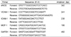
Total RNA was isolated using Trizol-reagent (Invitrogen, Carlsbad, CA, USA) and first strand cDNA was synthesized using the Superscript First Strand cDNA Synthesis kit (Invitrogen), according to the manufacturer's methods. The PCR primer sequences are shown in Table 1. The amplification profile consisted of an initial denaturation at 94℃ for 5 min followed by denaturation at 94℃, annealing from 58℃ to 63℃ and extension at 72℃ for 1 min. The total number of cycles for the best amplification profiles was 35, 22, 30, 22, and 20 for eNOS, VCAM-1, ICAM-1, MCP-1, and GAPDH, respectively. The intensity of the PCR products was densitometrically measured using the Gel Doc EQ System (Bio-Rad, Hercules, CA, USA). All signals were normalized to GAPDH house keeping gene mRNA levels and expressed as ratios.
NO production
Nitrite that accumulated in the culture medium was used as an indicator of NO production according to the Griess reaction [25]. One hundred µL of each medium supernatant was mixed with 50 µL of 1% sulfanilamide (in 5% phosphoric acid) and 50 µL of 0.1% naphthylenediamine dihydrochloride and incubated at room temperature for 10 min. The absorbance was measured at 550 nm against a NaNO2 serial dilution standard curve, and nitrite production was determined.
Electrophoretic mobility shift assay (EMSA)
Nuclear protein was extracted with a slight modification of the protocol from Dignam et al. [26]. Cells in 10 mm dishes were lysed with buffer, vortexed, kept on ice for 5 min, and centrifuged at 500 × g for 5 min at 4℃. Pelleted nuclei were resuspended in 50 µL extraction buffer. Following gentle mixing and an incubation on ice for 20 min, the samples were centrifuged at 500 × g for 5 min at 4℃. The supernatant fraction was transferred to new tubes and stored at -70℃. Protein concentration was determined by the Bradford assay. For the EMSA, NFκ B-specific oligonucleotide was end-labeled with [γ-32P]-ATP using T4 polynucleotide kinase (Promega, Madison, WI, USA) and purified using a Microspin G-25 column (GE Healthcare, Buckinghamshire, UK). EMSAs were performed according to the Promega instruction manual. Five µg of nuclear protein, binding buffer, 32P-labeled NFκB, and loading buffer were incubated for 30 min at room temperature. DNA-protein complexes were separated from unbound DNA probe by electrophoresis on 4% polyacrylamide gels with 0.5 × Tris-Borate EDTA buffer used as the running buffer. The gels were exposed to a phosphor screen (PerkinElmer, Waltham, MA, USA) for 2 hr at -20℃, and the bands were quantified with a phosphor imager (PerkinElmer).
Statistical analysis
All data are expressed as means ± SDs. Statistical analyses were performed using the SPSS program (SPPS, Inc., Chicago, IL, USA. A one-way analysis of variance and Duncan's multiple range test were used to examine the differences among treatments. P-values < 0.05 were considered significant.
Results
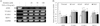
Effects of genistein and daidzein on MCP-1 and CAMs
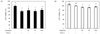
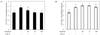
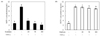
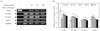
To examine the effects of genistein and daidzein on the expression of MCP-1 and CAMs, HUVECs were exposed to different concentrations (20-100 µM) of either genistein or daidzein and then stimulated with TNF-α. Cell viability was > 95% at the concentrations treated, as assessed by the neutral red assay (Fig. 1). Exposure of HUVECs to TNF-α induced significant MCP-1 and CAM expression, such as VCAM-1 and ICAM-1. Pre-incubation with genistein significantly (P < 0.05) decreased MCP-1 expression and production in a dose-dependent manner (Fig. 3A, 4A, and 4B). VCAM-1 production also decreased with genistein treatment (Fig. 2A), whereas VCAM-1 and ICAM-1 mRNA expression was not significantly decreased by genistein treatment (Fig. 4). However, daidzein did not affect VCAM-1 and ICAM-1 mRNA expression or VCAM-1 production (Fig. 2B, 5). Instead, MCP-1 production and expression were slightly lowered by daidzein treatment (Fig. 3B, 5), and its effect was smaller than that of genistein. These data suggest that genistein had a stronger effect on MCP-1 and CAM expression than that of daidzein.
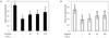
Effects of genistein and daidzein on NO production
Exposure of HUVECs to TNF-α downregulated eNOS expression and caused a significant decrease in NO production. Pre-treatment of genistein and daidzein with TNF-α significantly elevated eNOS expression and NO production (P < 0.05, Fig. 6).
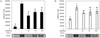
Effects of genistein and daidzein on NFκB transactivation
TNF-α induced NFκB transactivation in HUVECs. Pre-incubating HUVECs with 20 µM genistein or daidzein inhibited NFκB activity. However, the inhibitory effect on NFκB transactivation was slightly ameliorated at > 50 µM (Fig. 7).
Discussion
Early development of atherosclerosis is closely related with oxidative stress and inflammatory processes, such as the expression of chemokines and CAMs on endothelial cells. Several studies have reported that genistein and isoflavone supplementation in animal models improves endothelial dysfunction by regulating CAM and eNOS levels [2,5,6,8]. However, human trials do not consistently support the protective effects of higher isoflavone intake on cardiovascular disease risk [27], which might be due to the composition of the isoflavones used, study duration, or the supplementation amount. Therefore, this study was designed to compare the efficacy of genistein and daidzein, major soy isoflavones, on the regulation of chemokines, CAMs, and eNOS levels.
Genistein had a stronger inhibitory effect than daidzein on MCP-1 and CAM expression and production. Pro-inflammatory cytokines, such as TNF-α and IL-1β, are well known inducers of vascular adhesion molecules, including VCAM-1 and MCP-1 [10,11,28]. Tyrosine kinase inhibitors block transactivation of NFκB, a transcription factor that regulates CAMs and is induced by TNF-α and IL-1β [11,12,23]. Our study revealed that genistein, a tyrosine kinase inhibitor, selectively suppressed TNF-α-stimulated production of VCAM-1 and MCP-1 but not ICAM-1 mRNA expression. Furthermore, our study showed that genistein is not an effective VCAM-1 inhibitor compared to that of MCP-1. The inhibition of MCP-1 expression was dose-dependent and correlated with soluble MCP-1 concentrations. May et al. [11] found that genistein inhibits VCAM-1 expression induced by TNF-α or IL-1 in HUVECs, whereas it does not affect ICAM-1 expression. The selectivity of genistein for MCP-1 and VCAM-1 regulation may be desirable, as ICAM-1 is involved in leukocyte adhesion, whereas MCP-1 and VCAM-1 are primarily involved in adhesion of monocytes and lymphocytes found in atherosclerotic lesions [29]. However, CAM regulation may be affected by stimulus type. Weber et al. [28] reported that genistein does not affect CAM upregulation by phorbol myristic acid. Majewska et al. [13] also found that pre-treatment of HUVECs with genistein (10 µg/mL) for 30 min before IL-1 stimulation does not affect ICAM-1 and VCAM-1 expression. Chen et al. [30] found that isoflavoids have no obvious effect on neutrophilic CAM expression and concluded that isoflavoids protect the cardiovascular system through other mechanisms but not CAM regulation. Furthermore, genistein is ineffective for reducing LPS-stimulated E-selectin expression in HUVECs [12].
In contrast, daidzein showed very weak suppressive effects against MCP-1 expression and soluble protein excretion but no effect on ICAM-1 and VCAM-1 levels up to 100 µM. Gottstein et al. [23] also reported that daidzein decreases TNF-α-induced secretion of MCP-1 in HUVECs. However, we found that daidzein exerted weaker inhibitory effects than those of genistein for the expression of all CAMs. Our data agree with prior findings that genistein is a stronger suppressor of CAM expression than that of daidzein in TNF-α-stimulated HUVECs. Moreover, the present data contributes to our knowledge of the molecular mechanisms by which isoflavones may protect against coronary artery disease.
Genistein improves vascular reactivity by regulating eNOS, though the molecular mechanism is not clear. Our results showed that NO production and eNOS expression were elevated by preincubating HUVECS with genistein before TNF-α stimulation. Several studies have confirmed the stimulatory effect of genistein on NO and eNOS levels. Genistein acutely stimulates eNOS synthesis in vascular endothelial cells within a 10 minute incubation period through the cAMP/protein kinase A cascade without affecting PI3K/Akt or the ERK/MAPK signaling pathways [15]. Furthermore, genistein supplementation in spontaneously hypertensive rats shows a direct effect on transcription in vascular walls, leading to increased eNOS expression, thereby improving hypertension [5,7]. In contrast, soy isoflavone does not affect eNOS expression in Watanabe heritable hyperlipidemic rabbits [31]. These findings suggest that eNOS can be regulated by genomic and nongenomic actions.
Studies reporting the effect of daidzein on eNOS levels are scarce. In our study, the significance of genistein and daidzein on NO production and eNOS expression was compared. Genistein (20 µM) significantly elevated NO production and eNOS expression, whereas daidzein showed a significant difference only at 100 µM. Thus, daidzein was less effective at enhancing NO production and eNOS expression than genistein. The mechanism by which daidzein regulates eNOS seems to be somewhat different than that of genistein. Woodman et al. [32] proposed that daidzein enhances eNOS activity through an increase in calmodulin and a decrease in caveolin-1.
MCP-1 and CAM expression in endothelial cells is stimulated by IL-1β, TNF-α, and LPS, which serve as signals facilitating NFκB transactivation [10-12,33]. Our results indicated that both genistein and daidzein down-regulated CAM expression, and that this action was mediated, in part, through down-regulation of NFκB transactivation. Although we have demonstrated that TNF-α rapidly induced NFκB nuclear translocation in HUVECs, genistein and daidzein did not block NFκB translocation in a dose-dependent manner. In this study, the effects of genistein and daidzein on NFκB transactivation were biphasic; 20 µM of either isoflavone suppressed NFκB transactivation, whereas > 50 µM slightly increased NFκB transactivation. A similar trend was found by Guo et al. [34], who reported that daidzein stimulates cancer cell growth at low concentrations (1 µM) but inhibits cell growth at higher concentrations. This implies that regulation of CAM expression by genistein and daidzein is not solely dependent on the NFκB signaling pathway. Several researchers have reported that regulation of CAM expression does not necessarily require the NFκB signaling pathway but depends on the stimulus and cell type. That is, genistein as a tyrosine kinase cannot block NFκB translocation at the concentration that inhibits iNOS and COX-2 mRNA expression in rat mesangial cells [35]. IL-4-induced upregulation of the VCAM-1 gene in HUVECs does not appear to be involved with NFκB at the transcriptional level [36].
From the above results, it can be concluded that genistein and daidzein significantly suppressed MCP-1 and VCAM-1 production and elevated eNOS expression in TNF-α-stimulated HUVECs, although the genisteineffect was stronger than that of daidzein. Amelioration of NFκB transactivation might be, in part, related to MCP-1 and CAM downregulation. The ability to regulate CAM and NFκB expression, which are activated in atherosclerotic lesions, may explain the molecular mechanisms of genistein and daidzein to retard development of the initial stage of atherosclerosis. Genistein and daidzein enhanced eNOS expression and NO production, which are promising results in the search for treatments to maintain healthy blood pressure and prevent atherosclerosis.
 XML Download