

 PDF
PDF ePub
ePub Citation
 Print
Print


Introduction
Sheep and goats are frequently presented with different forms of hernias to veterinary clinics. Abdominal hernias may occur when the abdominal wall is severely traumatized and these hernias may be high or low in the flank, along the costal arch or between the last few ribs [12]. They are usually caused by violent force, such as from the impact of blunt objects, but they may result from overstretching of the abdominal muscles [27]. Various corrective procedures have been described elsewhere [2,5,8,9,11,13,14,22,24,26].
Umbilical hernias may be congenital or acquired, and they are seen in foals, calves, pups and pigs [7,26]. Many small umbilical hernias may appear to resolve spontaneously, but large or strangulated umbilical hernias will require surgical correction. Inguinal hernia is relatively common in bulls, rams and boars. Scrotal hernia is merely an extension of an inguinal hernia. Congenital inguinal hernia is rare in bulls, but it may result in evisceration at castration. Acquired inguinal hernias occur in mature bulls and rams [25,26].
The aim of the present study is to investigate the outcome of surgical treatment for abdominal, umbilical, inguinal and scrotal hernias in sheep and goats.
Materials and Methods
Animals
The present study was carried out on 58 clinical cases (44 sheep and 14 goats) that were presented to the Veterinary Teaching Hospital, College of Agriculture and Veterinary Medicine, Qassim University, Saudi Arabia from September, 2003 to September, 2006. These animals had abdominal (sheep = 30, goat = 10), umbilical (sheep = 6, goat = 4), inguinal (sheep = 7) and scrotal (sheep = 1) hernias.
The sheep were 18 males and 26 females, and they were classified as 3 local breeds (Nagdi = 34, Naimi = 9 and Sakni = 1). The ages of the sheep ranged from 1 month to 6 years. The histories of the cases indicated that the hernias were noticed at 10 days to up to 1 year before presentation to the hospital and the majority occurred 3 to 6 months before presentation.
Goats were 3 males and 11 females and they were classified as 2 breeds (Syrian = 9, Baladi = 5). The goats' ages ranged from 3 months to 6 years. Hernias were noticed in the goats at 1 month up to 8 months before admission to the clinic.
All the cases of hernias in the sheep and goats were subjected to full study, including the history of the case, classification of the hernias, the size of the hernial ring, surgical repair of the hernias, adhesions between the hernial sacs in each case (adhesions were graded from 1 to 4 with 1 as slight adhesions and 4 as severe adhesions), the postoperative care and the follow up of the cases, which was done by direct contact with or via phone calls to the owners. The data of the cases is summarized in Tables 1, 2 and 3.
Surgical treatment
Food was withheld for 24 h prior to surgery in each case. Surgical repair was conducted by aseptically preparing the site of operation after intramuscularly tranquilizing the fractious animals with 2% xylazine hydrochloride (Rompun 2%; Bayer, Turkey) at a dose rate of 0.05 mg/kg. The animal was restrained in the dorsal or lateral recumbent position, according to the type and position of the hernia.
In cases of abdominal and umbilical hernias, circular infiltration anesthesia was done using 2% lidocaine (Norbrook Laboratories, UK) at a dose rate of 10 mg/kg. An elliptical skin incision was performed and the adhesions between the parietal peritoneum and skin were freed with using both blunt and sharp dissection. The hernial ring was exposed and freshened before its suturing by simple interrupted or interrupted horizontal mattress sutures with using No. 2 chromic catgut (Ethicon, UK), polydioxanone (PDS; Ethicon, UK) or silk (Lukens Medical, USA) sutures. The subcutaneous tissue was then sutured by catgut or PDS, and the excessive skin was removed before its suture with using polypropylene (Ethicon, UK) or silk suture.
In cases of inguinal and scrotal hernias, linear infiltration anesthesia was applied at the site of the operation, which was lateral to the scrotum or the udder. A linear skin incision was made followed by sharp and blunt dissection to expose the hernial contents. The contents were reduced into the abdominal cavity through the inguinal canal and the external inguinal ring was narrowed by application of interrupted chromic catgut stitches (in 1 ram to keep the testis according to the owner's request). The testicles were removed whenever they appeared atrophied, and this was followed by complete closure of the external inguinal ring using catgut, PDS or silk sutures (in 7 cases).
Each animal was given postoperative therapy with penicillin-streptomycin at a dose rate of 30,000 IU/kg for the penicillin and 10mg/kg streptomycin for 5 days (Norbrook Laboratories, UK) and a prophylactic dose of anti-tetanus serum 1,500 IU subcutaneously.
Statistical analysis
The data was analyzed by a computer program and utilizing the SAS technique. Analysis of variance was used as the statistical method to evaluate the effects of breed, age, gender, the history of hernia, the size of the hernial ring, the type of the hernia, the degree of adhesions, the type of suture material and the outcome on the other variables. Multiple comparisons of means were determined using Tukey's method. Pearson correlation coefficient analysis was used to study the relationship between the different variables. The significant level was set at p < 0.05.
Results
Sheep
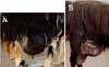
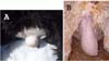
The sheep in the present study had 4 types of hernias; abdominal = 30, umbilical = 6, inguinal = 7 and scrotal = 1 (Fig. 1 & 2). The size of the hernial ring ranged from a finger breadth up to more than 2-hands breadth. All the cases were reducible hernias except for 1 ram that had a nonreducible abdominal hernia (Tables 1 & 2).
All the cases of hernias in the sheep were treated surgically (Fig. 3). Cesarean section was performed for 2 ewes before herniorrhaphy. The gravid uterus was the hernial content (hysterocele). Rumenotomy was done in 1 case to remove ruminal foreign bodies. In those 2 instances, open reduction was performed for which the parietal peritoneum was opened.
Outcomes
For the sheep, 26 out of 30 cases of abdominal hernia had good outcomes and their healing was excellent. There were postoperative complications in 4 ewes; abdominal hernia reocurred 1 month later, slight swelling in situ, muscular weakness at the site of operation and an abomasal fistula at the site of operation (Table 1).
The cases of umbilical hernias in sheep were surgically corrected without postoperative complications. However, a case of inguinal hernia, for which castration was not carried out upon the owner's request, had postoperative complications in the form of inflammation and swelling of the testis and scrotum of the affected side. This case necessitated castration at 2 weeks after the first operation. The other cases of inguinal and scrotal hernias had good healing without complications (Table 2).
For the goats, there were slight swellings at the site of operation in 2 out of 10 cases of abdominal hernia, while the remaining 8 cases had good outcomes. A case of umbilical hernia with an umbilical abscess had broken down with sepsis formation at the surgical site. The umbilical hernia reoccurred in this case (Table 3).
For the sheep, their age had no significant effect on the other parameters except its effect on the type of suture materials used (p = 0.006). Their gender only had an effect on the incidence of hernia. The incidence of abdominal hernias was higher in the females and the incidence of inguinal hernia was higher in the males. The history of hernia had no significant effect on the other variables except for its effect on the type of suture materials (p = 0.003). The size of hernia showed a significant effect only on the type of suture materials used (p < 0.001) and the outcome of surgery (p = 0.04). The other variables showed no significant effect on each other.
For the goats, there was no significant effect among the different variables except the effect of gender on the incidence of hernia. Both the abdominal and the umbilical hernias occurred more often in the females than in the males.
In this study, it was found that there was a positive correlation between the history of hernia and the degree of adhesion. There were no other correlations among the other variables.
Discussion
Hernias may be congenital or acquired; they may occur as isolated defects or they may be associated with defects of other parts of the body [7]. The results of the present study indicated that there were congenital umbilical hernias in sheep and goats, and these appeared just after birth; however, all the abdominal, inguinal and scrotal hernias in this study appeared to be acquired. Trauma due to horning from other animals appeared to be the most common cause of abdominal hernias. Increased intra-abdominal pressure during mounting for the males and pregnancy and the act of parturition in females are the probable causes of the inguinal and scrotal hernias. This study showed that abdominal hernia had the highest incidence in both sheep (68.2%) and goats (71.43%). The incidence of umbilical hernia was higher in the goats (28.57%) than in the sheep (13.6%), whereas the inguinal and scrotal hernias were seen only in sheep (18.2%). The present study showed that gender has an effect on the incidence of hernia in sheep and goats (females = 37, males = 21). In both species, females showed a higher incidence of abdominal hernia than the males. For sheep, the incidence of umbilical hernias was equal in both sexes. In goats, however, the incidence of umbilical hernias was higher in the females than in the males. Inguinal hernias mostly occurred in the males and particularly in rams. This may be due to slaughtering of the males at earlier ages than the females, which decreases the possibility of admission of the males to veterinary clinics.
The abdominal wall of a goat is relatively thin. Muscle tearing and separation often occur from blunt trauma during shearing, fighting or crowding through narrow doorways. Trauma or extreme abdominal distention in sheep occasionally leads to rupture of the ventral abdominal muscles caudal to the umbilicus [23].
The surgical treatment of hernias in the current study appeared to be a satisfactory treatment regimen for hernia repair. The success rates of surgical treatment for all types of hernias were very high and there were no significant difference in the success rate among the types of hernias in both sheep and goats. The success rate was approximately 93% (54 out of 58 cases). Very similar results were reported in another study in which 11 sheep with ventral hernias underwent surgical treatment [27]. The cases had an age range of 3 months to 4 years, and herniorrhaphy was done using chromic catgut No. 2 in an overlapping fashion. Slight swelling appeared 24 h postoperatively because of the accumulated fluid in the dead space and this disappeared spontaneously within 5-7 days. The ewes in this study were found to be 4 times more susceptible to this condition than the rams. Pregnancy would seem to be contributory factor, as was housing and the lack of proper management.
Displacement of the gravid uterus in ruminants occasionally occurs through rupture, and this most commonly happens on the right side of the abdominal floor. In most of these cases, a severe blow to the abdominal wall is the cause, although it may occur without trauma, resulting in weakening of the abdominal musculature so that the gravid uterus cannot be supported [1,15,19]. There were 2 ewes with hysterocele in the present study. Cesarean section was performed successfully just before herniorrhaphy and the ewes gave birth to their lambs. Successful repair has been described for goats. The late pregnant uterus can become trapped in the hernia in a subcutaneous location, making vaginal delivery difficult [23]. Moreover, a case of ventral metrocele in a pregnant goat has been previously reported [18]. The case was surgically corrected before the act of normal parturition. Umbilical hernia is a developmental defect. Although the size of the hernial ring in ewes with hysterocele in the present study was between 5 and more than 10 fingers breadth, the outcome of surgical repair was satisfactory.
The umbilical opening should close within a few days after birth. The failure of this opening to close properly is termed umbilical hernia. In addition to heredity, the etiology of umbilical hernia may be umbilical infection or abscess. Umbilical hernias are fairly commonly observed in young calves [11], they are rare in goats [23] and they are detected in pigs at between 9 and 14 weeks of age [17,26]. Most umbilical hernias in horses are reducible and the acquired hernias occur in the ventral or ventrolateral abdominal wall secondary to abdominal trauma or the stress of parturition, or they are due to previous abdominal surgery [13].
The cases of umbilical hernia in the present study were noticed as young as 4 months and up to 4 years in sheep; however, in goats, this malady was recorded in 3 month-old kids and in up to a 1.5 year-old goat. Two female kids had abscesses with their umbilical hernia, indicating that this is the probable cause of the herniation. Moreover, in the present study, there were younger sheep and goats as well as adult animals with umbilical hernia, indicated that umbilical hernia is probably a congenital or acquired hernia.
Various methods have been described in the literature for the treatment of umbilical hernia: counter irritation, clamping, transfixation sutures and even safety pins and commercially available rubber bands. The most popular of these techniques is the wooden or metal clamp technique. This method may result in infection, loss of clamps or premature necrosis of the hernial sac. The last complication can lead to an open wound, and possibly to evisceration or the formation of an enterocutaneous fistula. These methods are obviously unsuitable for the occasional strangulated hernia [28]. If the hernial ring is more than 1 finger in size or if it persists for more than 3 to 4 weeks, then surgical intervention is indicated [17]. Herniorrhaphy can be done by simply closing the abdominal wall with a horizontal mattress pattern of stitches with using absorbable sutures [17]. Surgery is required for older calves and this should be performed when there is an abscess along with the umbilical hernia [11]. The results of the current study indicated that the size of the umbilical hernia ring ranged from 2- to 7-fingers-breadth. Surgical repair was successful in 9 out of 10 cases of the sheep and goats. Herniorrhaphy was carried out using simple interrupted stitches with chromic catgut, PDS or silk sutures. One kid with a 7-fingers-breadth hernial ring recurred because of the presence of an abscess at the umbilicus that complicated the case, and this led to sepsis and break down of the hernia.
Inguinal hernia results when a defect permits intestines or other abdominal organs to pass into the inguinal canal. The hernia develops when an abnormally large and patent vaginal ring allows free communication between the vaginal tunic and the peritoneal cavities. The organs protrude into the scrotum to form a scrotal hernia, which is a more exaggerated form of this defect [25,26]. Congenital inguinal hernias are common in swine and they seem to occur due to a genetic influence [26]; they are rare in bulls, but they may result in evisceration at castration [25]. Although the incidence of inguinal hernia in sheep is not known with any certainly [16], acquired hernias have occurred in rams [25] and mature bulls [10,25] with the majority occurring on the left side of the scrotum, which is probably a result of the rumen's weight and also the mature bulls lying in a sternal position with the left rear leg abducted [10]. Acquired inguinal hernia is a problem in stallions, yet it is rare in Thoroughbred foals, but this is more common in the heavier breeds [6]. There is a predisposition towards acquired inguinal hernia in Standardbreds [21] and the hernias rarely become strangulated and they usually disappear spontaneously within a few weeks in newborn foals [13].
Old age may account for relaxation of and stretching of the rectus abdominis and internal and external oblique muscles [25]; however, the cause of scrotal hernia is almost certainly traumatic in origin [16]. Two 8 month-old Hampshire ram lambs with scrotal hernia were previously reported on [20], and a case of unilateral scrotal hernia in a 5-month-old ram lamb was corrected surgically [3]. The heritability of inguinal or scrotal hernia is not understood [3]. This condition is thought to be caused by trauma, and especially for group-housed rams [16]. Another possible influence on acquired scrotal hernia is the hormone concentration at the approach of breeding season [16]. Mounting during estrus increases the intra-abdominal pressure and contributes to herniation. Inguinal and scrotal hernias were recorded in 8 sheep (males = 7, female = 1) in the present study (inguinal = 7, scrotal = 1). The ages of the sheep ranged from 6 months to 5 years. The Nagdi breed seemed to have the vast majority of the inguinal hernias (Nagdi = 6, Sakni = 1, Naimi = 1). Unilateral inguinal hernia has been previously seen in ewes [25,29] and in a mature Merino ram [4]. The results indicated that surgical repair is successful when it is accompanied by castration of the testicle corresponding to the affected side. Castration on the affected side is preferred to minimize the chance of recurrent hernia and this is performed in most cases [13]. The only case that had postoperative complications had not undergone castration upon the owner's request. This case displayed postoperative swelling of the scrotum and testis due to inflammation. The cause might have been that the inguinal ring was too narrow, leading to ischemia of the testis.
Although inhalation anesthetics are not the best choice and other injectable anesthetics have not given ideal results for the treatment of inguinal hernias in sheep [16], 2 ewes with inguinal hernia were surgically corrected with the animals under halothane anesthesia [29]. This study indicated that local infiltration anesthesia with or without tranquilization was quite sufficient for performing surgical repair. However, positioning of the animal on surgical table was importance to facilitate reduction of the hernial contents and herniorrhaphy.
Nylon mesh was used successfully in the treatment of inguinal hernia in 1 ewe [29]. The results of the current study revealed that using a mesh was not necessary and herniorrhaphy is not difficult to perform in sheep and goats when simple interrupted stitches were preplaced, stretched and then tightened. The relaxed abdominal muscles in sheep and goats permitted favored herniorrhaphy, despite the size of the hernial ring. Statistically, the size of hernial ring in sheep had a significant effect on the outcome of the surgery (p < 0.05). The surgical success rate significantly increased with a smaller size of the hernial ring.
Interestingly, this study showed that either non-absorbable (i.e. silk) or absorbable (i.e. catgut or PDS) sutures can be used equally well to close the hernia rings. It is advisable to use silk when the hernial ring is large to give a chance for healing of the hernial ring. Chromic catgut and PDS were used when the hernial ring was relatively small. Moreover, the age of sheep had an effect on the types of suture materials used. Basically, the absorbable sutures can be used when the animals are less than 2 years old, whereas silk is recommended for the older animals (>2 years).
This study showed that the history of hernia in sheep had significant effects on the types of suture materials used. Generally, the nonabsorbable sutures (silk) should be used if the hernia was at least 8 months old. If the hernia is less than 8 months old, then it is acceptable to use the absorbable sutures (catgut or PDS). In addition, the size of the hernial opening has a significant effect on the types of suture materials used in sheep. With a large hernial opening (>4 fingers), silk should be used. PDS and catgut can be used in cases where the size of hernial ring is no more than 4 fingers. In fact, PDS can be used in cases where the size is between 3 to 4 fingers, and catgut can be used with small hernial rings (size <3 fingers). The types of suture materials and the types of hernias had no significant effect on the outcome of the surgical treatment.
The degrees of adhesion before surgery had no effect on the outcome of the surgical treatment. A positive correlation was found between the degree of adhesion and the history of hernia. This indicates that the degree of adhesion increased with the age of the hernia and vice versa.


 XML Download