

 PDF
PDF ePub
ePub Citation
 Print
Print


INTRODUCTION
Benign prostatic hyperplasia (BPH) is highly prevalent in older men [1]. Histologically, BPH is characterized by the presence of nonmalignant, unregulated overgrowth of the prostate gland. Clinically, BPH may be associated with lower urinary tract symptoms (LUTS) secondary to the ensuing prostate enlargement. About 60% of men aged >50 years have histologic evidence of BPH. This prevalence increases to 80% in patients aged ≥70 years [1]. Currently, BPH is the fourth most prevalent disease in men aged >50 years [2].
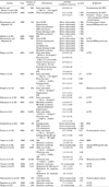
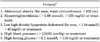
Despite the high prevalence of BPH and the socioeconomic burden related to its treatment [3], the pathogenesis of BPH is not well understood. In the past two decades, an increasing number of reports (Table 1) have suggested a possible relationship between BPH/LUTS and several metabolic disturbances, known collectively as the metabolic syndrome (MetS). MetS is a disease process associated with defective insulin-mediated glucose uptake, which is becoming an increasingly prevalent problem worldwide [4,5]. MetS involves a constellation of abnormalities including obesity, dyslipidemia, hypertension, and insulin resistance, with subsequent development of hyperinsulinemia and impaired glucose metabolism. Several definitions of MetS have been proposed by various organizations [6]. The Adult Treatment Panel III definition is the one used most today because it incorporates the key concepts of MetS and relies on routinely available clinical parameters (Table 2) [7].
The aim of the current report was to summarize and critically review the available literature that addresses the potential relationship between BPH/LUTS and MetS. Moreover, we aimed to clarify the underlying mechanisms that may link these two pathologic conditions and the changes in lifestyle that may decrease their prevalence.
BASIC SCIENCE AND EPIDEMIOLOGIC EVIDENCE
1. Obesity
Several anthropometric measures have been used to define obesity, including waist circumference, waist-to-hip ratio, and body mass index (BMI). Giovannucci et al examined the data of 25,892 men who participated in the Health Professionals Follow-up Study (HPFS) and observed that patients with an obese waist circumference (>109 cm) had a 2.4-fold higher risk (p<0.001) of being surgically treated for BPH than did those with a nonobese waist circumference (<89 cm) [8].
Dahle et al examined the relationship between waist-to-hip ratio (examined as quartiles) and LUTS in 502 Chinese men. In their analyses [9], individuals with a waist-to-hip ratio of ≥92% (highest quartile) had a 2.0-fold (p=0.01) higher risk of BPH (defined as symptomatic enlarged prostate requiring surgery) compared with their counterparts with a waist-to-hip ratio of ≤85% (lowest quartile). In the same report, no significant relationship was observed between BMI and BPH (p=0.1). The authors attributed this observation to the limited variation in BMI among study subjects (mean: 22.3 kg/m2 for BPH cases vs 21.9 kg/m2 for controls; p=0.1). In fact, only 4% of the study subjects were overweight. Nevertheless, even in such a lean population, the waist-to-hip ratio was an important independent predictor of BPH.
Parsons et al observed that men with BMI ≥35 kg/m2 had a 3.5-fold higher risk of harboring a large prostate (>40 ml) than did their counterparts with a BMI <25 kg/m2 [10]. Seim et al examined the data of 21,694 male residents of Norway and found that both BMI (odds ratio [OR]: 1.4) and waist-to-hip ratio (OR: 1.3) were significant predictors of moderate to severe LUTS [11], defined as an International Prostate Symptom Score (IPSS) of ≥8. Kristal et al further corroborated these observations in 5,677 participants of the Prostate Cancer Prevention Trial. In this cohort [12], men with BMI ≥35 kg/m2 had a 1.2-fold higher risk of developing LUTS (defined as IPSS ≥15 or a LUTS-specific treatment) compared with their counterparts with BMI <25 kg/m2. Several other investigators corroborated the significant direct relationship between obesity and BPH/LUTS [4,13-16].
Taken together, it appears that obesity predisposes patients to a higher risk of BPH/LUTS. This may be due to the development of insulin resistance and secondary hyperinsulinemia [17-19] or to the increased estrogen-to-androgen ratio [8]. It is noteworthy that the association between obesity and BPH/LUTS was stronger for patients with severe symptoms requiring surgery (OR: 2.0-2.4) [8,9] than for those who reported moderate-to-severe LUTS (OR: 1.2-1.4) and may indicate a dose-response relationship. Finally, the relationship between obesity and BPH/LUTS was observed in different races and ethnicities (i.e., American, European, and Asian), indicating its generalizability.
2. Dyslipidemia
Similar to obesity, the relationship between BPH and dyslipidemia has been documented in several studies. Rahman el al observed that prostate weight was significantly higher in hyperlipidemic rats than in controls (mean: 2.6 vs 1.4 g; p<0.001) [20]. Vikram et al conducted a longitudinal study over 12 weeks and reported that rats fed a high-fat diet had a significantly higher prostate weight than did controls [21]. These observations were further corroborated by Escobar et al in another rat model [22].
In addition to animal models, the relationship between BPH and dyslipidemia has been documented in epidemiologic studies. Hammarsten et al [14] examined the data of 158 men and reported that individuals with a low level of high-density lipoprotein (HDL) cholesterol (<1.18 mmol/l) had a larger prostate volume (mean: 49.0 vs 39.0 ml; p=0.002) and a higher annual BPH growth rate (mean: 1.02 vs 0.78 ml/y; p=0.006) than did individuals with a high level of HDL cholesterol (≥1.18 mmol/l). In a subsequent report, Hammarsten and Högstedt corroborated these findings in a larger sample (n=307) [4]. Nandeesha et al observed that men with BPH had a significantly higher total cholesterol (mean: 4.5 vs 4.2 mmol/l; p<0.05) and low-density lipoprotein (LDL) cholesterol levels (mean: 2.8 vs 2.2 mmol/l; p<0.001) than did men without BPH [23]. The same authors observed that the level of HDL cholesterol was significantly lower in men with BPH than in those without BPH (mean: 0.9 vs 1.2 mmol/l; p<0.001).
Conversely, in the Rancho Bernardo study, Parsons et al observed no overall association between LDL cholesterol and BPH (p=0.2) [24]. However, when men were stratified according to diabetes status, individuals with diabetes who had the highest LDL cholesterol had a 4-fold (95% confidence interval [CI]: 1.3-12.6; p=0.02) higher risk of reporting BPH (defined as a history of prostate surgery other than for cancer or physician-diagnosed BPH) than did those with the lowest LDL cholesterol (>133 mg/dl vs <110 mg/dl). This observation suggests that dyslipidemia may interact with other components of MetS to increase BPH risk [24]. This may at least partially explain why other investigators observed no significant relationship between dyslipidemia and BPH/LUTS [25-27].
3. Hypertension
Hypertension was also associated with BPH/LUTS in several animal models and epidemiologic studies. Golomb et al reported that spontaneously hypertensive rats develop BPH-like features with aging in the absence of any inductive exogenous agents [28]. Conversely, their normotensive counterparts did not develop such features. Hammarsten et al examined the data of 158 Swedish men referred to one center [14]. They reported that individuals with treated hypertension had a larger prostate volume (mean: 51.0 vs 44.0 ml; p=0.003) and higher annual BPH growth rate (mean: 1.06 vs 0.90 ml/y; p=0.002) than did controls. Similarly, Joseph et al reported that men with a history of hypertension had a 1.5-fold (95% CI: 1.1-2.1) higher risk of moderate-to-severe LUTS (defined as an American Urological Association Symptom Index [AUASI] of ≥8) compared with their counterparts without a history of hypertension [15]. Rohrman et al examined the data from 2,372 participants in the Third National Health and Nutrition Examination Survey (NHANES III) [29]. They reported that men with a history of hypertension had significantly higher odds of LUTS than did their counterparts without such a history (OR: 1.8; 95% CI: 1.2-2.6).
4. Insulin resistance
Insulin resistance represents another important component of MetS. To tackle the decreased responsiveness of insulin-sensitive tissues toward insulin, β-cells secrete more insulin and hence hyperinsulinemia is observed. These hormonal abnormalities may play an important role in the development of BPH/LUTS. Vikram et al reported an augmented prostatic epithelial cell proliferation in insulin-resistant rats [21]. The same authors showed that an induced status of hypoinsulinemia led to a dramatic decrease in the size of the prostate gland [30]. These observations were corroborated by Ikeda et al in another animal model [31].
At an epidemiologic level, Hammareston and Högstedt [4] addressed the relationship between fasting plasma insulin and BPH. In their cohort, men in the highest quartile of plasma fasting insulin (>13 mU/l) had a significantly larger prostate volume (mean: 61.0 vs 45.0 ml; p=0.009) and higher annual BPH growth rate (mean: 1.49 vs 0.84 ml/y; p=0.01) than did men in the lowest quartile of plasma fasting insulin (<7 mU/l). In the Flint Men's Health Study, individuals with a history of diabetes had twice the risk (OR: 2.0; 95% CI: 1.5-2.6) of suffering from moderate-to-severe LUTS (defined as an AUASI ≥8) as did their counterparts without a history of diabetes [15]. Seim et al evaluated the data of 21,694 individuals who participated in the second Nord-Trondelag Health Study and reported that men with diabetes were more likely to have an IPSS ≥8 than were their nondiabetic counterparts (OR: 1.3; 95% CI: 1.1-1.5) [11]. Similarly, Nandeesha et al reported that the level of fasting serum insulin was significantly higher in men with BPH than in controls (mean: 237.4 vs 134.7 pmol/l; p<0.001) [23]. In a recent report, Hammarsten et al confirmed the relationship between fasting serum insulin and prostate volume in a multicentric study (β coefficient: 0.2; p=0.02) [13].
5. Metabolic syndrome
Several criteria have been used to define MetS. Hammarsten et al defined MetS as having one or more of the following conditions: non-insulin-dependent diabetes mellitus, hypertension, obesity, high insulin level, and low HDL cholesterol [14]. They reported that men with MetS had a larger prostate volume (mean: 49.0 vs 28.5 ml; p=0.003) and a higher annual BPH growth rate (1.01 vs 0.69 ml/y; p=0.002) than did men without MetS. Rohrmann et al defined MetS as having at least three of the following five components [29]: (1) waist circumference >102 cm; (2) triglyceride concentration ≥1.69 mmol/l; (3) HDL cholesterol level <1.03 mmol/l; (4) systolic blood pressure ≥130 mmHg, diastolic blood pressure ≥85 mmHg, or current use of blood pressure medication; and (5) fasting glucose concentration ≥6.1 mmol/l or current use of oral diabetes medication or insulin. Additionally, they used a second, more specific definition that included components 1-3 but only in men who reported the use of blood pressure medication and men who used oral diabetes medication or insulin.
Rohrmann et al defined LUTS as having at least three of the following four urinary symptoms: nocturia, incomplete bladder emptying, weak stream, and hesitancy [29]. They reported that MetS was not statistically significantly associated with LUTS (OR: 1.2; 95% CI: 0.8-1.7). However, the OR was elevated in men with at least four components of MetS (OR: 1.6; 95% CI: 1.0-2.6) compared with men who had fewer components. Similarly, when the more specific definition of MetS was used, a significant association was evident between MetS and LUTS (OR: 1.8; 95% CI: 1.1-2.9). However, the cohort of this report [29] was restricted to men aged ≥60 years and included only four of the seven urologic symptoms that make up the AUASI.
To address the aforementioned limitations, Kupelian et al reexamined the relationship between MetS and LUTS in 1,899 men who participated in the Boston Area Community Health Survey [32]. They defined MetS according to the Adult Treatment Panel III guidelines as the presence of three or more of the following five characteristics: (1) waist circumference >102 cm; (2) systolic blood pressure ≥130 mmHg, diastolic blood pressure ≥85 mmHg, or antihypertensive medication use; (3) HDL cholesterol <40 mg/dl or lipid medication use; (4) self-reported type 2 diabetes or increased blood sugar or diabetes medication use; and (5) triglycerides >150 mg/dl. In multivariable analyses that adjusted for age, race, socioeconomic status, physical activity, alcohol consumption, smoking, and LUTS medication, MetS emerged as an independent predictor of moderate-to-severe LUTS, which was defined as an AUASI ≥2 (OR: 1.7; 95% CI: 1.2-2.4).
Although several investigators have observed a significant relationship between BPH/LUTS and MetS or one of its components, others have argued against these observations. Seitter and Barrett-Connor [33] reported no relationship between BMI and the rate of surgery for BPH. Meigs et al examined the data of 1,019 men who participated in the Massachusetts Male Aging Study and observed that obesity and blood pressure were not significantly related to subsequent clinical BPH development within a mean follow-up of 8.8 years [34]. Similarly, Hong et al observed no significant difference in the IPSS between men with and those without MetS (mean: 5.03 vs 5.40; p=0.3) [35].
In summary, it appears that MetS and its individual components may predispose patients to a higher risk of BPH/LUTS. However, this observation should be considered with caution for several reasons. The aforementioned reports defined the examined predictors in several different ways, and this variation may have affected the strength and significance level of their relationships with the examined endpoint. Moreover, the lack of consensus on the exact definition of MetS makes it more difficult to document the impact of this condition on the examined endpoint, namely, BPH/LUTS. The latter was also defined in several ways that ranged from a simple enlargement of the prostate gland to severe LUTS requiring surgery. Finally, different populations may have different genetic profiles, nutritional habits, and environmental risk factors. All of these variables may affect the potential relationship between MetS and BPH/LUTS.
HYPOTHESIZED MECHANISMS
1. Hyperinsulinemia and autonomic hyperactivity
Hyperinsulinemia is one of the components of MetS. Hyperinsulinemia is associated with increased sympathetic activity via enhanced glucose metabolism in ventromedial hypothalamic neurons [36]. This may contribute to an increase in the activation of the α-adrenergic pathway, which may in turn increase smooth muscle contraction throughout male genitourinary tract structures, including the prostate, the bladder neck, and the urethra, thereby contributing to LUTS [37,38].
This concept has been studied in both animal models and human subjects. In the rat model, McVary et al observed an association between autonomic neural input to the prostate and the prostatic growth rate such that the absence of this input resulted in regression of the gland volume [39]. In a subsequent report, the same authors investigated the relationship between autonomic tone and LUTS secondary to BPH in 38 human subjects of the Medical Therapy of Prostatic Symptoms trial [40]. They reported that markers of autonomic hyperactivity (blood pressure elevation, heart rate elevation, and elevated serum or urine catecholamines) were positively associated with subjective markers of LUTS (AUASI, quality of life score, and BPH Impact Index). Moreover, in multivariable analyses, plasma norepinephrine emerged as an independent predictor of the prostatic transition zone volume [40].
Using data from the same trial, Roehrborn et al showed that α-blockade alone may be sufficient in treating the symptoms of men with small prostate volume (<20 ml) [41], whereas men with larger prostates benefited more from a combination of α-blockade and 5α-reductase inhibitor therapy. These findings corroborate what has been reported by McVary et al and imply that autonomic hyperactivity may be an important determinant of LUTS in men with BPH [39,40]. Several other investigators confirmed the relationship between increased autonomic activity and BPH/LUTS [42,43].
Hyperinsulinemia is also associated with an increase in the level of free insulin-like growth hormone 1 (IGF-1) [44]. Several studies reported that the increase in the level of IGF-1 predisposes patients to a higher risk of BPH [45-47]. Consequently, hyperinsulinemia may also indirectly increase the risk of BPH by increasing the level of IGF-1.
2. Impaired nitric oxide and nitric oxide synthase activity
MetS has been associated with elevated levels of C-reactive protein (CRP) as well as other inflammatory markers [48-50]. This may reduce nitric oxide (NO) synthesis in endothelial cells [51]. Similarly, other components of MetS, namely, hyperglycemia and dyslipidemia, are associated with increased free radical production [52]. The latter may inhibit NO synthesis via activation of the protein kinase C pathway that may ultimately lead to decreased NO synthase (NOS) activation [52].
The diminished prostatic NO/NOS activity may lead to increased smooth muscle proliferation, prostatic enlargement, and subsequent LUTS [37]. Takeda et al examined tissue preparations of human and canine prostates and observed that NO plays an important role in mediating smooth muscle relaxation within the gland [53]. Consequently, the impairment of NO activity may contribute to voiding dysfunction in LUTS patients by impairing muscular relaxation.
Bloch et al performed a histochemical and ultrastructural examination of the prostate tissue with the intent of determining the location of endothelial and neuronal NOS in human prostate [54]. Their study resulted in several important findings. First, they observed that NO plays an important role in the autonomic innervation of all compartments of prostatic tissue. Second, they found that in obstructive BPH, the nitrergic innervation is reduced compared with that in normal prostate tissue. They concluded that the increased muscular tone in patients with BPH may be attributed to strengthened α-adrenergic stimulations as well as to decreased nitrergic innervation.
Apart from smooth muscle relaxation, NO has crucial functions in maintaining vascular health. It defends against the initiation of atherosclerosis by inhibiting adhesion of platelets and leukocytes to the vascular wall. It decreases the proliferation of vascular smooth muscles [52]. Bloch et al [54] suggested that NO has an important role in the regional circulation of the prostate gland; thus, impaired NO/NOS activity may lead to endothelial dysfunction and atherosclerosis. Numerous animal studies have linked atherosclerosis and chronic ischemia to bladder and prostatic changes [37]. Rabbits with induced pelvic atherosclerosis developed bladder fibrosis, smooth muscle atrophy, and decreased bladder compliance [55] as well as chronic prostatic ischemia with resultant stromal and capsular fibrosis, glandular cystic atrophy, impaired smooth muscle relaxation, and increased prostatic weight [56,57].
3. The Rho kinase system
The Rho kinase system plays an important role in the maintenance of tonic contraction or high basal tone and may contribute to prostate contractility [58]. Specifically, the Rho kinase system causes smooth muscle contraction by modifying the calcium sensitivity of the contractile machinery [37,59]. This appears to be mediated by the inhibition of myosin-light chain phosphatase, which promotes myosin-light chain phosphorylation and contraction through actin-myosin interaction [37,60,61]. NO counteracts this by favoring the active form of myosin-light chain phosphatase [61]. In contrast, α-adrenergic activity stimulates the Rho kinase pathway [37]. Similarly, the higher levels of interleukin (IL)-8 and the vasoconstrictor endothelin-1, which are usually observed in men with MetS, may lead to an increased Rho kinase system activity [37,50,52,58,59,62-64]. All of these MetS components (ie, impaired NO activity, autonomic hyperactivity, elevated IL-8 level, and elevated endothelin-1 level) may favor the activity of the Rho kinase system and may in turn result in a higher prostate contractility and LUTS.
4. Proinflammatory status
Metabolic syndrome is associated with proinflammatory status. This is demonstrated by elevated levels of CRP, IL-1β, IL-6, and tumor necrosis factor α (TNF-α) in men with MetS [48-50,62]. Obesity induces adipose cell enlargement and chemokine release, leading to macrophage infiltration of adipose tissue [65]. Macrophage infiltration further perpetuates the proinflammatory state and may account for the adipose secretion of adipokines such as IL-1β, IL-6, IL-8, CRP, and TNF-α [65-68]. Macrophage and T-lymphocyte infiltrates are commonly found in prostate tissue removed during open prostatectomy and transurethral resection of the prostate [69]. Similarly, several proinflammatory cytokines are upregulated in BPH, suggesting an immunologic etiology for the inflammation [69]. Cytokines IL-6 and IL-8, for example, are elevated in MetS and may contribute to inflammation in BPH/LUTS, as both can be secreted by stromal cells with cytokine stimulation and both result in proliferation of prostatic tissues [70]. In patients with BPH, seminal IL-8 levels were positively correlated with LUTS via the IPSS [71].
5. Abnormalities of sex hormones
Sex hormonal changes may further contribute to the linkage of BPH/LUTS and MetS. Men with MetS as well as those with LUTS/BPH may have lower androgen and higher estrogen levels. Rohrmann et al analyzed 260 individuals from NHANES III and reported that elevated estrogen levels and molar estradiol/testosterone ratios as well as lower androstanediol glucuronide (a metabolite of dihydroxytestosterone [DHT]) levels were associated with greater LUTS risk [72]. Similarly, Schatzl et al found a direct correlation between elevated estradiol levels and prostatic volume determined with transrectal ultrasound [73]. In the Physicians' Health Study, elevated estradiol level was an independent predictor of BPH surgery in men with low testosterone or DHT levels [74]. Haider et al studied hypogonadal men and observed an improvement in IPSS and postvoiding volume after a period of treatment with testosterone undecanocate [75]. Similarly, Kalinchenko et al treated 30 hypogonadal men with either testosterone gel or testosterone undecanoate and reported IPSS improvement in both groups [76]. However, the regulation and impact of sex hormones on prostatic growth and BPH/LUTS is quite complex, and not all authors found evidence of such definitive relationships [77,78].
In addition to the aforementioned mechanisms, other unknown mechanisms may contribute to the relationship between MetS and BPH/LUTS. Moreover, the aforementioned mechanisms may interact and articulate with each other. The mechanisms that regulate the potential relationship between MetS and BPH/LUTS are quite complex and require further scientific inquiry.
PHYSICAL ACTIVITY, METABOLIC SYNDROME, AND BENIGN PROSTATIC HYPERPLASIA
If MetS is indeed associated with an increased risk of BPH/LUTS, then it can be hypothesized that physical activity, which is known to decrease the cardiovascular morbidity risk associated with MetS, may also decrease the risk of BPH/LUTS. Several epidemiologic data support this hypothesis. Platz et al examined the data of 30,634 men who participated in the HPFS and observed that physical activity was associated with lower risk of total BPH (OR: 0.75; 95% CI: 0.64-0.90; p<0.001), surgery for BPH (OR: 0.76; 95% CI: 0.64-0.90; p<0.001), and symptomatic BPH (OR: 0.75; 95% CI: 0.64-0.87; p<0.001) [79].
Similarly, Rohrmann et al analyzed the data of 2797 individuals who participated in NHANES III [80] and reported that men with the highest rate of physical activity (highest quartile) had almost half the risk of suffering from LUTS (defined as having three of the following four urinary symptoms: nocturia, incomplete bladder emptying, weak stream, and hesitancy) as did their counterparts who reported no physical activity (OR: 0.49; 95% CI: 0.29-0.84; p=0.05). In an interesting report, Dal Maso et al observed that "heavy" occupational activity (eg, farmers, construction workers) led to a lower risk of developing BPH than did sedentary occupational activity [81]. However, not all investigators observed a significant inverse relationship between physical activity and BPH/LUTS [82]. Nonetheless, a recent meta-analysis that examined the data from eight reports, with a total of 35,675 patients included, confirmed that physical activity reduces the risk of BPH/LUTS [83].
In summary, it appears that sustained physical activity reduces the overall risk of BPH/LUTS in men. However, none of the aforementioned studies exclusively addressed patients with MetS. Consequently, the real benefit of physical activity in patients with MetS and how it may modify the risk of BPH/LUTS in these patients has yet to be clarified. In addition to physical activity, other lifestyle changes, such as dietary strategies to reduce postprandial glucose and triglyceride spikes and to increase the intake of dietary antioxidants, potassium, and omega-3 fatty acids may be helpful in reducing the incidence of metabolic syndrome [84] and its association with BPH/LUTS.
CONCLUSIONS
Numerous reports have documented a direct and significant relationship between components of MetS and the BPH/LUTS complex. However, differences in the strength and the statistical significance level of this relationship were observed. These differences may originate from the use of different descriptions to define the examined risk factors and endpoints as well as differences in the characteristics of the examined populations. Although the association between these two pathologic conditions seems plausible, the underlying mechanisms that create such an association need to be clarified.
Further study of the interplay between MetS and BPH/LUTS may provide new insight into the cause of these diseases and novel targets for their treatment and prevention. In the meantime, obesity, dyslipidemia, hypertension, and diabetes should be considered risk factors when advising patients with LUTS. Modifications of lifestyle, such as increased physical activity and dietary strategies, may be of benefit for these individuals to improve their LUTS.
 XML Download