

 PDF
PDF ePub
ePub Citation
 Print
Print


INTRODUCTION
Adipose tissue acts as a central regulator of energy metabolism and vascular homeostasis by secreting a diverse range of adipokines, such as adiponectin, leptin, and proinflammatory mediators (1). Excessive expansion of white adipose tissue (WAT), the hallmark of obesity, is a potent risk factor for type II diabebetes (T2D) and cardiovascular diseases (2). Inflammation in WAT, characterized by macrophage infiltration and elevated production of adipokines such as leptin and proinflammatory cytokines has played a key role in linking obesity with insulin resistance and metabolic dysfunction (3,4).
AMP-activated protein kinase (AMPK) is a phylogenetically conserved intracellular energy sensor that plays a central role in the regulation of glucose and lipid metabolism. AMP-activated protein kinase is a heterotrimeric complex composed of a catalytic subunit and two regulatory subunits, and is activated when cellular energy is depleted (5). Recently, it has been found that AMPK plays an important role in inflammation (6) and accelerates ATP-generating catabolic pathways, including glucose and fatty acid oxidation (7,8). In vitro and in vivo studies have demonstrated that the AMPK agonist, 5-aminoimidazole-4-carboxy-amide-1-D-ribofuranoside (AICAR), enhances insulin-mediated glucose transport and insulin action in muscle and liver of insulin-resistant rats fed a high fat diet (9,10).
Aloe species have been used for centuries for their anti-inflammatory (11), immunostimulant, antiseptic (12), burn healing (13), and antitumor activities (14). Fifty years ago, Mertz and coworkers proposed that chromium (Cr) was an essential trace element (15,16) and that it was required for normal carbohydrate and lipid metabolism (17-19). However, it remains unclear as to whether Aloe mediates the activation of AMPK, especially in muscles and WAT.
In the present study, we examine whether the Aloe QDM complex does indeed down-regulate inflammatory responses and adipogenesis and further, how it exhibits anti-inflammatory responses in WAT. We demonstrated that Aloe QDM complex reduced the expression of scavenger receptors and the expression of proinflammatory cytokines in obese mice in an AMPK-dependent manner. These results suggested that the Aloe QDM complex would exhibit advantageous effects in cases of obesity-related metabolic disorders through an AMPK stimulatory mechanism.
MATERIALS AND METHODS
Chemicals and reagents
Processed Aloe vera gel (PAG) (20), Aloesin (ALS), Aloe QDM, and Aloe QDM complex (21) were provided by Univera, Inc. (Seoul, Republic of Korea). Pioglitazone (Actos®) was purchased from Eli Lilly (Toronto, Canada); chromium (Cr) was purchased from Lallemand Inc. (Montreal, Canada); and leupeptin, aprotinin, and phenylmethylsulfonyl fluoride (PMSF) were purchased from Sigma Chemical Co. (St. Louis, MO, USA). Antibodies of Hypoxia induced factor 1 alpha (HIF-1α) and NF-κB p65 were purchased from Novus Biologicals (Littleton, CO, USA) and Santa Cruz Biotechnology Inc., (Santa Cruz, CA, USA), respectively. Anti-mouse AMP-activated protein kinase alpha (AMPKα) and phospho-AMPKα (Thr172) were purchased from Cell Signaling Technology (MA, USA), and all other chemicals and reagents used in this study were reagent grade.
Animals and diets
Male C57BL/6NCrjBgi mice were purchased from the Charles River Laboratory of Animal Science (Orient Co., Seoul, Republic of Korea) at four weeks old and fed a normal diet for one week. Animals were housed in individual cages with free access to water and food in a temperature-controlled animal facility under a 12 h light-dark cycle at 22±2℃ and 55±5% humidity. Mice were fed either a high-fat diet (HFD) (Open Source diets #D12492; Research Diets Inc., New Brunswick, NJ) to induce obesity, or a regular diet (RD; Open Source diets #D12450B; Research Diets Inc.). The nutritional contents of the HFD were similar to those on the regular diet except for low carbohydrate content and a high level of fat.
At 26 weeks of age, mice exhibiting blood glucose levels >160 mg/dl were selected as non-insulin-dependent diabetes mellitus (NIDDM) animals and were divided into five groups of 20 animals per group. One group was administrated PBS only and served as diabetic controls; four groups received daily 100 mg/kg of PAG, 2 mg/kg of Aloesin (ALS), 100 mg/kg of PAG containing 2% ALS (Aloe QDM), and Aloe QDM plus 500 mg/kg of Cr-enriched yeast containing 0.2% Cr (Aloe QDM complex) respectively, and the fifth group was administered pioglitazone (PGZ, 2.5 mg/kg), an anti-diabetic drug currently in clinical use. Mice were weighed and blood samples were collected weekly by tail bleeding into heparin-coated tubes after 4 h fasts.
At the end of the experimental period, mice were sacrificed and WAT were immediately removed from periepididymal and perirenal fat for morphological examination. Mice were treated in accordance with the guidelines issued by Sahmyook University for the care and use of laboratory animals.
Histological analysis
Adipose tissue was isolated from mice, fixed in 10% formalin, and embedded in paraffin. Four-micrometer thin sections were obtained, mounted on two glass-slides, and stained with hematoxylin-eosin. Sections were viewed with an Olympus microscope (Olympus, Tokyo, Japan) at 400X magnification.
Isolation of total RNA and reverse transcription polymerase chain reaction (RT-PCR)
Twenty mice were selected and used for this analysis. WAT and muscles were immediately frozen in liquid nitrogen and stored at -70℃ for the determination of gene transcripts. Total RNA was extracted from tissues using the RNeasy Mini kit (QIAGEN, Valencia, USA) in an RNase-free environment. RNA was then quantified by reading the absorbance at 260 nm. The reverse transcription of 1µg RNA was carried out using M-MLV reverse transcriptase (Promega, USA), an oligo (dT) 16 primer, dNTP (0.5µM), and 1 U RNase inhibitor. After incubation at 65℃ for 5 min and 37℃ for 60 min, M-MLV reverse transcriptase was inactivated by heating at 70℃ for 15 min. The polymerase chain reaction (PCR) was performed in 50 mM KCl, 10 mM Tris-HCl (pH 8.3), 1.5 mM MgCl2, and 2.5 mM dNTPs with 5 units of Taq DNA polymerase, and 10 pM of each primer set for CD36, scavenger receptor A (SR-A), interleukin (IL)-1β, and interleukin (IL)-6. The cDNA was amplified by 35 cycles of denaturing at 94℃ for 45 s, annealing at 62℃ for 45 s, and extension at 72℃ for 1 min. Final extension was performed at 72℃ for 5 min. The PCR products were then electrophoresed on 1.5% agarose gels and stained with ethidium bromide. The primers selected were 5' AGT AGG CGT GGG TCT GAA GG 3' (forward) and 5' CTT GCT TGC CCA GTC ACA GG 3' (reverse) for CD36, 5' GGA GAC AGA GGG CTT ACT GG 3' (forward) and 5' GTT GAT CCG CCT ACA CTC CC 3' (reverse) for SR-A, 5' CAG GAT GAG GAC ATG ACA CC' (forward) and 5' CTC TGC AGA CTC AAA CTC CAC3' (reverse) for IL-1β, 5' GTA CTC CAG AAG ACC AGA GG 3' (forward) and 5' TGC TGG TGA CAA CCA CGG CC 3' (reverse) for IL-6, and 5' CAA CTT TGG CAT TGT GGA AGG 3' (forward) and 5' ATG GAA ATT GTG AGG GAG ATG C 3' (reverse) for GAPDH, which was used as an internal control.
Preparation of nuclear extracts
After homogenized tissues were collected and washed twice with cold PBS, they were resuspended in ice lysis buffer (20 mM Trizma base, 50 mM NaCl, 250 mM sucrose, 50 mM NaF, 5 mM Na4P2O7·10H2O, 1% Triton-X100, 5µg/ml leupeptin, 1 mM phenylmethylsulfonyl fluoride (PMSF), and 5µg/ml aprotinin). After 15 min incubation on ice, the cells were lysed by the addition of 0.1% NP-40 and vigorous vortexing for 1 min. The nuclei were pelleted by centrifugation at 12,000×g for 1 min at 4℃ and resuspended in high salt buffer (20 mM HEPES, pH 7.9, 25% glycerol, 400 mM KCl, 1.5 mM MgCl2, 0.2 mM EDTA, 0.5 mM DTT, 1 mM NaF, 1 mM sodium orthovanadate). The supernatant fluid was stored in aliquots at -70℃.
Western blot analysis
Frozen epididymal adipose tissue and muscles were homogenized in 3 volumes of ice lysis buffer (20 mM Trizma base, 50 mM NaCl, 250 mM sucrose, 50 mM NaF, 5 mM Na4P2O7·10H2O, 1% Triton-X100, 5µg/ml leupeptin, 1 mM phenylmethylsulfonyl fluoride (PMSF), and 5µg/ml aprotinin). Twenty micrograms of protein from cell lysates were applied to 8~12% SDS-polyacrylamide gels and then transferred to nitrocellulose membranes. The membranes were blocked with 5% skim milk in TBST solution for 1 hr. They were then incubated with anti-HIF1α, anti-AMPK, anti-p-AMPK, and anti-p65 monoclonal antibodies for 2 hrs and washed 3 times with TBST. After incubation with alkaline phosphatase-labeled secondary antibody for 2 hrs, the bands were visualized using a Western Blot Kit with an alkaline phosphatase substrate (Vector, Burlingame, USA).
RESULTS
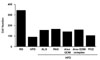
Reduction in adipocyte size via the suppression of scavenger receptors on macrophages in the WAT of obese mice
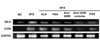
CD36 and SR-A are scavenger receptors for oxidized low-density lipoprotein (oxLDL) and cellular transporters of long-chain fatty acid (22). In the present study, we examined changes in the level of expression of scavenger receptors and their affect on the amount of adipose tissues in obese mice. We obtained WAT, stained with H&E, then counted the adipocytes. Numerous adipocytes completely disappeared after aloe formula supplementation, especially after Aloe QDM complex supplementation (Fig. 1). In agreement with this, levels of scavenger receptors such as SR-A and CD36 in the WAT were significantly lower in obese mice suppplemented with dietary aloe formula than in the HFD group (Fig. 2). These findings indicated that dietary aloe formula supplementation suppressed fat size through inhibition of scavenger receptors in ATM.
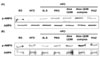
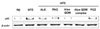
Aloe QDM complex enhances AMPK phosphorylation in the both WAT and muscles
We demonstrated that aloe formulas increased AMPKα phosphorylation in both WAT and muscles. Since the AMPK cascade has been shown to activate insulin signaling (10), we hypothesized that AMPK might be involved in the effect of lipolysis on WAT. The Aloe QDM complex enhanced phosphorylation of AMPK in tissues (Fig. 3), a finding indicating that dietary aloe formula reduced fat levels through mitochondrial biogenesis.
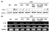
Reduction of inflammatory responses via suppression of NF-κB in WAT
To examine whether the dietary aloe formula modulated obesity-induced inflammation, we measured a proinflammatory cytokine gene and its transcription factor, NF-κB p65, and protein expression in WAT by RT-PCR and western blot. As shown in Figs. 4A and B, HIF1α protein and proinflammatory cytokines, IL-1β and IL-6 mRNA were lower in the obese mice supplemented with aloe formula than in the HFD group. The transcription factor, NF-κB p65 was also inhibited by aloe formulas, and in particular, the Aloe QDM complex (Fig. 5). These findings indicated that aloe formulas, and especially the Aloe QDM complex reduce WAT inflammatory responses through inactivation of NF-κB p65.
DISCUSSION
We previously demonstrated that the Aloe QDM complex reduced fat via a suppression of the expression of scavenger receptors and obesity-related inflammatory cytokines through activation of AMPK and inhibition of translocation of NF-κB p65 proteins in WAT and muscles.
Herbal prescriptions have been recognized as potentially valid by the scientific medical establishment, and their use has been increasing. Since traditional herbal prescriptions are generally prepared from a combination of crude drugs, on the basis of oriental prescriptions, they may exert combined effects that differ from the sum of the effects of the individual constituents (22).
Dietary aloe formula has been demonstrated to affect inflammation by virtue of an immunosuppression. Recent studies have suggested that inflamed adipocytes in the obese trigger the development of obesity-related metabolic disorders such as insulin resistance and T2D, indicating that the reduction of tissue inflammation may be beneficial in obesity-related metabolic diseases. Our previous in vivo study demonstrated a potential effect of PAG, an aloe formula, on hypoglycemia and hypolipidemia (20,21). In particular, our results demonstrated that the administration of aloe formulas including PAG, ALS, Aloe QDM, and Aloe QDM complexes to these mice prevented the development of T2D-related symptoms. However, the prevention or a therapeutic effect on obesity-induced metabolic disorders has never been fully established.
The most important finding in this study was that aloe formulas reduced body fat via activation of AMPK in the both WAT and muscle (Figs. 1 and 3). Other parameters such as proinflammatory activity also supported a reduction of inflammation via the inhibition of translocation of NF-κB p65 by aloe formula supplementation in obese mice (Figs. 4B and C). These results reveal that the aloe formulas increased mitochondrial biogenesis in the both WAT and muscles by activation of AMPK and further, reduced body fat by down-regulating the expression of scavenger receptors in the WAT. This implies that aloe formulas, especially the Aloe QDM complex oppose the development of an inflammation state and adipogenesis.
Scavenger receptors, specifically SR-A and CD36 have played crucial roles in the pathogenesis of atherosclerotic lesions by identifying and facilitating the uptake of oxLDL (23). In phagocytes, CD36 primarily functions as a scavenger receptor, recognizing specific self and nonself molecular patterns and triggering internalization and inflammatory signaling pathways to eliminate pathogens and altered self components, such as apoptotic cells (24,25). CD36 cooperates with toll-like receptor (TLR)-4 and -6 to mediate the sterile inflammatory response to altered self components oxLDL (26) and also acts as a coreceptor with TLR2 and -6 in the recognition of microbial diacylglycerides (27,28). Interestingly, TLR4 recently has been suggested to mediate lipid-induced inflammation (29,30). Despite the important role of adipose tissue-resident macrophages in the pathogenesis of insulin resistance, to our knowledge this is first study measuring the expression of scavenger receptor genes in diet-induced obesity (DIO) mice supplemented with aloe. Our data demonstrated that the aloe formulas decreased protein expression of scavenger receptors in WAT of obese mice (Fig. 2), and indicated that the Aloe QDM complex suppressed the scavenger receptor expression in WAT leading to reduced total fat mass in obese mice.
Obesity is associated with insulin resistance and T2D. Massive development of the adipose tissue leads to the formation of hypoxic areas. As adipose tissue expands, some adipocytes become too distant from the vasculature to be correctly oxygenated (31). In humans, the existence of hypoxia in the adipose tissue of obese patients is supported by the observation that although obese patients have more adipose tissue than lean patients, the cardiac output and blood flow directed to adipose tissue do not increase (32). Moreover, HIF-1 and HIF-1 target genes are over-expressed in the adipose tissue of obese individuals and subsequently, decrease after weight loss (33). Finally, obesity has been associated with hypertrophic adipocytes in which size prohibits a correct diffusion of oxygen within the tissue. Our data demonstrated that the aloe formulas decreased protein expression of HIF1α in WAT of obese mice (Fig. 4A), and indicated that Aloe QDM complex suppressed the HIF1α expression in WAT leading to reduced obesity-related inflammation in obese mice.
In nearly all data, the Aloe QDM complex has been demonstrated to significantly improve inflammation and adipogenesis in WAT and muscles, suggesting that Cr supplementation with an aloe formula may regulate inflammation homeostasis in DIO mice.
In conclusion, aloe formulas suppressed obesity-induced inflammatory responses by reducing levels of the HIF1α, proinflammatory cytokines, and related transcription factor in WAT and also improved lipid dysfunction via the suppression of the expression of scavenger receptors leading to reduced fat levels. The beneficial effects of aloe formula supplementation with respect to obesity-induced inflammation have been associated with its action on AMPK. In the current study, we demonstrated that the Aloe QDM complex was a useful dietary phytochemical for improving not only obesity-induced inflammation but also obesity-related metabolic disorders.
 XML Download