Abstract
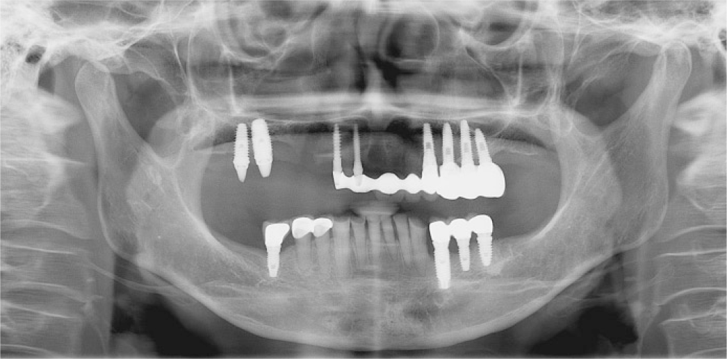
Fixed implant prosthesis and removable implant overdenture are the main treatment options for treating edentulous maxilla with implants. If clinicians select one of the treatment options without accurate diagnosis and evaluation, this may lead to unfavorable treatment result and one would not be able to guarantee successful long term prognosis. In this case, 69 year-old female presented with failed fixed implant prosthesis that was treated in private dental clinic. Since the patient did not want additional insertion of implants and considering factors such as oral hygiene maintenance, splinting effect, and esthetics, the patient was treated with removable implant bar type overdenture using pre-existing implants. The clinical results were satisfactory in the aspect of esthetics and masticatory function, oral hygiene maintenance. (J Korean Acad Prosthodont 2016;54:393-400)
REFERENCES
1.Byun JS., Cho LR., Kim DG., Huh YH., Park CJ. Various factors influencing on the satisfaction of complete denture wearers. J Dent Rehab Appl Sci. 2014. 30:53–63.

3.Cabello G., Gonza′lez DG., Fa′brega J. The edentulous maxillary arch: A novel approach to prosthetic rRehabilitation with dental implants, based upon the combination of optimum mechanical resources. Dent. 2014. 4:217–21.
4.Sadowsky SJ. Treatment considerations for maxillary implant overdentures: a systematic review. J Prosthet Dent. 2007. 97:340–8.

5.Zitzmann NU., Marinello CP. Treatment plan for restoring the edentulous maxilla with implant-supported restorations: removable overdenture versus fixed partial denture design. J Prosthet Dent. 1999. 82:188–96.

6.Goodacre CJ., Bernal G., Rungcharassaeng K., Kan JY. Clinical complications with implants and implant prostheses. J Prosthet Dent. 2003. 90:121–32.

7.Cavallaro JS Jr., Tarnow DP. Unsplinted implants retaining maxillary overdentures with partial palatal coverage: report of 5 consecutive cases. Int J Oral Maxillofac Implants. 2007. 22:808–14.
8.Schneider AL. Use of guide planes and implant supported bar overdentures: a case report. Implant Dent. 1998. 7:45–9.
9.Kang JK., Nam GH. Case report: Application of implant supported removable partial denture due to multiple dental implant loss of the fixed implant supported prosthesis. J Korean Acad Esthet Dent. 2014. 23:34–40.

10.Pasciuta M., Grossmann Y., Finger IM. A prosthetic solution to restoring the edentulous mandible with limited interarch space using an implant-tissue-supported overdenture: a clinical report. J Prosthet Dent. 2005. 93:116–20.

11.Murphy T. Compensatory mechanisms in facial height adjustment to functional tooth attrition. Aust Dent J. 1959. 4:312–23.

12.Dawson PE. Functional occlusion: from TMJ to smile design. St. Louis: Mosby;2007. p. 430–52.
Fig. 1.
Intraoral view at first visit. (A) Frontal view, (B, C) Occlusal view of maxilla and mandible.

Fig. 3.
(A) Removal of pre-existing fixed implant prosthesis, (B) Occlusal view of maxilla after prior fixed implant prosthesis removal, placement of healing abutment on maxillary first and second molar implantation site and residual abutment preparation.

Fig. 4.
Provisional denture delivery (A) Right lateral view, (B) Frontal view, (C) Left lateral view, (D) Composite resin filling of mandible anterior teeth.
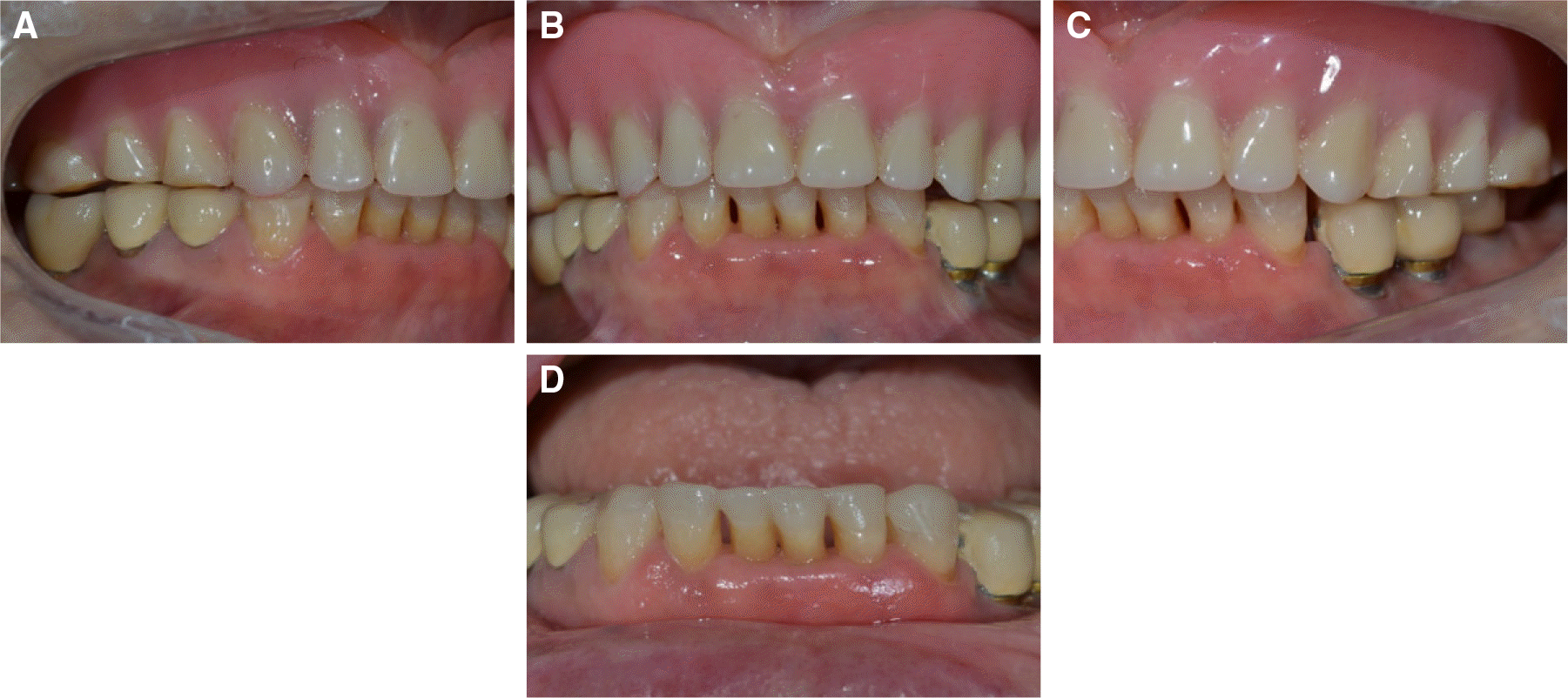
Fig. 7.
Wax denture try-in. (A) Occlusal view of maxilla, (B) Extraoral view, (C) Right lateral view, (D) Frontal view, (E) Left lateral view.
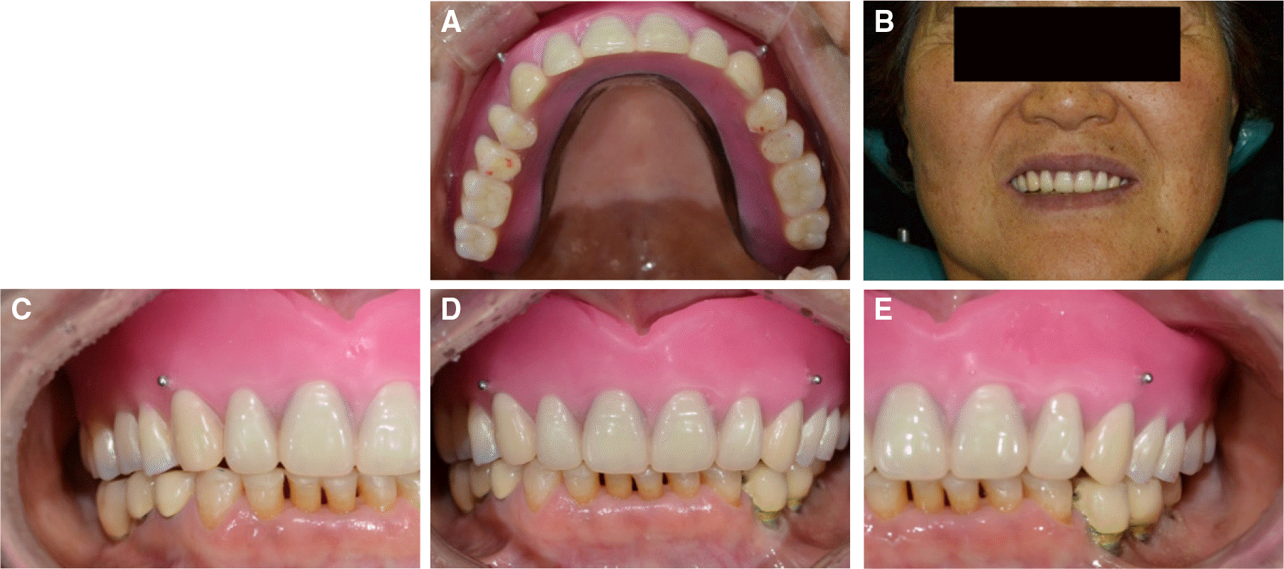
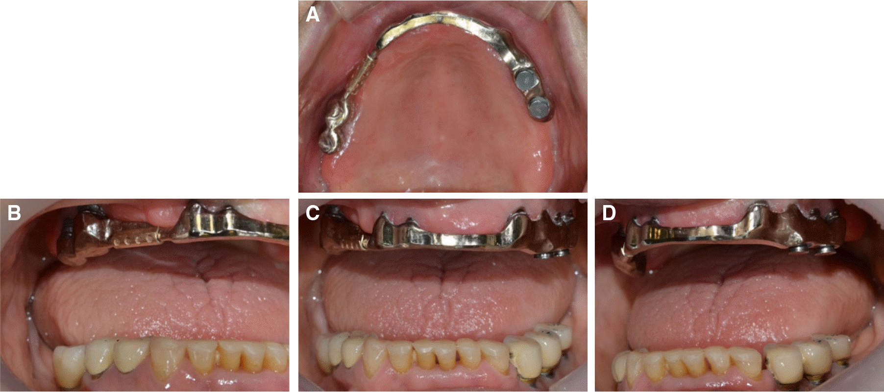
Fig. 8.
Cementation of bar. (A) Occlusal view of maxilla, (B) Right lateral view (C) Frontal view, (D) Left lateral view.


 PDF
PDF ePub
ePub Citation
 Print
Print





 XML Download