

 PDF
PDF ePub
ePub Citation
 Print
Print


INTRODUCTION
Natural esthetic appearance and longevity of the restoration combined with an efficient workflow in the laboratory and dental practice are major aims in restoring teeth or implants in prosthetic dentistry. With increasing capabilities and acceptance of computer-aided design and computer-aided manufacturing (CAD/CAM) especially for single restorations, new machinable esthetic materials have been introduced. While lithium disilicate and zirconia ceramics have been successfully clinically applied for many years,1234567 new classes of zirconia-reinforced lithium silicate ceramics, ceramic-network materials and, recently, lithium-disilicate-strengthened lithium aluminosilicate glass ceramics have been introduced. While most previously used ceramic materials required time-consuming working steps performed in the dental laboratory (milling, sintering/crystallization), chairside ceramics that are easy to mill in a fully crystallized state may represent an advantageous option. Some new classes of ceramics offer chairside fabrication without subsequent crystallization, while esthetics and mechanical properties may be comparable to lithium disilicate. With a complete chairside workflow, the clinical procedure of the surface finish gains importance. Chairside polishing may be an alternative to glazing in the laboratory.
The advantages of a digital workflow (intraoral scanning, CAD/CAM) in combination with choosing an appropriate dental material may not only be relevant for tooth restorations but also for implant-supported crowns. The success of chairside cemented implant restorations may be limited by gingival and peri-implant inflammation effects caused by residual cement.8 Labside bonding of implant crowns to a titanium base and leaving a screw hole for chairside fixation may resolve this problem, but the strength of the crown may be affected by the presence of the screw hole.910111213 For both the chairside and the labside procedures of implant-supported crown fabrication, an altered loading situation with increased masticatory forces by rigid implant bearing exists, and therefore a higher fatigue and fracture resistance may be required for implant-supported restorations.141516 Previous studies have shown that CAD/CAM materials may perform differently depending on their use as implant- or tooth-supported restorations.911
Clinical evidence for survival of new chairside CAD/CAM materials is very limited.1718 First in vitro results about performance and material characterization of recently introduced CAD/CAM materials (e.g. zirconia-reinforced lithium silicate ceramics, hybrid materials) are promising.1920212223 However, further in vitro investigations are necessary as advanced ceramic materials as lithium disilicate strengthened lithium aluminosilicate glass ceramics are constantly launched on the market.
Prior to routine clinical application, in vitro tests may facilitate a contemporary evaluation of new materials and restorations by combining reproducible laboratory conditions with basic requirements (occlusal loading, thermocycling) of the clinical situation.
The hypothesis of this study was that the in vitro performance and fracture resistance of lithium-disilicate-strengthened lithium aluminosilicate glass ceramic crowns
MATERIALS AND METHODS
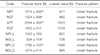
The study investigated molar crowns fabricated of a novel lithium-disilicate-strengthened lithium aluminosilicate glass ceramic (Group N, Li2O-Al2O3-SiO2, Nice, Straumann, Freiburg, Germany) in comparison to a well-known lithium disilicate glass ceramic (Group E, Li2Si2O5, IPS e.max CAD, Ivoclar Vivadent, Schaan, Liechtenstein) as a reference material. Further details on the materials used are given in Table 1. The lithium disilicate strengthened lithium aluminosilicate glass-ceramic was manufactured by a co-crystallization of lithium disilicate and lithium aluminosilicate upon controlled firing of a starting glass.
The molar crowns (n = 64; n = 8/group) were tested with two different surface modifications (P = polished, G = glazed) on human teeth (T) or implant-abutment analogues (I). For the implant groups, either a chairside (C) or labside (L, with screw channel) clinical procedure was simulated. The following eight groups were defined:
NPT: Nice polished crowns tested on human teeth.
NGT: Nice glazed crowns tested on human teeth.
EPT: E.max CAD polished crowns tested on human teeth (reference group).
EGT: E.max CAD glazed crowns tested on human teeth (reference group).
NPI_L: Nice polished crowns tested on implant abutments with labside procedure.
NGI_L: Nice glazed crowns tested on implant abutments with labside procedure.
NPI_C: Nice polished crowns tested on implant abutments with chairside procedure.
NGI_C: Nice glazed crowns tested on implant abutments with chairside procedure.
For the tooth groups (T), extracted caries-free human molars (mandibular right first molar, n = 32) were collected and stored in 0.5% chloramine solution for no longer than 4 weeks. The variability of human molars was respected by preselecting teeth with comparable size and shape and by randomly dividing the teeth to the subgroups. The teeth were prepared according to ceramic guidelines respecting a circular and occlusal anatomical reduction of 1.5 mm and a preparation angle of 4 degrees. A 1 mm deep cervical circular shoulder with rounded inner angles was prepared. All teeth were prepared by one person with identical preparation equipment. Standardized preparation was performed on basis of an original model, and the preparation design was controlled with a gauge.
The roots of the teeth were coated with a 1 mm polyether layer (Impregum, 3M, Seefeld, Germany) to simulate the human periodontium and the resilience of the teeth. Therefore, the roots of the teeth were dipped in wax, which was replaced by polyether in a subsequent fabrication process 242526 before the teeth were vertically fixed in sample holders (Palapress Vario, Kulzer, Hanau, Germany).
For the implant groups (I), implant-abutment analogues (n = 32; Straumann, Freiburg, Germany, titanium grade IV, implant diameter 4.1 mm, implant length 12 mm, abutment length 6 mm, 6°) were vertically positioned in resin blocks (Palapress Vario) in order to simulate a posterior implant situation replacing the mandibular right first molar.
All abutments and prepared teeth were digitalized (Cerec Omnicam, Sirona, Bensheim, Germany) and full-contour crowns of the lithium-disilicate-strengthened lithium aluminosilicate glass ceramic (group N, n = 48) and the lithium disilicate glass ceramic (reference group E, n = 16) were milled (Cerec, MCXL, Sirona, Bensheim, Germany). As the lithium-disilicate-strengthened lithium aluminosilicate glass ceramic blocks were milled in a fully crystallized state, only the lithium disilicate glass crowns were crystallized after milling.
Half of the specimens were polished (OptraFine, Ivoclar Vivadent, Schaan, Liechtenstein) or glazed (group N: Vita Akzent Plus, Vita Zahnfabrik, Bad Säckingen, Germany; group E: IPS e.max ceram glaze, Ivoclar Vivadent, Schaan, Liechtenstein) according to the manufacturers' instructions.
Crown dimensions on human teeth (groups T) were: circular and occlusal wall thickness 1.5 mm and cervical wall thickness 1 mm. All crowns were etched (5% hydrofluoric acid, 20 seconds) and silanized (silane coupling agent Monobond Plus, Ivoclar Vivadent, Schaan, Liechtenstein, 60 seconds). Before adhesive bonding (Variolink Esthetic DC, Ivoclar Vivadent, Schaan, Liechtenstein; Elipar Trilight, 3M, Seefeld, Germany, 3 × 60 seconds), the prepared teeth were treated with the bonding system Syntac classic (Syntac Primer/Syntac Adhesive/Heliobond, Ivoclar Vivadent, Schaan, Liechtenstein) according to the manufacturer's instructions.
For the implant groups (I), all abutments were sandblasted (110 µm Al2O3, 1.5 bar) and the crowns were conditioned analogous to the tooth groups. Identical crowns were fabricated for two situations: a) chairside procedure: the crowns were directly bonded onto the implant-abutment analogue and the excess luting material was removed; b) labside procedure: a screw channel was manually drilled into the central fossa of the crown with a diamond bur (red/fine, diameter: 1.5 mm, water cooling). The crowns were bonded onto the implant-abutment analogue, the excess luting material was removed, and the screw channel was restored with composite (Filtek Supreme; 3M, Seefeld, Germany; Elipar Trilight, 40 seconds).
A combined thermal cycling and mechanical loading (TCML: 3000 × 5℃/ 3000 × 55℃, 2 minutes each cycle, H2O distilled; 1.2 × 10⁶ cycles à 50 N, 1.6 Hz) was performed in a chewing simulator (eGo Kältesysteme, Regensburg, Germany). Extracted human molars were used as antagonists in three-point-contact for crowns on human teeth. Steatite balls (diameter: 12 mm, CeramTec, Plochingen, Germany) were used to standardize antagonists in three-point-contact on identical implant crowns. Parameters were chosen on data of zirconia and ceramic restorations simulating a maximum of five years of oral service.2728
During TCML all crowns were controlled daily for failures and failed crowns were excluded from further simulation and testing. After TCML all crowns were investigated in detail for failure analysis with optical microscopy (light microscope SV8, Olympus, Hamburg, Germany, magnification ×10) and scanning electron microscopy (SEM, Quanta FEG 400, FEI, Hillsboro, Oregon, USA, low vacuum, 10 keV, magnification ×20–100). Wear facets of the tooth groups were characterized. All crowns that survived TCML were loaded to fracture (universal testing machine Zwick 1446, Zwick, Ulm, Germany). The force was applied on the centre of the crowns using a steel ball (diameter: 12 mm, velocity: 1 mm/min) with a 1 mm tin foil (Dentaurum, Ispringen, Germany) that was inserted between crown and ball to prevent force peaks. The failure determination was set to 10% decrease of the maximum force or acoustic signal failure (crack). Power calculation (G*Power 3.1.3, HHU Düsseldorf, Germany) provided an estimated power of > 90% using eight specimens per group. Force distribution was controlled with the Kolmogorow-Smirnov-test. Mean values and standard deviations (SD) were calculated and analysed by one-way analysis of variance (ANOVA) and the Bonferroni multiple comparison test for post hoc analysis (SPSS 23, IBM, Armonk, NY, USA, α = .05).
RESULTS
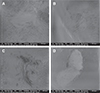
No crowns failed during TCML. All crowns showed small wear traces in their individual contact points. No chipping or fractures were found, but small cone cracking was partly observed both for polished and glazed crowns. For glazed crowns, worn inhomogeneous glaze material was found in the marginal areas of the wear facets. No strong differences were found between the lithium-disilicate-strengthened lithium aluminosilicate glass ceramic and the reference material. Individual surface inhomogeneities with a diameter smaller than 1 mm were found for all materials. Enamel antagonists showed scratches and inhomogeneities, with some cracks and fractures. Exemplary SEM pictures of worn ceramic surfaces are given in Fig. 1.
Fracture forces in the tooth groups (T) were 1214 ± 293 N (NPT) and 2044 ± 302 N (EPT) for polished crowns, and 1324 ± 498 N (NGT) and 1550 ± 317 N (EGT) for glazed crowns. Statistical comparison revealed significant (P = .000, ANOVA) differences between the materials. Individual differences (Bonferroni) were found between EPT and NPT (P = .001) and NGT (P = .003). For both materials, no significantly (P > .066) different fracture results were found between glazed or polished crowns. Polished reference crowns showed significantly (P = .001) higher fracture results.
Fracture values for the crowns on implants varied between 934 ± 154 N (NGI_L) and 1782 ± 153 N (NPI_C). Chairside crowns for both types (glazed and polished) provided higher fracture results than the respective labside crowns. For both clinical procedures (L, C) lower fracture values were found for glazed crowns, but the results were not statistically different (P > .152) (Table 2).
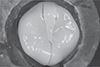
Failures were characterized by fracture of the crown in all cases, partly combined with a complete debonding of the crown. An exemplary light microscope picture is given in Fig. 2.
DISCUSSION
Lithium-disilicate-strengthened lithium aluminosilicate glass ceramic crowns showed similar performance during in vitro TCML, but lower fracture resistance than the lithium disilicate reference crowns, rejecting the first part of the hypothesis with regard to fracture resistance. Polishing or glazing did not significantly influence the results. Therefore, the second part of the hypothesis was rejected. The third part of the hypothesis was confirmed with restrictions since only chairside implant-supported crowns partly showed higher fracture resistance in comparison to labside implant- and tooth-supported crowns.
A CAD/CAM lithium disilicate ceramic has been chosen as reference material in this study, as it has been proven to be suitable for single tooth restorations both in vivo1242930 and in vitro19313233 for many years. Fracture forces of these reference crowns are comparable to previous results, ranging between about 1000 N to 2500 N,193134 depending on the testing conditions. The present results showed that the lithium-disilicate-strengthened lithium aluminosilicate glass ceramic crowns had comparable resistance against fatigue as the reference crowns, but lower fracture resistance in the respective tooth groups. Nevertheless, as the fracture values exceeded maximum chewing forces, which are reported to be up to 900 N,35 all groups of molar crowns are able to withstand occlusal forces applied in the posterior region. Differences in fracture resistance between the two materials may be explained by the material composition and the mechanical properties like fracture toughness and modulus of elasticity (Table 1). The chairside machining of fully crystallized lithium disilicate strengthened lithium aluminosilicate glass ceramic crowns may also influence fatigue and fracture resistance. A previous study about the machinability of CAD/CAM materials has found a higher susceptibility to edge chipping induced by milling for ceramic-based materials compared to polymer materials.36 Another study investigating implant-supported zirconia-reinforced lithium silicate crowns reported significantly lower fracture resistance if milled in a fully crystallized condition compared to milled in a presintered condition.37 These results may not be applied to other ceramic materials, but it may be assumed that any surface damage induced by hard milling can reduce fracture resistance. However, SEM evaluation did not indicate any superficial defects. The particular crystal morphology (co-crystallization of lithium disilicate and aluminosilicate) embedded in a glassy matrix may be responsible for favorable post-firing milling properties of chairside ceramics like Nice. Nevertheless, further investigations on the machinability of fully crystallized chairside ceramics and potential detrimental effects are recommended.
The surface state of ceramics gains high importance for chairside fabrication without subsequent firing and surface finish (glazing) in the dental laboratory. Smooth and glossy surfaces might be important not only for esthetic reasons but also longevity of the restoration. Any surface irregularities, roughness, or superficial defects induced by milling may act as stress concentration sites, increase wear, and cause potential origins of cracks that might propagate by water-assisted subcritical crack growth2338 during clinical service time. Due to repeated load cycles, fatigue and wear lead to the formation and proceeding of subsurface cracks39 that may finally result in chipping failures or fracture. Because of their microstructure, complex interactions between glass and crystalline phases, and minor mechanical properties, glass ceramics are obviously more prone to microploughing and microcracking than high-strength ceramics as zirconia. In the present study, wear facets with small cone cracking were observed both for polished and glazed crowns irrespective of the glass ceramic material. Cone cracks are described as defects that appear when the antagonist slides across the ceramic surface.4041 These cracks might not necessarily result in fatigue failures of the crown, which is in accordance with the present observations as all crowns survived TCML without failures. However, to keep wear and mechanical degradation of the material as low as possible, surface finish after milling is mandatory. The designated workflow for chairside ceramics like Nice includes polishing after milling. As an alternative, ceramics may still be glazed in the dental laboratory. Glaze firing is supposed to be advantageous in terms of healing cracks that might have been induced by hard milling.42 Glaze may also seal deep grooves or imperfections that might not be completely reached by polishing. From roughness aspects, polishing has proven to be effective in reducing high surface roughness of glass ceramics to similar values as measured for glaze layers.4143 Therefore, the use of chairside polishing kits is a reasonable and time-saving alternative in clinical practice and might justify the description of Nice as genuine chairside ceramic. Present results did not indicate lower fracture resistance of polished Nice crowns compared to glazed ones. Quite the contrary, for polished implant-supported crowns, fracture forces were even higher than for their respective glazed crowns, even though not significant. Irrespective of polished or glazed surfaces, ongoing wear in contact areas results in removal of glass/glaze and exposure of crystalline phases (lithium disilicate, aluminosilicate) that may superpose the original surface state and enforce wear of material and antagonist. Therefore, it is recommended to control and if necessary to repolish roughened glass ceramic surfaces at annual recall sessions at dental practice.
Different fracture values for the two clinical procedures (chairside/labside) for implant restorations were found, showing higher values for the chairside crowns. However, this difference was only significant for the polished chairside crown (NPI_C). A weakening effect of the screw channel in labside crowns therefore existed, but it did not critically affect fracture resistance. This is in accordance with previous studies that did not report any significant influence of the screw channel on the failure loads of most ceramic materials (zirconia, lithium disilicate, zirconia-reinforced lithium silicate).91144 The presence of a screw channel may therefore be a critical factor for the success of implant-supported crowns only if materials with lower strength, for example composites, are applied.911 The focus for brittle materials has to be put on the drilling process; if done carefully, no pre-damaging of the crystalline material structure might occur and no negative effects on strength might be expected for glass ceramics. Of course it has to be considered that the present study design eliminated any potential failures related to the screw in the implant-abutment connection, as an one-piece implant-abutment analogue without screwing joint was used. While it has been confirmed earlier that screwed connections are critical parts in implant-supported restorations,4546 the present study focused on investigating a new chairside CAD/CAM material in its application as crown with regard to clinical procedures. With fracture values up to about 1700 N, Nice implant-supported crowns showed comparable fracture resistance as previously reported1137 for zirconia-reinforced lithium silicate implant-supported crowns that were also milled in a fully crystallized state. Nevertheless, fracture resistance of both chairside CAD/CAM materials is still inferior to lithium disilicate in implant-supported restorations.1137
Although crowns of the tooth groups were not absolutely identical in shape and geometry due to inevitable individual differences of the human teeth, similar fracture values were found between tooth and implant groups of the lithium-disilicate-strengthened lithium aluminosilicate glass ceramic crowns. Only one implant group (NPI_C) provided significantly higher fracture values. Influences of the different geometry resulting from smaller occlusal support or higher crown thickness in the case of the implanted-supported crowns were not apparent, but may have affected the results. Assuming an altered loading situation with increased chewing forces by rigid implant bearing, a higher stability of the materials is generally required for implant restorations. 14154748 Against these expectations, the modulus of elasticity of the abutment (tooth, implant) did not negatively influence the results. In contrary, group NPI_C even showed significantly higher fracture resistance than NPT. Further tests are recommended for investigating these influences.
Failure analysis after fracture testing indicated contact-induced cracks resulting in complete crown fractures in all groups. It has to be considered that fracture testing does not reflect any clinically observable failure modes and the found fracture forces exceeded physiological chewing forces and force peaks in vivo. Crown fractures were partly combined with debonding. Considering that none of the crowns failed during TCML, surface activation/treatment of the crown materials is considered good. As all crowns were bonded adhesively in this study, the influence of the luting material has to be discussed. The mechanical strength of the crowns may have enabled conventional cementation. Shock absorbing capacity might have been improved although a previous study did not show any significant difference in fracture strength among different luting agents used with different types of ceramics.37
Further aspects of the study design have to be evaluated critically. The use of steatite spheres and human molars as antagonists during TCML might be discussed controversially. Since dental ceramics ideally achieve wear behaviour similar to that of enamel, their wear properties should be characterized in relation to those of human tooth antagonists. Therefore, characterization of wear facets was only applied for the tooth groups. By contrast, steatite spheres guaranteed a standardized antagonistic situation but might have caused different wear and damage. Nevertheless, steatite spheres have proven their suitability for testing implant-supported restorations in previous studies.114649 While fatigue and wear phenomena of the antagonist are wishful during TCML, fracture results may not be influenced by damage or deformation of the antagonist. Therefore, steel spheres of identical diameter were chosen for fracture testing.
The artificial periodontal mobility only allows an approximation of the clinical situation, and the design did not simulate feedback and control of the applied loading forces as under clinical conditions.50 Considering that the contact situation during TCML is influenced by wear effects and flexible bearing in the tooth groups, a shift of contact points may occur. However, no different failure patterns, wear traces or fatigue effects were found between the groups in the failure analysis.
CONCLUSION
Survival of lithium-disilicate-strengthened lithium aluminosilicate glass ceramic crowns (Nice) during TCML was comparable to lithium disilicate crowns. Fracture values of all restorations were in a range where clinical application seems not restricted. Fracture resistance was partly influenced by the tooth or implant situation and the applied clinical procedure, showing higher values for the chairside situation without screw channel.
The type of surface finish (polishing versus glazing) did not significantly influence the results; therefore polishing seems to be a time-saving and promising alternative to enable a complete chairside workflow of the new CAD/CAM ceramic.

 XML Download