

 PDF
PDF ePub
ePub Citation
 Print
Print


Dear Editor,
Propriospinal myoclonus (PSM) is characterized by myoclonic jerks arising from thoracic or abdominal muscles and spreading to caudal and rostral segments.12 The PSM phenotypes have been classified into symptomatic, idiopathic, functional, and at sleep onset.1 PSM at sleep onset is characterized by jerks at the transition from wakefulness to sleep.13 Here we report a patient with the unusual presentation of PSM observed during stable sleep.
A 25-year-old woman with involuntary jerks in her trunk that had impeded her sleep for 1 year visited our sleep clinic. The jerky movements started 4 years previously when she was falling asleep after sleep deprivation. The involuntary jerks continued for a few days and then disappeared spontaneously. However, her movements restarted 3 years later when she started to work at night. The jerky movements occurred once or twice per night within 1 hour after sleep onset. The jerky movements were not associated with dreaming. Frequent interruptions of stable sleep by jerky movements, which resulted in nonrefreshing sleep and daytime sleepiness, continued after she stopped working at night. She had no past medical or family history including of sleep disorders such as circadian rhythm disorders, parasomnia, periodic limb movement disorder, or restless legs syndrome.
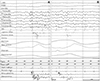
The findings of a neurological examination were normal, and the movements were not provoked by lying down or by sensory stimuli. Her laboratory parameters were normal. Whole-spine MRI revealed incidental 0.9-cm-long focal hydromyelia at C6–7 in the spinal cord without enhancement. Simultaneous overnight polysomnography with video-electroencephalography revealed eight episodes of jerky truncal flexions during REM sleep (Fig. 1) (Supplementary Video 1 in the online-only Data Supplement), and once during stage-N1 sleep at 15 minutes after sleep onset (Supplementary Video 2 in the online-only Data Supplement). EMG bursts initially started in the abdominal muscles and propagated to the muscles of the lower extremities (Fig. 1). The duration of the EMG bursts ranged from 500 to 2,000 ms, and the latency of intermuscular bursts ranged from 50 to 150 ms. During these events, no interictal or ictal epileptiform discharges were observed in simultaneous electroencephalography recordings using the international 10–20 system. Clonazepam was applied at 0.5 mg once daily before bedtime to control her nocturnal movements. The frequency of involuntary movements as estimated by herself decreased significantly from once or twice daily to once a week after clonazepam treatment.
The etiology of PSM is idiopathic in approximately 80% cases, and a clear correspondence between the level of the spinal symptom generator and the level of the spinal lesion is very rare even among PSM cases involving spinal lesions.12 Although incidental focal hydromyelia was found at C6–7 in our case, the anatomical location of the lesion was unlikely to be correlated with the jerks starting in abdominal muscles and it could not explain the 3-year symptom-free period.
According to third edition of the International Classification of Sleep Disorders diagnostic criteria for PSM at sleep onset, PSM appears at the sleep-wake transition and disappears upon mental activation, and with a stable sleep onset.4 However, all of the PSM events in our case occurred during stable sleep, including REM sleep and non-REM sleep, and not at sleep onset or during the sleep-wake transition. Typical EMG findings of PSM include simultaneous and progressive propagation from a thoracic spinal source at a slow rate of 3–15 m/s with intermuscle latencies of 16–200 ms and burst durations of 100–1,000 ms, which can be longer.123 The initial involvement of the thoracic muscles and the propagating pattern of EMG bursts were compatible with PSM in our case despite the small number of EMG electrodes.123
We have presented an interesting case of PSM occurring during stable sleep independently of the sleep stages. The pathomechanism of jerks during stable sleep is unclear, and further cases need to be followed to determine whether the current diagnostic criteria for PSM at sleep onset need to be revised.

 XML Download