

 PDF
PDF ePub
ePub Citation
 Print
Print


INTRODUCTION
External-beam radiation therapy (EBRT) with intracavitary high-dose-rate (HDR) brachytherapy is the standard treatment modality for advanced cervical cancer. Although long-term survival rates in patients with cervical cancer treated with definitive radiotherapy (RT) have improved considerably in recent years, the incidence of late gastrointestinal complications has also increased. The rectum is a major organ at risk during treatment planning because of its close proximity to the pelvic organs and relative immobility [1]; radiation proctitis has been reported in 5% to 11% of patients treated for gynecological cancer [2]. Sigmoid complications such as obstructive sigmoiditis and perforation are rarely reported, although some previous studies have estimated they occur in 0.6-6.4% of cases [3,4]. Herein we report two uncommon cases of radiation sigmoiditis mimicking sigmoid colon cancer after EBRT with intracavitary HDR brachytherapy for uterine cervical cancer with dosimetric consideration. We also discuss the importance of radiation dose to the sigmoid colon in three-dimensional image-based brachytherapy.
CASE REPORTS
1. Case 1
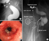
A 79-year-old woman received definitive RT for International Federation of Gynecology and Obstetrics (FIGO) stage IIIB cervical cancer. EBRT was performed at a total dose of 50.4 Gy in 1.8 Gy fractions for five days each week using a 15-MV photon. Intracavitary HDR brachytherapy (microSelectron-HDR) of 24 Gy in six fractions was conducted twice each week using a tandem and colpostat. Following the completion of RT, the cervical cancer completely disappeared. After one year, the patient returned to the hospital complaining of intermittent rectal bleeding. A barium study revealed a 3-cm-long severe narrowing of the sigmoid wall, which was suspicious for sigmoid colon cancer (Fig. 1A). The patient was referred to our hospital. Sigmoidoscopy revealed luminal narrowing with a friable hyperemic nodular mucosal change 15 cm from the anal verge (Fig. 1B). Biopsies were performed, and the results showed focal chronic active inflammation. The patient was diagnosed as having radiation sigmoiditis and was managed conservatively.
2. Case 2
A 58-year-old woman received definitive RT with concurrent chemotherapy for FIGO stage IIIA cervical cancer. EBRT was performed at a total dose of 50.4 Gy in 1.8 Gy fractions for five days each week using a 15-MV photon. Intracavitary HDR brachytherapy (microSelectron-HDR) of 24 Gy in six fractions was conducted twice each week using a tandem and colpostat. Six cycles of weekly cisplatin (30 mg/m2) were given during RT. The patient has been followed with no evidence of recurrence. After five years, the patient visited our emergency department with complaints of poor oral intake, lower abdominal pain and no stool passage over ten days. Sigmoidoscopy revealed an approximately 5-cm-long section of diffuse luminal narrowing in the sigmoid colon and hyperemic mucosal nodularities with mucosal edema (Fig. 2A). The endoscopist suspected primary sigmoid colon cancer or local invasion of recurrent cervical cancer. Pathologic examination reported diffuse active inflammation with inflamed granulation tissue. A computed tomography (CT) scan also revealed thickening of the wall of the rectosigmoid colon probably caused by radiation or inflammation; however no evidence of any cancer recurrence was found (Fig. 2B). She has been managed conservatively with laxatives and has been clinically followed with improved symptoms.
3. Dosimetric analysis
We were only able to restore the old simulation film in case 1. The simulation film of the HDR brachytherapy is shown in Fig. 1C. The radiation dose was prescribed at point A in 4 Gy fractions. We estimated an irradiated dose of brachytherapy of 518.7 to 642.3 cGy at the sigmoid colon point, which was 29.5% to 60.5% higher than prescribed dose at point A per fraction.
DISCUSSION
Acute and late GI complications are a major concern during and after RT. Radiation sigmoiditis is not well-known compared to other GI complications such as proctitis and enteritis because it has been rarely reported. Galland and Spencer [5]. reported 23 cases of sigmoid complications among 80 cases of radiation colitis induced by pelvic RT. Ramirez et al. [4] reported that a total of 35 patients developed sigmoid perforation among over 5,000 patients who were treated with RT for stage IB-IIIB cervical cancer.
In this report, we described two cases of late sigmoid complication after pelvic RT. The sigmoiditis occurred one year after RT completion for one patient and five years after RT completion for the other patient. Both cases showed obstructive symptoms, which make clinicians suspicious of sigmoid colon cancer or recurrent cancer. During the follow-up of cancer patients who received RT previously, clinicians need to keep the possibility of RT complications in mind.
Holloway et al. [6] reported that the sigmoid-to-tandem distance is significantly related to sigmoid dose using 3D imaging techniques. Concordantly, the simulation films of case 1 showed close proximity between the sigmoid colon and the tandem, which suggests the possibility of a high sigmoid dose during brachytherapy (Fig. 1C). The estimated dose at the sigmoid colon point during brachytherapy was 518.7 to 642.3 cGy, which was 29.5% to 60.5% higher than prescribed dose at point A (400 cGy) per fraction.
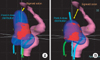
The International Commission on Radiation Units (ICRU) 38 did not describe this hot spot in the sigmoid colon. The rectal and bladder reference dose points are only standard for reporting normal tissue doses with brachytherapy. Indeed, these points are based on plain films and two-dimensional (2D) brachytherapy planning. As EBRT is moving from conventional to conformal planning, the incorporation of three-dimensional (3D) sectional imaging such as CT and MRI into brachytherapy planning is under active investigation [6,7]. Recently, there has been a proposal to establish image-guided intracavitary brachytherapy guidelines for cervical carcinoma. In the Group European de Curietherapies and European Society for Therapeutic Radiology and Oncology (GEC-ESTRO) recommendations, the rectum and bladder, as well as the sigmoid colon, are highlighted as organs at risk [8]. Many studies have reported a sigmoid dose in 3D image-based planning for the treatment of cervical carcinoma with HDR brachytherapy [6,7]. Kim et al. [7] reported that the sigmoid dose was higher than the rectal dose in 8 of 13 (61%) patients and its volume was discontinuously located as multiple "hot spots" in 9 of 13 (69%) patients on dosimetric analysis of 3D image-based planning of intracavitary brachytherapy for cervical cancer. Currently at our institution, 3D image-based brachytherapy is implemented to decrease the radiation dose to organs at risk, including the sigmoid colon (Fig. 3).
3D image-based brachytherapy planning can be helpful, especially for patients who have risk factors. Patients who have a history of abdominal surgery, pelvic inflammatory disease, or diverticulosis are at a greater risk of developing radiation-induced GI complications because such conditions may create adhesions to the uterus, which prevent the loops of the bowel from moving during RT [9].
In conclusion, radiation sigmoiditis is characterized by obstructive symptoms and its radiological findings can mimic sigmoid colon cancer. Radiation oncologists should acknowledge the sigmoid colon as well as rectum and bladder as organs at risk. 3D image-based brachytherapy could be an optimal modality to reduce the radiation-toxicity associated with treatment for uterine cervical cancer.

 XML Download