

 PDF
PDF ePub
ePub Citation
 Print
Print


Clear-cell meningioma is a rare variant of meningioma that differs from the ordinary type in that it affects younger patients, arises more often in spinal canal or cerebellopontine locations, and shows a higher recurrence rate (1). Cytologically, it has whorled, syncytial architecture and spindled-to-polygonal, bland-appearing nuclei. Immunohistochemistry shows that tumor cells are positive for vimentin and epithelial membrane antigen (2). Because its biological behavior is frequently aggressive in spite of its benign histology, clear-cell meningioma should be differentiated from other meningiomas of the central nervous system. We report two cases of clear-cell meningioma, one arising from the thoracolumbar spine in a young child and the other from the cerebellopontine angle in a young adult.
CASE REPORTS
Case 1
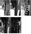
A 14-month-old girl presented with a 3-month history of bilateral leg weakness and poor micturition. MR imaging of the thoracolumbar spine demonstrated a 4cm-sized, well-demarcated, ovoid mass located in the intradural extramedullary spinal canal at the level of T12 L2. Both T1- and T2-weighted images showed that the mass was isointense to the spinal cord and contrast enhancement was strong and homogeneous (Figs. 1A-C). Intradural exposure through lumbar laminectomy of T11 L2 disclosed a yellowishpink, well-encapsulated, partly nodular elliptical mass adherent to the dura on the right side. Gross total resection was performed.
Microscopically, the tumor was characterized by sheets of polygonal cells with moderately abundant clear cytoplasm and round-to-oval, bland nuclei. The cytoplasm was heavily laden with granular periodic acid Schiff-positive material. Assessment of cell proliferation with Ki-67 labeling showed an index of 3%, and immunohistochemical studies were positive for vimentin and epithelial membrane antigen. On the basis of these histologic findings, the differential diagnosis was clear-cell meningioma or metastatic clear-cell tumor. Other imaging work-ups including ultrasonography of the abdomen and pelvis and RI bone scanning provided no new information, and the mass was diagnosed as clear-cell meningioma.
Eight months after initial sugery, the patient showed sudden general irritability, and T2-weighted images of the spine revealed a recurrent intradural mass at exactly the same site (Fig. 1D). A second operation disclosed the presence of a fungating intradural mass which caused sinistrad displacement of the cauda equina and conus. The histologic findings were the same as for the previously resected tumor.
Seven months after the second operation, the patient experienced voiding difficulty and weakness in both legs. MR imaging again showed a recurrent, well-enhanced, intradural mass at the site of previous surgery (Fig. 1E). A third operation revealed the presence of a dura-based mass encompassing adjacent nerve roots. Following its gross total removal, its histologic composition was found to be the same as those previously resected, and radiation therapy was subsequently recommended.
Case 2
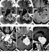
A 17-year-old girl presented with chronic headache and hearing loss in the left ear. CT scans showed a huge, irregular, mixed solid and cystic mass in the left cerebellopontine angle. The cystic portion of the mass was expansile and occupied its anterior aspect. The clivus was eroded by the mass, the solid portion of which was seen as slightly hyperdense on precontrast CT scans and contained multiple, small, irregular areas of low attenuation that suggested cystic change or necrosis (Fig. 2A). Contrast-enhanced axial CT scanning revealed mild, homogeneous enhancement of the solid portion of the mass (Fig. 2B), and MR imaging indicated that a large, multilobulated, mixed solid and cystic mass with a dorsal exophytic component was present in the upper medulla. The mass extended superiorly to the pons, and the brainstem was displaced to the right and posterosuperiorly. The solid portion of the mass showed an isointense signal, with a central area of high intensity, and was strongly and homogeneously enhanced after gadolinium injection. There was associated mild hydrocephalus (Figs. 2C-F).
Midline suboccipital craniotomy revealed a well-demarcated, lobulated mass, enveloped entirely by the leptomeninges and with no evidence of attachment to the dura mater or adjacent neural tissue. Because of its location, only partial resection was performed.
Histologically, the tumor was found to be moderately cellular and composed of sheets of polygonal cells. The cytoplasm of these was heavily laden with granular periodic acid Schiff-positive and diastase-sensitive material representing glycogen. Immunohistochemical studies were positive for vimentin and epithelial membrane antigen, and Ki-67 labelling showed an index of 1%. The final diagnosis was clear-cell meningioma.
DISCUSSION
According to the World Health Organization (WHO) classification of tumors of the central nervous system, clear-cell meningioma is a benign variant of meningioma (3). It is rare, and since it may recur, spread locally, and even metastasize, is clinically aggressive despite its bland histologic appearance (4).
The clinical characteristics of this tumor are reported to be somewhat different from those of ordinary meningiomas. While these are likely to occur during the fifth and sixth decades, with female predominance, clear-cell meningioma occurs at any age and without gender predilection (5). The average age at the time of surgery is reported to be less in clear-cell meningioma cases than in those of ordinary meningioma (2): in previously studied patients, a mean age of 29.8 years (range, 22 months -67 years) was reported (6). Our first patient, aged 14months at presentation, is the youngest so far mentioned in the literature, and our second is younger than average.
As seen in our Case 1, clear-cell meningioma is prone to recur. According to Jellinger and Slowik (7), the recurrence rates in cases ordinary spinal and intracranial meningioma are 4.8% and 14.2%, respectively; in contrast, the recurrence rates in spinal and intracranial clear-cell meningioma are 80% and 46%, respectively, with an overall recurrence rate of 60.9% (6). The most likely reason for multiple recurrences is that the extensive infiltrative growth pattern of the tumor hinders complete microscopic surgical resection. Cytologic examination reveals a sheet-like proliferation of polygonal cells, with cytoplasm which at hematoxylin staining is clear due to glycogen accumulation (5). Subtle whorl formation is reported to be a diagnostic clue. Since cellular anaplasia was not present, and during the first operation and all subsequent recurrences the growth fraction was low, histologic parameters were not predictive of recurrence (8).
A review of previous radiological reports shows that the features of most primary clear-cell meningiomas were not different from those of ordinary meningiomas (6). The signal intensity of a spinal meningioma is usually similar to that of the spinal cord at both T1- and T2-weighted imaging, and after the injection of gadolinium, enhancement is usually intense and homogeneous (9). In our Case 2, the mass was solid and had a prominent cystic portion, a composition somewhat different from that of the tumors described in previous reports. To our knowledge, a case in which a clear-cell meningioma has a solid, prominent cystic component has not been reported. In the solid portion, however, the signal was iso-intense to gray matter on both T1- and T2-weighted images, a circumstance common to both ordinary and clear-cell meningiomas.
In summary, although clear-cell meningioma is a rare subtype, it should be considered in the differential diagnosis of a central nervous system mass with neuroimaging findings of meningioma, especially in young patients. The tumor is characterized by its aggressive nature and high rates of recurrence.
 XML Download