

 PDF
PDF ePub
ePub Citation
 Print
Print


Abstract
Purpose
To understand the usefulness of the lumbar MRI studies to establish therapeutic plans for cauda equina syndrome (CES) including the management of rectal and bladder dysfunction symptoms.
Materials and Methods
We retrospectively reviewed the lumbar MRI studies of 10 patients with CES. Their diagnoses included four adhesive arachnoiditis of cauda equina (CE), three conus medullaris atrophies, three spinal canal stenoses, one tuberculous leptomeningitis, one metastatic tumor on the sacral canal, and one dural arteriovenous fistula with venous congestion of the conus medullaris.
Results
In 6 of the 10 total cases the symptoms of rectal and bladder dysfunction were resolved by decompression laminectomies (n=2), irradiation (n=1), glue embolization (n=1), anticholine and steroid infusion (n=1), and anti-tuberculous medication (n=1) within at least 5 days. The 4 other cases were settled by lumboperitoneal shunting and neural stem cell implants.
Figures and Tables
Fig. 1
66-year-old male with metastatic tumor on the 2nd sacral spine complained of cauda equina syndrome. Sagittal contrast enhanced T1 weighted MR image reveals bony destruction of the 2nd sacral spine body (arrow).
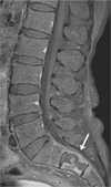
Fig. 2
31-year-old male with dural arteriovenous fistula at the level of the L2 spine. Sagittal contrast enhanced T1-weighted MR image reveals multifocal enhancing high signal area in the conus medullaris (white arrow) due to venous congestion and multifocal dark signal void area in the cauda equina (black arrow) due to rapid blood flow of arteriovenous fibula.
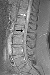
Fig. 3
59-year-old male confirmed to tuberculous leptomeningitis with caudal equina syndrome including acontractile neurogenic bladder, constipation, and right lower leg pain. Sagittal (A) and axial (B) contrast enhanced T1-weighted MR images reveals ringlike peripheral enhancement lesions (arrows) in the conus medullaris.
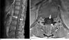
Fig. 4
63-year-old female with segmental cord atrophy at the level above the conus medullaris (CM). Sagittal T2-weighted MR image reveals abnormal slender portion (white arrows) at the cord above the CM consistent with cord atrophy required neural stem cell implant therapy.


References
1. Fraser S, Roberts L, Murphy E. Cauda equina syndrome: a literature review of its definition and clinical presentation. Arch Phys Med Rehabil. 2009; 90:1964–1968.
2. Ba B, Wu H, Jia LS, Yuan W, Shi GD, Shi JG. Cauda equina syndrome: a review of clinical progress. Chin Med J. 2009; 122:1214–1222.
3. Kerslake RW, Mitchell LA, Worthington BS. Case report: CT and MRI of the cauda equina syndrome in ankylosing spondylitis. Clin Radiol. 1992; 45:134–136.
4. Coscia M, Leipzig T, Cooper D. Acute cauda equina syndrome: diagnostic advantage of MRI. Spine. 1994; 19:475–478.
5. Bell DA, Collie D, Statham PF. Cauda equina syndrome: what is the correlation between clinical assessment and MRI scanning? Br J Neurosurg. 2007; 21:201–203.
6. Tait MJ, Chelvarajah R, Garvan N, Bavetta S. Spontaneous hemorrhage of a spinal ependymoma: a rare cause of acute cauda equina syndrome. Spine. 2004; 29:E502–E505.
7. Kostuik JP, Harrington I, Alexander D, Rand W, Evans D. Cauda equina and lumbar disc herniation. J Bone Joint Surg Am. 1986; 68:386–391.
8. Gleave JR, Macfarlane R. Cauda equina syndrome: what is the relationship between timing of surgery and outcome? Br J Neurosurg. 2002; 16:325–328.
9. Mangialardi R, Mastorillo G, Minoia L, Garofalo R, Conserva F, Solarino GB. Lumbar disc herniation and cauda equina syndrome: considerations on a pathology with different clinical manifestations. Chir Organi Mov. 2002; 87:35–42.
10. Storm PB, Chou D, Tamargo RJ. Lumbar spinal stenosis, cauda equina, and multiple lumbosacral radiculopathies. Phys Med Rehabil Clin North Am. 2002; 13:713–733.
11. Agur AMR, Lee MJ. Grant's atlas of anatomy. 9th Ed. Philadelphia: Williams & Wilkins;1991. p. 246–254.
12. Olcay L, Aribas BK, Gokce M. A patient with acute myeloblastic leukemia who presented with conus medullaris syndrome and reviewth conusliterature. J Pediatr Hematol Oncol. 2009; 31:440–447.
13. Lamer AJ, Pall HS, Hockley AD. Arrested progression of cauda equina syndrome of ankylosing spondylitis after lumboperitoneal shunting. J Neurol Neurosurg Psychiatry. 1966; 61:115–116.
14. Spector LR, Madigan L, Rhyne A, Darden B, Kim D. Cauda equina syndrome. J Am Acad Orthop Surg. 2008; 16:471–479.
15. Lee JW, Myung JS, Park KW, Teom JS, Kim KJ, Kim HJ, et al. Fluoroscopically guided caudal epidural steroid injection for management of degenerative lumbar spinal stenosis: short-term and long-term results. Skeletal Radiol. 2010; 39:691–699.
16. McCarthy MJ, Aylott CE, Grevitt MP, Hegarty J. Cauda equina syndrome: factors affecting long-term functional and sphincteric outcome. Spine. 2007; 32:207–216.
17. Jellema K, Tijssen CC, van Gijn J. Spinal dural arteriovenous fistulas: a congestive myelopathy that initially mimics a peripheral nerve disorder. Brain. 2006; 129:3150–3164.
18. Gupta RK, Gupta S, Kumar S, Kohli A, Misra UK, Gujral RB. MRI in intraspinal tuberculosis. Neuroradiology. 1994; 36:39–43.
19. Ruff CA, Fehlings MG. Neural stem cells in regenerative medicine: bridging the gap. Panminerva Med. 2010; 52:125–147.
 XML Download